
Charles Teo
INTRODUCTION
The supraorbital approach is a minimally invasive keyhole technique in which a small anterolateral craniotomy is used to reach a wide range of anterior and midline skull base pathologies. This technique provides effective surgical access to the anterior fossa floor, the parasellar region, the proximal Sylvian fissure, the circle of Willis, the basal frontal lobe, and the ventral brainstem. The versatility of the supraorbital keyhole makes it a frequently used approach for providing access to lesions in these anatomic sites. The supraorbital keyhole technique is a classic example of a minimally invasive, maximally effective approach; when practiced correctly, it offers excellent exposure with a small cosmetically acceptable incision and minimal bone drilling. These features make it one of the most efficient and practical approaches in neurosurgery.
HISTORY
It is important to obtain a history of previous surgery in this area. Recurrent tumors have poorly defined planes and may require more extensive cranial access. With tumors involving the cribriform plate, obtaining a history of olfaction is mandatory if one is attempting to preserve the sense of smell.
PHYSICAL EXAMINATION
The width of the eyebrow will determine how good the result will be cosmetically. The presence of frontal protuberances occasionally offers assistance in determining the location of the adjacent frontal sinuses. Also, in the elderly, the skin creases in the forehead may be prominent enough to “hide” a scar. If so, the eyebrow incision may be replaced with a forehead skin crease incision. Physical examination may reveal scarring in the area of the operative site, which may change the plan of management.
INDICATIONS
Indications for the supraorbital approach include the presence of a surgical lesion involving the floor of the anterior cranial fossa, the suprasellar or parasellar regions, the ipsilateral circle of Willis, the basal frontal lobe, or the interpeduncular cistern. Supplementing this approach with neuroendoscopy augments the exposure to include the contralateral circle of Willis, the sella, the anterior third ventricle, the anterior interhemispheric fissure, the midline anterior portion of the anterior cranial fossa, the superior third of the clivus and interpeduncular cistern, and a portion of the ipsilateral middle cranial fossa. In many cases, the supraorbital approach is an effective less-invasive alternative to the standard pterional or orbitozygomatic craniotomies in providing access to these anatomic regions.
When selecting a keyhole supraorbital approach, care must be taken in reviewing pertinent imaging and in assessing the anticipated surgical trajectory. Although selection of the surgical technique must be individualized to the unique features of each case, in general, the supraorbital approach provides effective access to the following lesions:
· Aneurysms of the anterior communicating artery
· Aneurysms of the ipsilateral internal carotid artery
· Aneurysms of the ipsilateral middle cerebral artery
· Aneurysms of the ipsilateral posterior communicating artery
· Anterior clinoid meningioma
· Anterior cranial fossa cerebrospinal fluid (CSF) leaks
· Basal frontal gliomas
· Craniopharyngioma
· Intraorbital lesions approaching the roof of the orbit
· Intraorbital lesions that are superolateral to the optic nerve
· Olfactory groove meningioma (see Contraindications)
· Pituitary adenoma
· Tuberculum sellae meningioma
· Tumors of the ventral midbrain
CONTRAINDICATIONS
The primary contraindications to the supraorbital approach refer to the limits of its anatomic access. Because the supraorbital approach provides a flat trajectory along the roof of the orbit, it provides poor access to the lateral cavernous sinus and middle cranial fossa. In addition, although it can be enhanced by a supplemental orbitotomy, the supraorbital approach provides limited cephalad trajectory. Lesions with significant superolateral extension or those involving a substantial portion of the middle cranial fossa are better approached using a standard pterional craniotomy, with or without removal of the orbital rim. Midline lesions with a significant superior component may be better approached using the endonasal transsphenoidal corridor. In addition, because the craniotomy of the supraorbital approach is small, broad-based superficial lesions, such as a frontal meningioma with a wide dural tail, may require a more extensive craniotomy for adequate exposure.
The common anatomic contraindications to the supraorbital approach include the following:
· Substantial middle fossa extension of the lesion of interest
· Significant superior or lateral extension of the lesion of interest
· Superficial extension (e.g., dural tail) beyond the limits of the craniotomy
· Lesions confined to the anterior olfactory groove (especially with underlying bone involvement such as meningiomas)
Because the supraorbital craniotomy adheres to the keyhole principle, whereby a small cranial opening provides wider access to deeper structures, the allowable position of the craniotomy and the trajectory it provides must be carefully considered. One important concern in planning a supraorbital craniotomy is the position and size of the frontal sinus. A large frontal sinus requires a more lateral craniotomy that may further limit the surgical working area and alter the achievable surgical trajectory. The use of image-based frameless stereotaxy is helpful in assessing the operative trajectory in each case. Furthermore, knowledge of alternative approaches, such as a traditional pterional or orbitozygomatic craniotomy, is important if the exposure provided by the supraorbital approach proves to be inadequate.
PREOPERATIVE PLANNING
Careful preoperative review of a recent MRI is critical prior to planning a supraorbital craniotomy, both in assessing involvement of surrounding neurovascular structures and in determining the surgical accessibility of the lesion of interest (Fig. 16.1). Close examination of axial sequences will demonstrate the lateral and posterior extent of the lesion in relation to the sella, the carotid arteries, and the sphenoid ridge. Coronal sequences will also provide important details regarding the lateral extent of the lesion into the adjacent middle cranial fossa, involvement of the cavernous and supraclinoid carotid arteries, and the position of the optic nerves and chiasm. A careful examination of sagittal sequences will demonstrate potential intrasellar extension, the position of the infundibulum and optic apparatus, and the cephalad extent of the lesion. A thoughtful assessment of the radiographic features of the lesion is crucial before one selects the supraorbital approach.
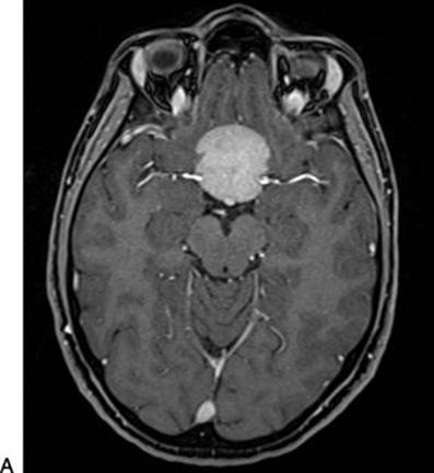
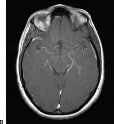


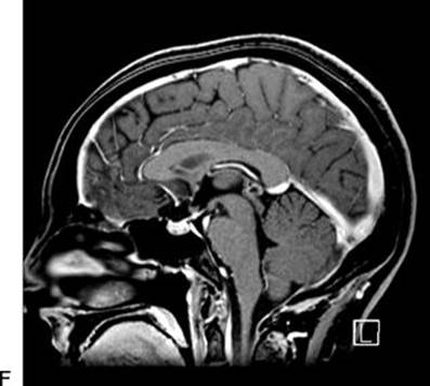
FIGURE 16.1 A–F. Preoperative (A, C, E) and postoperative (B, D, F) MRIs of a tubercular meningioma resected via a right supraorbital keyhole approach. The large size of the lesion and involvement of neurovascular structures were not contraindications to this keyhole approach. The postoperative MRI demonstrates a gross total resection.
Before surgery, patients for whom a supraorbital approach is selected should be counseled regarding the risk of numbness of the forehead, frontalis palsy, and nasal CSF leak although the rates of these complications are very low in experienced hands. In our series of over 450 cases, our CSF leak rate is negligible, and only two patients experienced permanent frontalis palsy.
SURGICAL TECHNIQUE
After intubation under general endotracheal anesthesia, the patient is placed in the supine position. The head is fixed in a 3-point rigid head holder. The table is placed in approximately 20 degrees of reverse Trendelenburg to optimize venous drainage. After pinning, the head is gently extended to bring it above the level of the heart, and 20 degrees of neck extension is applied to facilitate the natural “retraction” of the frontal lobe by gravity. This maneuver is crucial in opening the subfrontal corridor through which microsurgery will proceed and in avoiding the use of retractors. The head is then rotated slightly to the contralateral side to bring the malar eminence to the uppermost position. The degree of contralateral rotation may depend on the site of the surgical pathology, with 15 to 30 degrees used to address ipsilateral lesions and up to 45 to 60 degrees of rotation required to approach contralateral lesions. The ipsilateral eye is lubricated and closed with a temporary nylon tarsorrhaphy suture to prevent surgical prep solution from contacting the cornea.
Following positioning, the image-based frameless stereotaxic system is registered. Use of image guidance prior to incision is helpful to assess the position of the frontal sinus and to ensure an optimal trajectory to the lesion of interest. The lateral margin of the frontal sinus is marked, and the supraorbital notch is palpated. The supraorbital notch represents the medial limit of the skin incision. When the supraorbital notch is lateral to the frontal sinus, it also marks the medial limit of the craniotomy. In cases where a large frontal sinus extends lateral to the supraorbital notch, the lateral margin of the frontal sinus marks the medial limit of the craniotomy.
After prepping and draping, a skin incision is made within the eyebrow in its superior half (Fig. 16.2), extending from the supraorbital notch medially to the lateral aspect of the brow. The subgaleal layer is undermined, and the soft tissue is retracted superiorly with fishhooks. A U-shaped pericranial flap is fashioned by incising the pericranium as superiorly as possible and reflecting it inferiorly to the orbital rim. This pericranial flap is held inferiorly with sutures to the drapes. Laterally, the superior portion of the temporalis muscle is dissected to allow placement of a burr hole below the superior temporal line in the keyhole region. Any further temporalis dissection is unnecessary and should be avoided. A craniotomy is fashioned as low on the frontal floor as possible. The supraorbital craniotomy is typically 2 to 3 cm wide and 1.5 to 2 cm high (Fig. 16.3). While a small craniotomy can provide versatile access, the opening in the bone must be wide enough to accommodate a fully spread bipolar instrument. Particular lesions, such as those with a wide superficial component, may require a wider opening. Care is taken to preserve the supraorbital nerve medial to the craniotomy and to avoid violation of the frontal sinus. If the frontal sinus is breached, it can be packed with Betadine-soaked Gelfoam and sealed with bone wax, or repaired with the pericranial flap.

FIGURE 16.2 Proper positioning for the supraorbital craniotomy with eyebrow skin incision. The right eyelid temporary suture and skin incision, extended from the supraorbital notch to the lateral aspect of the brow (yellow line), are demonstrated in this photograph. The patient's head is extended to allow the frontal lobe to fall away. The degree of head rotation may vary, depending on the size and location of the lesion of interest. In this case, minimal contralateral rotation was used. Rotation of the operating table during surgery, however, allows for the angle to be tailored during the course of the procedure.
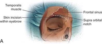


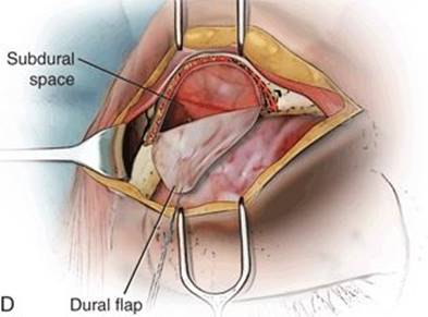
FIGURE 16.3 A. A skin incision is made within the eyebrow in its superior half, extending from the supraorbital notch medially to the lateral aspect of the brow. B. After the subgaleal layer is undermined and the soft tissue is retracted superiorly with fishhooks, a U-shaped pericranial flap is fashioned by incising the pericranium as superiorly as possible and reflecting it inferiorly to the orbital rim. C. The superior portion of the temporalis muscle is dissected and retracted laterally to allow placement of a burr hole below the superior temporal line in the keyhole region. Two osteotomies are made with a craniotome, both starting from the burr hole and meeting medially just lateral to the supraorbital notch. The inferior osteotomy is fashioned as low on the frontal floor as possible, flush with the orbital roof. D.After the inner cortex of the supraorbital bone is drilled, a U-shaped dural opening is made and reflected inferiorly to give access to the intradural space.
The dura is dissected off the roof of the orbit prior to opening the dura. Protuberances of the orbital roof are drilled flat, as is the inner table of the inferior ledge of the craniotomy; this maneuver is critical in maximizing visualization of the skull base. The dura is then opened in a U-shaped fashion with the base of the flap at the inferior aspect near the orbital rim (Fig. 16.4A). Immediately after opening the dura, a small arachnoid incision is made to initiate CSF drainage, while the surgical microscope is brought into position.

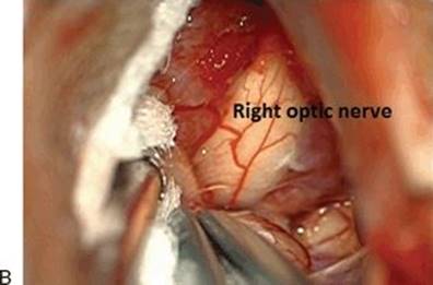


FIGURE 16.4 A–D. Intraoperative photographs of the case depicted in Figure 16.2. Following craniotomy and dural opening, the base of the frontal lobe is exposed (A). Identification of the ipsilateral optic nerve is a crucial first step as tubercular meningiomas tend to push the optic nerve superiorly (B). The involvement of the anterior cerebral arteries and ACOM complex are clearly demonstrated (C). Following tumor removal, both optic nerves and the pituitary stalk are clearly visible (R A1 = right proximal anterior cerebral artery) (D). This final photograph allows one to appreciate the anatomic reach of the supraorbital approach.
Without retractors, the subfrontal corridor is dissected under microscopic visualization. The ipsilateral optic nerve and carotid artery are identified. The arachnoid of the interchiasmatic, opticocarotid, and carotid–oculomotor cisterns, as well as the proximal Sylvian fissure, may be opened. With dissection of arachnoid adhesions and CSF egress, the frontal lobe falls away to further aid in the surgical approach. The pathology is identified and treated using the appropriate lesion-specific microsurgical techniques (Fig. 16.4B–D). Care is taken in assessing involvement and/or displacement of the optic nerves, the optic chiasm, the ipsilateral and contralateral carotid arteries, the anterior cerebral arteries and anterior communicating artery (ACOM), and the pituitary stalk. Neuroendoscopy with a 30-degree endoscope may augment visualization of the sella, interpeduncular cistern, interhemispheric cistern, contralateral circle of Willis, and middle cranial fossa, if necessary.
After the lesion of interest has been addressed, meticulous hemostasis is established. The dura is closed in a watertight fashion and sutured to the lower rim of the craniotomy. Adequate repair of a breached frontal sinus is confirmed. A noncompressive layer of Surgicel is placed in the epidural space, and the bone is replaced with low-profile titanium plates. Care is taken to minimize the gap between the skull and bone flap at the more cosmetically noticeable superior margin. Surgicel is placed in the remaining craniotomy line. The pericranium, frontalis muscle, and galea are closed in layers. The skin of the eyebrow is reapproximated with a subcuticular nylon suture with no knots.
Proper placement of this final skin suture is essential in obtaining a cosmetically acceptable result. The skin must be approximated under slight tension to ensure healing by primary intention. Prior to tying of the suture, its free ends are flossed back and forth to ensure that it can be freely removed in 5 to 7 days. The free ends of this suture are tied so that the skin edges are reapproximated under gentle tension. A nonabsorbable dressing is placed under the tied ends of the extradermal portion of the suture to prevent it from burrowing into the incision. Immediately after the skin is closed, gentle continuous pressure is held over the incision until the patient is extubated and breathing comfortably. This maneuver prevents formation of a pseudomeningocele that could be promoted by coughing or straining as the patient emerges from anesthesia. The skin suture is removed 5 to 7 days postoperatively.
 Video 16.1 demonstrates the surgical nuances of the case depicted in Figures 16.2 and 16.4. The right supraorbital craniotomy is drilled flush with the orbital roof. The subfrontal corridor is dissected without the use of retractors. The arachnoid of the ipsilateral opticocarotid cistern is opened with an 11 blade, and the surgeon patiently allows egress of CSF to occur. This step is critical in achieving adequate brain relaxation. The tumor extending from the tuberculum sellae along the planum sphenoidale is addressed. The lesion is devascularized along its base with bipolar electrocautery and resected with a combination of suction and sharp dissection. The involvement of the anterior cerebral arteries, ACOM complex, and both optic nerves is appreciated. Once the tumor is freed from these surrounding structures, a large portion is removed en bloc. Following complete removal, both optic nerves and the pituitary stalk are clearly visualized.
POSTOPERATIVE MANAGEMENT
The supraorbital keyhole approach is usually associated with minimal discomfort and a short hospital stay. In most cases, patients are discharged home on the 1st postoperative day. Some degree of periorbital edema is to be expected if the periorbita is violated during pericranial dissection or placement of the craniotomy. This generally resolves within 5 days. All patients should be assessed clinically for possible CSF leaks and, in cases of involvement of the pituitary stalk, endocrinopathies.
COMPLICATIONS
Aside from general surgical complications, the following complications are specific to the supraorbital approach:
· Numbness of the forehead (caused by injury to the supraorbital nerve)
· Frontalis palsy (caused by injury to the frontalis branch of the facial nerve)
· Nasal CSF leak (caused by violation of the frontal sinus and inadequate repair)
· Poor cosmetic appearance. This may be the result of poor skin closure, partial or full thickness burns from the surgical microscope set at greater than 75% intensity for any length of time, infection, or persistent pseudomeningocele.
Overall, because the supraorbital approach requires only a small skin incision with minimal temporalis dissection, scalp pain, swelling, and difficulty with mastication are observed with less frequency than with standard pterional or orbitozygomatic approaches.
RESULTS
I have operated on over 450 patients using the supraorbital keyhole approach in over 450 cases. This includes more than 430 tumors, eight aneurysms, and three cases of trauma to the anterior skull base. In no case was conversion to a larger craniotomy required. The frequency with which I use this approach is a testament to its versatility. My 0% incidence of conversion to a larger craniotomy demonstrates that, with proper selection and execution, the supraorbital keyhole does not compromise necessary anatomic access.
PEARLS
· Carefully assess preoperative imaging. Close inspection of the preoperative MRI is the most important step in properly selecting the supraorbital approach and ensuring that the lesion of interest can be reached.
· Extend the head to allow the frontal lobe to fall away. This simple positioning maneuver makes gravity work in the surgeon's favor. Mild extension of the head allows the frontal lobe to fall away, naturally opening the subfrontal corridor and obviating the need for retractors.
· Avoid the frontal sinus. The frontal sinus can be easily avoided with image-based frameless stereotaxis. Successful avoidance of entrance into the sinus eliminates the risk of nasal CSF leak.
· Position the craniotomy flush with the orbital roof. Because the supraorbital craniotomy is a keyhole approach, each millimeter of bone left superficially above the orbital roof disproportionately limits the surgical trajectory to deeper targets. The goal is to achieve a low anterior-to-posterior working trajectory along the base of the frontal lobe, directly along the roof of the orbit.
· Drill the roof of the orbit flat to achieve a flat trajectory along the orbital roof.
· Patiently allow CSF egress to relax the brain.
· In experienced hands, the anatomic limits can be pushed with the use of angled endoscopes, and a 30-degree endoscope may provide access to the sella, the contralateral circle of Willis, the interhemispheric fissure, and a portion of the middle cranial fossa.
· A watertight closure minimizes the risk of pseudomeningocele and offers additional protection against a nasal CSF leak.
· A cosmetic closure is of utmost importance. The following steps are key in performing an optimal cosmetic closure:
o Close with a subcuticular suture with no buried knots.
o Floss the skin stitch.
o Tie the loose ends under gentle tension.
o Place a nonabsorbable dressing under the tied ends.
o Hold pressure until the patient is extubated.
PITFALLS
· Inadequate review of the preoperative imaging or an incomplete understanding of the anatomic limits of the supraorbital approach may lead to inadequate surgical access.
· Breach of the frontal sinus can be avoided by identifying the lateral edge of the frontal sinus with image guidance prior to performing the craniotomy.
· Generally, the supraorbital nerve may be preserved by palpating the supraorbital notch to identify the medial limit of the skin incision.
· The risk of injury to the frontalis branch of the facial nerve is reduced by limiting the extent of the lateral incision and minimizing lateral dissection of the subcutaneous tissue.
· Pseudomeningocele may be avoided with a watertight closure of the dura and by holding pressure on the closed incision until after extubation.
INSTRUMENTS TO HAVE AVAILABLE
· “Matchstick” drill for leveling the orbital roof
· 30-degree endoscope to expand the field of view
· Angled instruments for endoscopic-assisted surgery
SUGGESTED READING
Paladino J, Pirker N, Stimac D, et al. Eyebrow approach in vascular neurosurgery. Minim Invasive Neurosurg 1998;41:200–203.
Reich R, Perneczky A. Ten year experience with the supraorbital subfrontal approach through an eyebrow skin incision. Neurosurgery 2005;57 (4 Suppl):242–255.
Teo C. Application of neuroendoscopy to the surgical management of craniopharyngiomas. Childs Nerv Syst 2005;21:696–700.
Brydon HL, Akil H, Ushewokunze S, et al. Supraorbital microcraniotomy for acute aneurysmal subarachnoid hemorrhage: results of first 50 cases. Br J Neurosurg 2008;2:40–45.
Little AS, Gore PA, Darbar A, et al. Supraorbital eyebrow approach: a less invasive corridor to lesions of the anterior cranial fossa, parasellar region, and ventral brainstem. In: Cappabianca P, et al., eds. Cranial, cranio-facial and skull base surgery. Italia: Springer-Verlag, 2010.