
Roy R. Casiano
INTRODUCTION
Although en bloc craniofacial resection is the traditional teaching in oncologic surgery of the anterior cranial base, there has been a growing acceptance of alternative minimally invasive transnasal endoscopic techniques. In select patients, the endoscopic approach for resection of the anterior skull base, when compared to the historical “gold standard” of external craniofacial resection, has been shown to be oncologically sound. This approach is not only less invasive and cosmetically appealing, obviating the need for a lateral rhinotomy or sublabial incision, but also more cost-effective in select patients.
HISTORY
The traditional anterior skull base resection involves removal of the entire anterior skull base bilaterally (Fig. 17.1A and B). In very select patients, a “hemi” anterior skull base resection, sparing one olfactory nerve and cribriform plate, has been performed (Fig. 17.1C). However, the latter is more controversial.
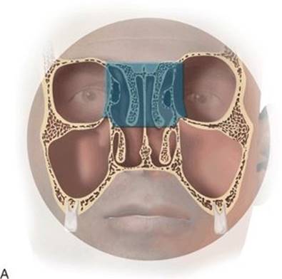
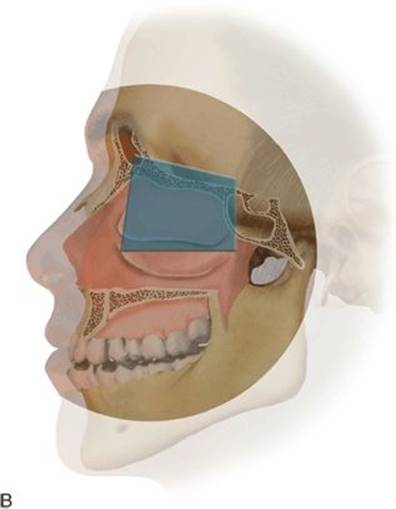
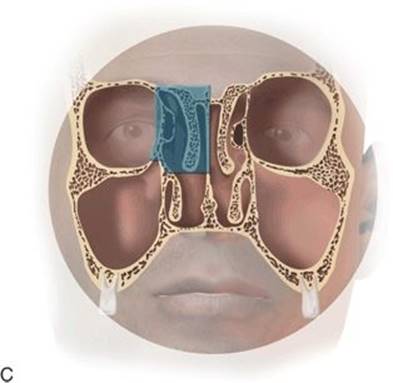
FIGURE 17.1 Coronal (A) and sagittal (B) images illustrating the anatomical limits of a more traditional anterior skull base resection. In select cases, a “hemi” (C) anterior skull base resection may be more appropriate, in order to spare an olfactory bulb.
The anterior skull base is limited laterally, by the medial orbital walls; by the planum or roof of the sphenoid bone, posteriorly; and by the posterior table of the frontal sinus, anteriorly. The midline structures of the anterior skull base include the crista galli and cribriform plates, which are lined intranasally by olfactory neuroepithelium that perforates the cribriform plate, allowing for the transmission of olfactory nerve fibers to the nasal cavity. Lateral to the cribriform plate lies the roof of the ethmoid sinus or fovea ethmoidalis.
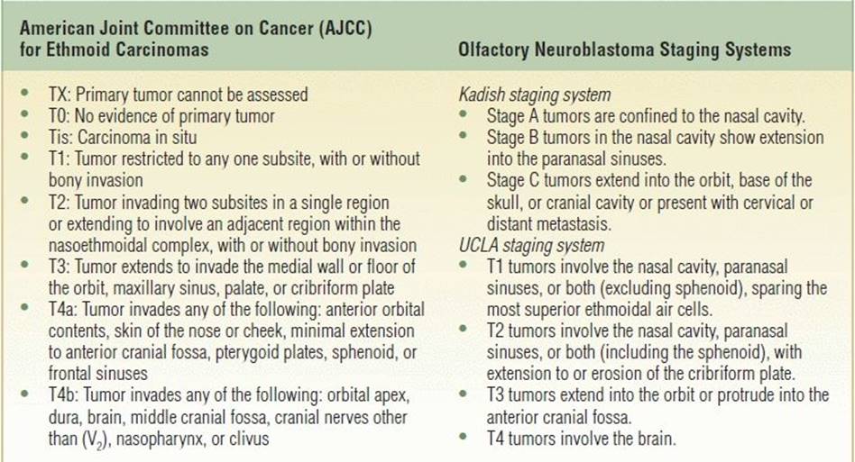
Depending on the stage and location of the neoplasm, resection of the anterior skull base may be required to obtain negative superior margins in the surgical management of many sinonasal malignancies (Tables 17.1 and 17.2). However, it is not necessarily the pathology but rather the extent of disease that determines the appropriateness for a purely endoscopic approach versus a craniofacial resection.
Table 17.1 Common Sinonasal Malignancies
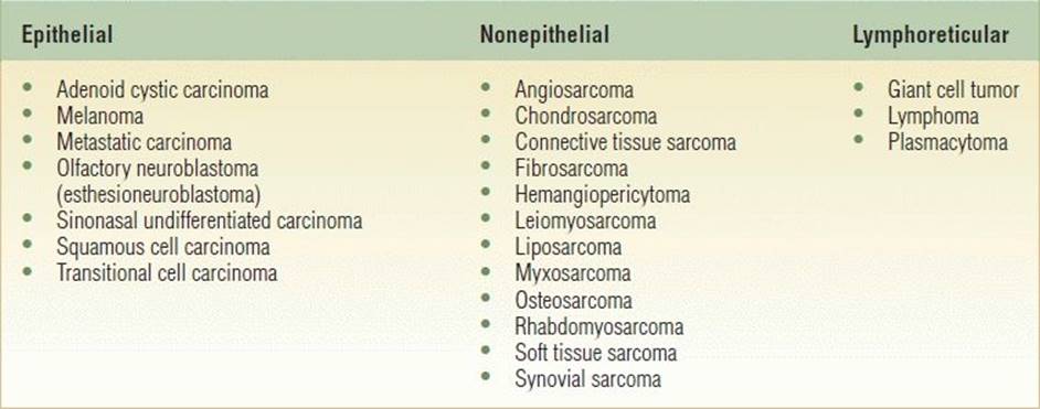
Table 17.2 Common Sinonasal Cancer Staging Systems
When confined to the nose, the most common early symptoms are unilateral nasal obstruction and/or epistaxis. These symptoms are nonspecific and usually confused with chronic rhinosinusitis. One must exercise a high index of suspicion in these patients. Depending on the type of pathology and/or the aggressiveness of the neoplasm, additional orbital or neurologic symptoms may ensue. These include proptosis, facial swelling, cranial neuropathy, ophthalmoplegia, visual loss, and/or mental status changes.
PHYSICAL EXAMINATION
Nasal endoscopy, and a complete examination of the head and neck, including cranial nerve assessment, should be performed on all patients. Physical examination may reveal proptosis; extraocular muscle impairment; mass effect of the cheek, gingiva, or gingivobuccal sulcus (e.g., ill-fitting dentures); and/or loose dentition. Numbness of the skin of the cheek or upper lip, or hypesthesia of the infraorbital (V2) branch of the maxillary nerve, strongly suggests malignant invasion of the nerves. Transnasal endoscopic biopsy of the lesion is commonly performed in the office under topical or local anesthesia, when possible. Alternatively, the sampling can be performed in the more controlled environment of an operating room when a deep biopsy is required, when the lesion is difficult to access due to its location (i.e., dependent sinus or skull base), or if profuse bleeding is anticipated.
INDICATIONS
Most benign and malignant neoplasms originating in the nose and paranasal sinuses, which are determined to be free of extensive orbital, intracranial, lateral maxillary, or palatal invasion, may be considered candidates for endoscopic anterior skull base resection as a stand-alone procedure. A combined external/endoscopic approach is otherwise indicated, during the same setting, or staged for a later date. Therefore, patients should be appropriately counseled and provided informed consent.
CONTRAINDICATIONS
Not all lesions are amenable to this technique, due to the extent of supraorbital, orbital, lateral maxillary, or palatal involvement. In such cases, the endoscopic-assisted external approach or more traditional craniofacial resection still affords many of the benefits of a purely endoscopic resection. Tumors in these locations may necessitate an orbital exenteration, a palatectomy, and/or a radical maxillectomy, respectively.
PREOPERATIVE PLANNING
Computed tomography (CT) scan and magnetic resonance imaging (MRI) are considered complementary to each other for the evaluation of neoplastic disease of the paranasal sinuses and adjacent skull base and orbit (Fig. 17.2). CT scan is superior for the evaluation of the bony architecture, assessing for bony erosion or remodeling in critical areas of the skull base and orbit. The use of contrast also reveals tumor vascularity and its relationship to the carotid artery. Disadvantages of CT include its inability to differentiate tumor borders from the surrounding soft tissue and exposure to ionizing radiation. MRI is the best modality for defining soft tissue detail. It can differentiate adjacent tumor from soft tissue (e.g., gadolinium enhances tumor diffusely to an intermediate degree, whereas inflamed mucosa enhances more intensely in a peripheral fashion), differentiate tumor from secretions in an opacified sinus, demonstrate perineural spread (especially adenoid cystic carcinoma), and demonstrate invasion of the dura, orbit, or brain parenchyma. Additional testing, to evaluate or embolize vascular structures (i.e., MRA or angiography), or to evaluate for metastatic disease (i.e., PET/CT), may be necessary in select cases.
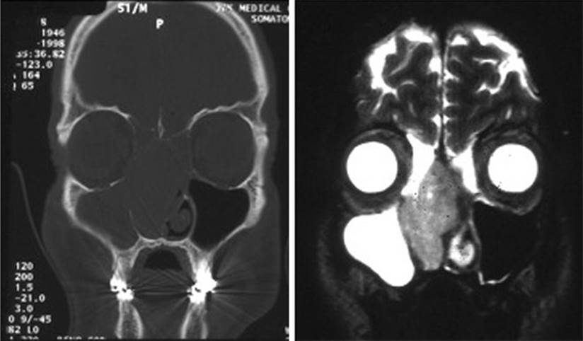
FIGURE 17.2 CT and MRI of an esthesioneuroblastoma. Obstructive changes are denoted by the high signal intensity in the maxillary sinus and frontal infundibular area, relative to the low signal intensity of the intranasal and ethmoidal tumor displacing the septum to the left.
Intrathecal fluorescein is not routinely used for elective skull base resections but may be instilled prior to taking the patient back to the operating room to assist the surgeon with skull base reconstruction. The absence of fluorescein along the margins of the reconstructed dural defect denotes absence of cerebrospinal fluid (CSF) leak. Also, intraoperative computer-assisted navigation is not used routinely for anterior skull base resections. However, it may be useful when resection margins are extended posteriorly around the carotid artery and adjacent neurovascular structures. If used, the patient would have placement of fiducials preoperatively and registration performed once the patient is intubated, prior to prepping.
SURGICAL TECHNIQUE (VIDEO 17.1)
Intraoperative Patient Positioning and Prepping
The patient is placed in the supine position on the operating table. Following intubation, the endotracheal tube is secured to the left side of the oral cavity. As with all endonasal procedures, a right-handed surgeon typically stands on the right side of the patient's head. When a four-handed technique is required, typically during the anterior skull base resection portion of the procedure, the assistant stands to the left of the operating surgeon. Both have simultaneous visualization of the video monitor(s). Ointment is placed in each eye, and each eyelid is secured in the closed position with a transparent dressing, in order to detect the presence of an expanding orbital hematoma. Oxymetazoline-soaked, one-half-by-three-inch, neurosurgical Cottonoids are placed in both nasal cavities. The head of the bed is then rotated 180 degrees. Placing the bed in reverse Trendelenburg helps to decrease venous congestion and intracranial pressure throughout the procedure, if necessary. The patient is prepped with Betadine paint and draped in a sterile fashion, including the nose and both eyes in the surgical field.
After removal of the oxymetazoline-soaked Cottonoids, a 30-degree rigid nasal endoscope is used for visualization and exchanged for a 70-degree rigid nasal endoscope when needed during the course of the procedure. A 70-degree endoscope is useful when inspecting the nasoseptal angle and anterolateral frontal and maxillary sinus margins. One percent lidocaine with 1:100,000 epinephrine is used to inject the septal mucosa, the attachment of the middle turbinate, and the area adjacent to the sphenopalatine foramen when exposure permits. However, depending on the size and extent of the lesion, endoscopic debulking of intranasal tumor may first be required.
Intranasal Tumor Debulking and Determination of the Tumor Epicenter
The surgery begins on the ipsilateral side of the tumor. A microdebrider is used to resect the intranasal portion of the mass, taking care to identify and preserve the point(s) of mucosal attachment and the tumor's “epicenter.” Although noncutting forceps can be used as an alternative, the microdebrider system has the added benefit of specimen collection into a filtration sock placed on the suction apparatus. Once the mass is adequately debulked, the contents can then be sent for confirmatory pathologic evaluation. The sock is changed as often as needed and labeled appropriately, based on anatomical site. It is not uncommon for the middle and superior turbinates to be involved in the disease process and therefore are removed during debulking. Even if the structure is grossly uninvolved, turbinate resection is performed in order to improve visualization of the anterior skull base. However, it is still sent as a separate specimen for permanent sections.
Tumor Mapping and Clearance of Sinonasal Margins
Once the site of origin or epicenter of the tumor is identified, a complete resection of all involved structures of the nose and paranasal sinuses is performed in a stepwise manner, sending separate surgical specimens for permanent section for the purpose of postoperative tumor mapping. This helps to differentiate which cavities or structures are involved with tumor versus inflammation. In most endoscopic anterior skull base resections, this typically involves sending 15 to 20 separate specimens for permanent section, each specifying a different and detailed anatomical area, bilaterally. During this portion of the procedure, close endoscopic inspection and frozen sections from critical areas, known to necessitate further external procedures, such as deep involvement of the periorbita, lateral involvement of the maxillary sinus bone, palatal involvement, pterygomaxillary space involvement, or extensive nasopharyngeal involvement, are determined. Prior to proceeding with the anterior skull base resection, the surgeon needs to decide during this portion of the procedure whether to proceed endoscopically, stage the procedure for a future date (endoscopic and/or external approach), or proceed with a combined external approach at this time.
Once the nasal patency is reestablished, a wide maxillary antrostomy is performed. The posterior fontanelle is entered posteriorly with a frontal curette and removed, along with the remaining soft tissue and bone of the middle meatus, all the way anteriorly to the convexity of the nasolacrimal duct. Conversely, if the uncinate process is detectable, then a standard antrostomy is created anteroposteriorly in the standard fashion, starting at the maxillary natural ostium. Any areas suspicious for neoplastic involvement are sent for frozen section. In the absence of lateral involvement of the maxillary sinus mucosa and bone, the procedure begins with a total radical sphenoethmoidectomy with exenteration of all mucosal elements. When the inferior turbinate or medial wall of the maxillary sinus appears to be involved with tumor, then an endoscopic medial maxillectomy is performed, including resection of the nasolacrimal duct. Once the total ethmoidectomy is completed, the sphenoid sinus is entered and the anterior wall removed, incorporating the sphenoid cavity into the total ethmoid cavity. A Kerrison rongeur is used to widen the osteotomy, initially in the medial and inferior directions, until the underlying pneumatized sinus is visualized. Mucosal elements are exenterated and sent for frozen section. The roof of the sphenoid sinus, or planum sphenoidale, indicates the vertical level of the anterior skull base at its most posterior aspect and is closely inspected. Following along the continuation of the skull base in a posterior-to-anterior fashion, the roof of the ethmoid or fovea ethmoidalis is carefully inspected as well. Any residual bony ethmoid septations not destroyed by the disease process are then resected flush to the medial wall of the orbit and ethmoid fovea. Abnormal pitting of bone, bone erosion, or abnormal looking dura is noted throughout this endoscopic skull base exposure part of the procedure.
With a curved ostium seeker or probe, the infundibulum of the frontal sinus is identified, and the septations of the agger nasi cell are then fractured anteroinferiorly and removed. The lateral wall of the nose over the nasolacrimal sac is inspected at this point and if suspicious, removed for frozen section. Before proceeding with the extended frontal and sphenoid sinusotomies described below, a contralateral sphenoethmoidectomy and frontal sinusotomy are performed, thereby creating a pathologic map of the contralateral side.
Medial and anterior enlargement of the frontal sinus ostium is then undertaken using bone curettes and angled cutting burs, and a Draf III (modified Lothrop procedure) is performed, demarcating the anterior margin of resection (Fig. 17.3). Additional burring of the nasal side of the nasal bone is performed, if tumor extends close to this area. The lateral margin of resection on each side can be extended to include the lamina papyracea and/or periorbita, as dictated by the extent of the lesion. A superior septectomy (including bone and cartilage) is performed as far inferiorly as necessary to clear the margins. This may include extending the septectomy to the nasal floor. A caudal and dorsal strut of septal cartilage can usually be preserved, minimizing the chance of dorsal nasal collapse and subsequent saddle nose deformity.
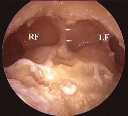
FIGURE 17.3 Completed modified Lothrop (Draf type III frontal sinusotomy), delineating the anterior margin of an anterior skull base resection. Left (LF) and right (RF) frontal sinus cavities are examined. Small arrows denote the remnant of the frontal intersinus septum.
An extended sphenoid sinusotomy is performed (Fig. 17.4). This includes removal of the sphenoid rostrum, intersinus septum, and exposure of the anterior planum or roof of the sphenoid sinus in the midline. The nasopharyngeal and sphenopalatine mucosal margins are sampled at this point and sent for frozen section.
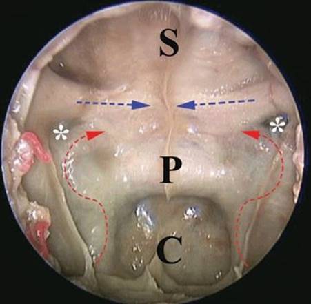
FIGURE 17.4 Completed extended sphenoid sinusotomy after removal of the rostrum and sphenoid intersinus septum and superior septectomy. Sphenoid planum or roof (P); sella turcica (S); clivus (C); opticocarotid recess (asterisk). Dotted curved arrows denote the course of the intrasphenoidal internal carotid artery. Dotted straight arrows denote the optic nerves coursing through the canalicular portion of the optic canal toward the optic chiasm.
This completes the exposure of the anterior skull base. As mentioned previously, up to this point, the procedure can be staged for a later date without the need for reconstruction or proceed with the anterior skull base resection part of the procedure below. Regardless, all sinonasal and septal margins must be cleared pathologically prior to proceeding with the anterior skull base resection of the procedure below.
Anterior Skull Base Resection
Although neurosurgical support should be available from this point on, neurosurgical involvement for most anterior skull base cases is rarely used, unless extensive dural or parenchymal invasion is encountered. An ultrasonic bone curette or aggressive diamond burr is used to thin the bony margins of the anterior skull base (i.e., fovea ethmoidalis, anterior planum, and posteroinferior frontal sinus posterior table). Once eggshell thin, any remaining bone is gently mobilized from the overlying dura, removed, and sent for permanent pathologic evaluation. In order to facilitate reconstructive efforts and further dural resection (if necessary), the dura overlying the orbits is gently elevated at this time with a neurosurgical Cottonoid. After cauterizing the anterior and posterior ethmoid arteries with a monopolar suction cautery at 10 W of energy, the dura is incised with a sickle knife and resected in a four-hand technique with careful suction traction using endoscopic scissors, starting as far laterally as possible, but leaving a cuff of dura for frozen section, circumferentially. Alternatively, a small tru-cut forceps may be used to resect and sample the dura simultaneously, therefore assuring clear margins on frozen sections, while the resection continues below. Dural incisions are begun over the fovea first and then connected anteriorly over the posterior wall of the frontal sinus. As the incisions are created, the specimen is retracted inferoposteriorly into the nasopharynx, allowing visualization of the overlying intracranial vasculature and brain parenchyma. Any bleeding intracranially from dural vessels is controlled with bipolar cautery. Venous bleeding may also be controlled with gentle pressure for a minute or two minutes using a neurosurgical Cottonoid and powdered Gelfoam. Superiorly, the specimen must be separated from the crest of the crista galli and falx cerebri. The crista galli can be thinned with the ultrasonic bone curette, or burr, and down-fractured as high as possible. The falx cerebri is incised bilaterally, over the crista galli, and the specimen is slowly dissected free in a posterior direction. The final dural cuts over the planum sphenoidale, as well as transection of both olfactory nerves, are performed at this time, allowing the specimen to drop into the nasopharynx. At this point, the anterior skull base specimen is removed en bloc through the nostril (Fig. 17.5). If the olfactory neuroepithelium is involved with the disease process (i.e., esthesioneuroblastoma), the olfactory nerves are sampled and sent for frozen section to be certain that this margin is clear.
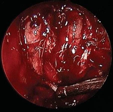
FIGURE 17.5 Completed anterior skull base resection and skull base defect with brain parenchyma and cut edge of falx cerebri. Suction tip is on the cut end of the right olfactory nerve.
Anterior Skull Base Reconstruction
Once the dural and intracranial margins are negative, reconstruction of the skull base defect may begin. Inability to adequately clear the intracranial dural margins requires neurosurgical intervention with possible conversion to a combined craniofacial resection. Reconstruction is performed with hydrated acellular dermal graft of approximately 1 mm average thickness (Alloderm, LifeCell Corporation, Branchburg, NJ), placed to span the dural defect of the anterior skull base circumferentially, like a hammock (Fig. 17.6). It is not important whether the graft is placed epidurally, subdurally, or both. What is more important is that a large enough sheet of graft material is available and placed intracranially as described below. Typically, one can measure the anteroposterior and lateral (superior orbit to orbit) size of the skull base defect and add at least 2 cm circumferentially, for the final graft size. That would be a total of 4 cm on top of the final defect side, for the anteroposterior, as well as the lateral, dimensions. For example, if the final skull base defect ranges from 3 cm orbit to orbit, by 4 cm anteroposteriorly, then a 7 cm × 8 cm sheet of graft material is used. It is critically important that the edges of the graft are infolded onto itself circumferentially, so that an intranasal portion overlies the de-epithelialized orbital, planum, and posterior frontal bony margins (Fig. 17.7). In order to maintain graft position around the periphery and prevent intracranial or intranasal migration, 1 to 2 cm2 pieces of moistened compressed gelatin sponge (Gelfoam, Pfizer, New York, NY) are then placed into the pocket created by this infolding, circumferentially, over the orbital roof, planum sphenoidale, and posterior table of the frontal sinus. This step helps to tightly seal the defect, reducing the risk of postoperative CSF leak and pneumocephalus. The Gelfoam is further dehydrated and firmed up by tucking it into the pocket using a curved suction tip over a neurosurgical Cottonoid. The remaining graft spanning the central defect is then overlaid with moistened compressed gelatin sponge and secured with two polyvinyl alcohol–compressed nasal packs (Merocel, Medtronic Xomed, Jacksonville, FL), suspended between the common frontal and sphenoid sinus cavities, to provide counter pressure against brain parenchyma and restored CSF pressure levels in the immediate postoperative period (Fig. 17.8).
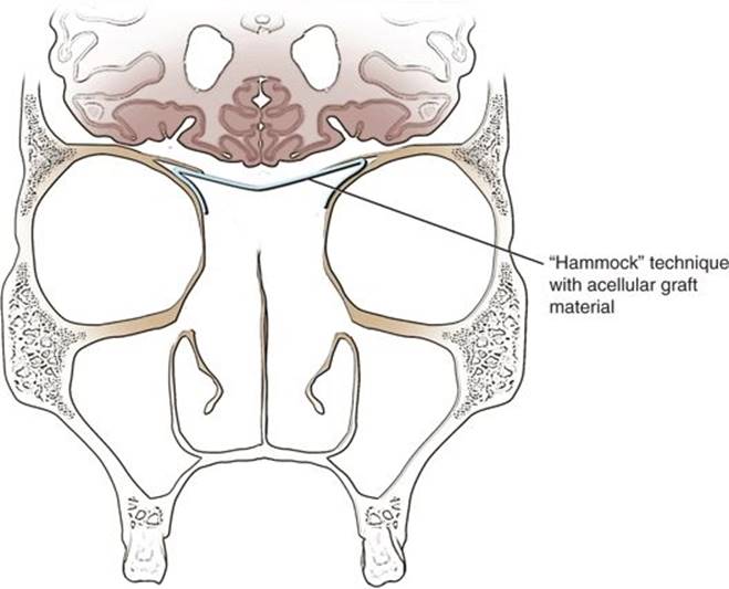
FIGURE 17.6 “Hammock” technique for anterior skull base reconstruction with acellular dermal graft or lyophilized dura.
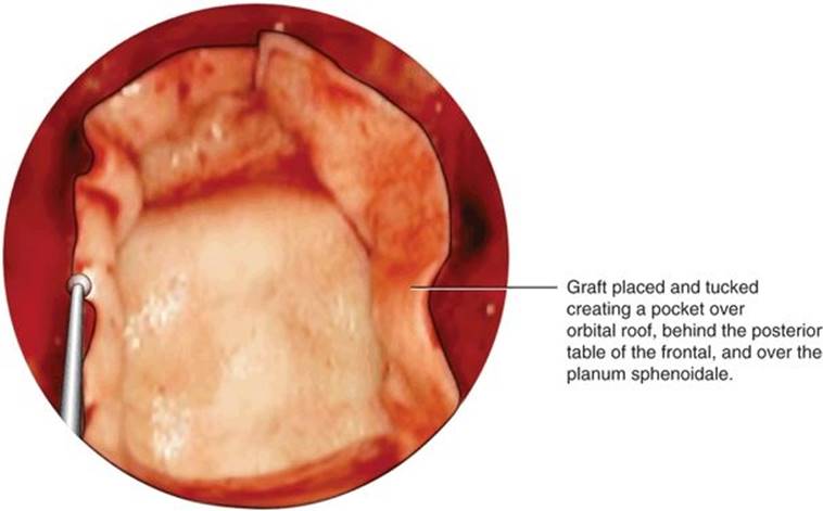
FIGURE 17.7 Acellular dermal graft placed into the defect and tucked circumferentially, creating a pocket that is filled with Gelfoam. The free nasal edge of the graft is seen circumferentially over the medial orbital wall, posterior edge of the common frontal sinusotomy, and bony edge of the planum defect. Care is taken not to obstruct the common frontal opening with the graft.
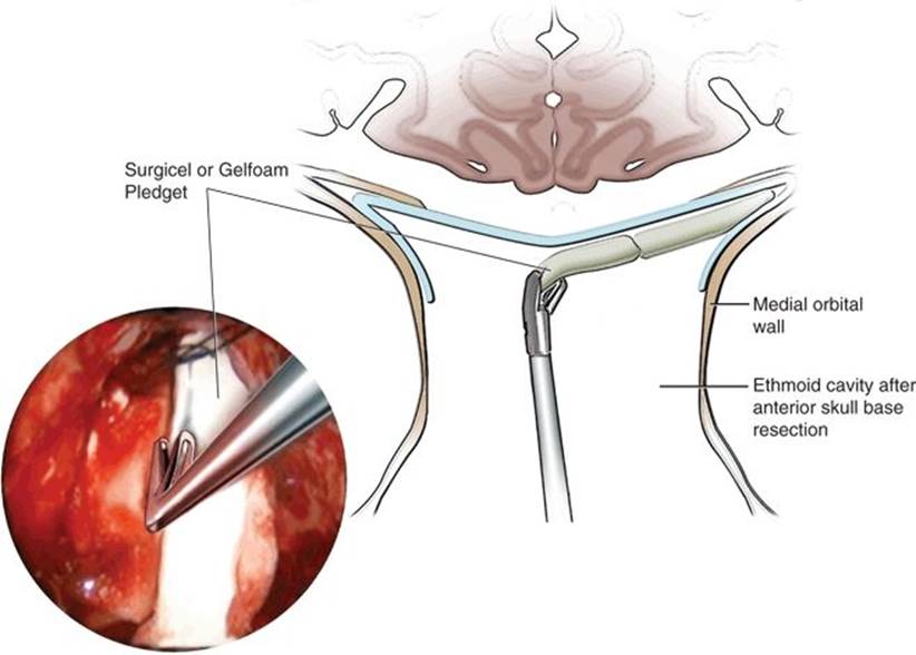
FIGURE 17.8 Merocel nasal tampon placed over the Gelfoam bed overlying the graft, between the common sphenoid and frontal sinus cavities.
POSTOPERATIVE MANAGEMENT
Lumbar drains are not used routinely. The patient is kept in a monitored bed overnight, before being transferred to a regular bed. The presence of CSF leak is checked daily by having the patient sit up in bed and then gently bend forward, without straining. Any clear watery fluid emanating from the nostril(s) is noted. The patient is monitored for any mental status or visual changes, along with routine vital signs. In the absence of fever, CSF leak, mental status change, or visual changes, the patient typically is discharged from the hospital by the 3rd postoperative day. A return clinic appointment is given for approximately 7 days postoperative.
Postoperative prophylactic antibiotics are prescribed until packing removal in the office approximately 1 week after surgery. However, the underlying layer of Gelfoam is left undisturbed and left to fall off over time and with the help of irrigations. Nasal irrigations with isotonic saline are begun at this time. The patient is instructed to refrain from straining, and if indicated, use stool softeners. Pain medication is prescribed as needed.
Prolonged crusting is inversely proportional to the degree and rapidity of mucosalization of the sinonasal and anterior skull base cavities. Mucosalized cavities crust less than do cavities that need to heal by secondary intention and formation of granulation tissue. Prolonged crusting of the reconstructed skull base area may be a slight disadvantage with acellular dermal graft (Alloderm) or lyophilized dura, as compared to mucosalized grafts or flaps. However, the ease of repair, and rapid availability of these grafting materials, along with their consistent results in providing excellent closure of the anterior skull base defect, outweighs this minor issue in our hands. Also, donor site crusting may still occur in cases where pedicled mucosal flaps are used. Periodic endoscopic debridements, daily postoperative irrigations, and culture-directed topical and oral antibiotics, as needed, help mitigate the crusting if it occurs. Radiation therapy, when indicated, may be begun within 4 weeks after surgery. Complete remucosalization may be expected within 6 months (Fig. 17.9).
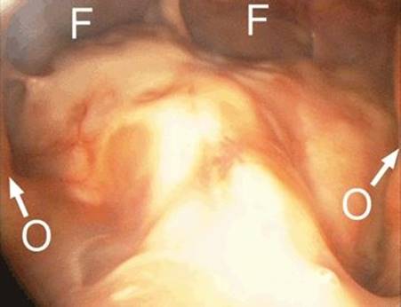
FIGURE 17.9 Mucosalized anterior skull base approximately 1 year postoperatively. Common frontal sinus cavity (F) is inspected. Medial orbital walls (O, arrows) are also in view.
As with all oncologic patients, postoperative long-term endoscopic and radiologic follow-up is imperative. At a minimum, patients are followed every few months, with a repeat brain MRI at least once per year, or sooner if symptoms warrant (Fig. 17.10). Follow-up CT is used to address any symptomatic obstructive sinus disease. The management of symptomatic chronic sinonasal obstructive disease is identical to that for patients with chronic rhinosinusitis.
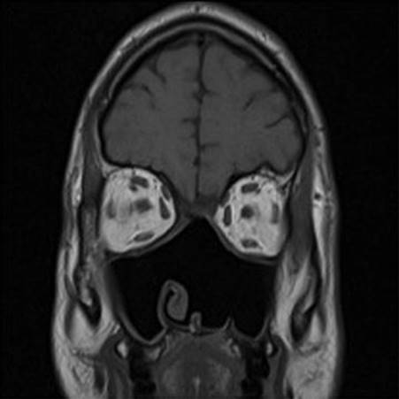
FIGURE 17.10 Postoperative MRI 3 years after endoscopic anterior skull base resection for an esthesioneuroblastoma. Radiation therapy was started 1 month after surgery. This patient also had a concomitant endoscopic left medial maxillectomy.
COMPLICATIONS
The most common reported complications of endoscopic anterior skull base resection are CSF leak and epistaxis. Sinus obstruction due to fibrosis, crusting, or mucosal thickening may occur at any point and can account for persistent inflammatory disease, discolored mucous, headaches, and crusting. Altered mental status, hypoesthesia, orbital hematoma, diplopia, loss of vision, cellulitis, vestibular stenosis, and deep vein thrombosis have also been documented. However, these are more common in patients undergoing the more traditional craniofacial resection.
RESULTS
The endoscopic approach, when compared to craniofacial resection, has been demonstrated to be both safe and effective with comparable rates of operative complications and overall survival in select patient populations. Endoscopic techniques do not impact one's ability to obtain negative surgical margins. Decreased overall morbidity and shorter hospital stays are routine. An added benefit of the technique is the dynamic quality and superior visualization afforded by angled nasal endoscopes, thus improving the accuracy of tumor resection while sparing uninvolved vital structures.
PEARLS
· The care of each patient is individualized based on the stage of neoplastic disease and the specific histopathology.
· Intraoperative navigation and intrathecal fluorescein is not used routinely for anterior skull base resection and is usually unnecessary for anterior skull base resections, unless significant resection is extended posteriorly into the middle or posterior cranial fossa, where identification of the carotid artery and adjacent neurovascular structures is critical.
· Meticulous intraoperative mapping and intraoperative frozen sections are imperative, not just to determine the extent of disease and clearance of the resection margins but also to determine close or unresectable margins. This helps guide postoperative adjuvant radiation therapy protocols or allows for further surgical planning at the same sitting or as a staged surgical procedure (endoscopic or external).
· When using Alloderm for reconstruction of the anterior skull base
o Use 1 mm thickness. Too thin may not give enough support. Too thick is not pliable enough to create the circumferential “pocket” over the orbit, posterior table of the frontal sinus, and planum sphenoidale.
o The pocket must extend at least 1 cm circumferentially.
o Make sure to tuck sufficient Gelfoam into the circumferential pocket created by infolding the graft back on itself. This keeps the infolded part firmly against the bone and allows the cranial portion of the graft to move superiorly or inferiorly. The intranasal edge of the pocket must clearly be visible after placement of Gelfoam into this pocket.
PITFALLS
· Tucking the graft circumferentially without creation of a pocket risks superior displacement particularly if there is low intracranial pressure, brain atrophy, or CSF levels are slow to reestablish in the immediate postoperative period.
· It is not necessary to use free mucosal grafts or flaps to cover the graft in the majority of the cases. Alloderm becomes quickly replaced with granulations, followed by mucosalization within months. However, this is at the expense of increased crusting in the postoperative period.
INSTRUMENTS TO HAVE AVAILABLE
· Standard endoscopic sinus surgery trays
· Powered instrumentation
· Ultrasonic bone emulsifiers
· Long fine endoscopic skull base scissors, forceps, probes , suction tips, tissue dissectors and bipolar grasping forceps
SUGGESTED READING
Folbe A, Herzallah I, Duvvuri U, et al. Endoscopic endonasal resection of esthesioneuroblastoma: a multicenter study. Am J Rhinol Allergy 2009;23(1):91–94.
Mantravadi A, Zender C. Craniofacial approaches to the anterior skull base. Oper TechOtolaryngol 2010;21:181–187.
Casiano RR, ed. Endoscopic sinonasal dissection guide. New York, NY: Thieme Medical Publishers, 2012.
Tessema B, Eloy JA, Folbe AJ, et al. Endoscopic management of sinonasal hemangiopericytoma. Otolaryngol Head Neck Surg 2012;146(3):483–486.
Wood JW, Eloy JA, Vivero RJ, et al. Efficacy of transnasal endoscopic resection of malignant anterior skull base tumors. Int Forum Allergy Rhinol 2012;2(6):487–495.