
Aldo C. Stamm
INTRODUCTION
Angiofibroma is a rare benign tumor making up only 0.05% of tumors of the head and neck. It typically affects young males, between 10 and 24 years of age, with an incidence between 1:5,000 and 1:60,000. First described by Chaveau in 1906, angiofibromas originate from the sphenopalatine foramen and may occupy the pterygopalatine space and the infratemporal fossa. As the tumor enlarges, it may extend to the middle fossa via various neurovascular foramina (i.e., the carotid canal, jugular foramen, foramen spinosum, foramen ovale, and foramen lacerum) and extend to the orbit via the inferior orbital fissure.
Histologic studies revealed myofibroblasts as the cells of origin. Fibrous connective tissue with abundant endothelium-lined vascular spaces, a pseudocapsule of fibrous tissue and blood vessels lacking a complete muscular layer are other histologic features.
Although benign and slow growing, these tumors are locally aggressive and cause symptoms of nasal obstruction and epistaxis in 63% of patients.
Surgery following embolization is the standard treatment of angiofibroma. Depending upon the size, extension, and structures involved, the tumor can be removed by either an open or endoscopic approach (Fig. 29.1).
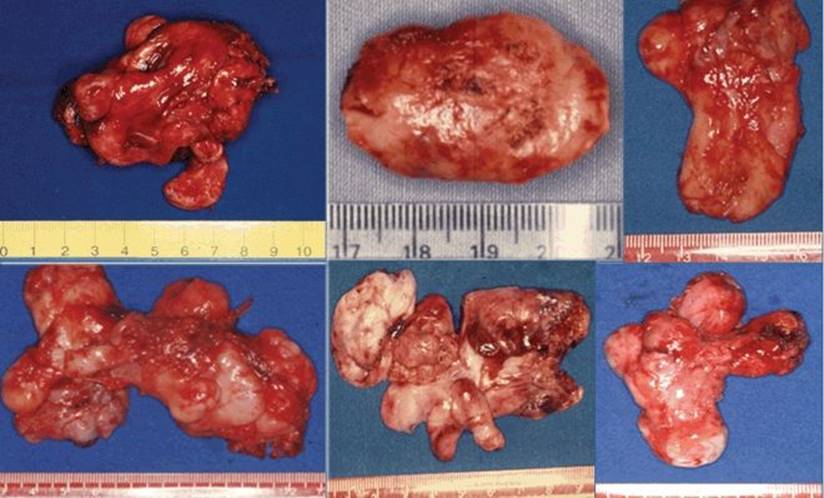
Figure 29.1 Angiofibromas with different size and configurations.
Until the 1980s, the surgical technique for removal of tumors involving the infratemporal fossa involved only external (preauricular, postauricular (transtemporal), anterior transfacial, transorbital) approaches.
With the improvement in endoscopic techniques and angiography with embolization, a large number of angiofibromas, especially the early stages, are being removed endoscopically.
HISTORY
The main clinical presentation is unilateral nasal obstruction in 91% and epistaxis in 63% of patients. Headaches and facial pain may be present secondary to the blockage of the paranasal sinuses. Compression of the Eustachian tube orifice in the fossa of Rosenmüller produces middle ear effusion with a conductive hearing loss. Other related symptoms include facial deformity and ocular symptoms of proptosis, diplopia, and, less often, visual loss.
PHYSICAL EXAMINATION
Nasal endoscopy reveals a smooth lobulated mass in the nasopharynx or lateral nasal wall: pale, purplish, red-gray, or beefy red (Fig. 29.2). Although compressible, it is important to avoid manipulating the tumor, due to the high risk of bleeding. Patients in more advanced stages may have proptosis, diplopia, and facial deformity.
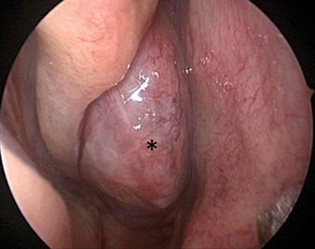
Figure 29.2 Endoscopic view of an angiofibroma (asterisk) in the right nasal cavity.
INDICATIONS
Surgical excision, with or without preoperative embolization, is the primary treatment option for angiofibromas. Before choosing the approach, the tumor must be evaluated based on the classification proposed by Andrews and modified by Fisch (Table 29.1).
Table 29.1 Andrews’ Classification (Modified Fisch) of Angiofibromas
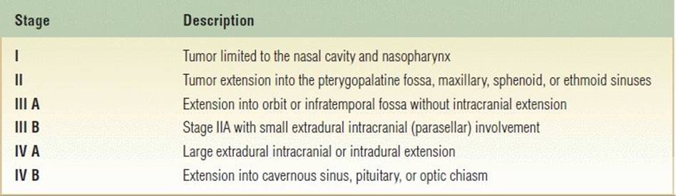
Lesion Grades I, II, IIIA, and IIIB can be resected through an endoscopic transnasal approach. This technique in experienced hands has the advantage of decreasing intraoperative blood loss, reducing hospital stay, and producing equal or reduced recurrence rates compared with open approaches. The use of the endoscope may be difficult depending on the lateral extension, mainly to soft tissues of the zygomatic region and cheek.
The open surgical treatment is most frequently reserved for grade IV angiofibroma. A great number of open approaches have been described (transpalatal, lateral rhinotomy, midface degloving, medial maxillectomy, transantral, infratemporal fossa, and frontotemporal craniotomy). I often use the midface degloving approach under microscopic visualization since it does not produce external scars.
CONTRAINDICATIONS
In patients who present with encasement of the internal carotid artery or cavernous sinus extension and blood supply from branches of the ICA, a gross total removal by the endoscopic and even the open approach may be more difficult.
PREOPERATIVE PLANNING
Preoperative imaging includes CT and MRI scans. The bone window of the CT scan without contrast shows classic signs such as enlargement of the anteroposterior diameter of the sphenopalatine fissure, anterior bowing of the posterior maxillary wall (Holman-Miller Sign), and erosion of the basisphenoid in the axial view and provides the bony landmarks for the surgery. Soft tissue window with contrast demonstrates a lesion with homogenous enhancement (Fig. 29.3). CT scans are also commonly used for intraoperative navigation to confirm the extent and resection of tumor. MRI with gadolinium is important in assessing tumor enhancement, intralesional features, and the relation of the tumor to critical structures such as the internal carotid artery, cavernous sinus, periorbita, dura mater, and pituitary gland and its extension. Moreover, it helps in differentiating tumor from secondary chronic rhinosinusitis.
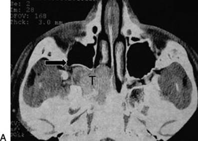
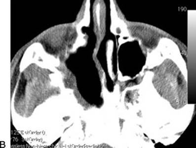
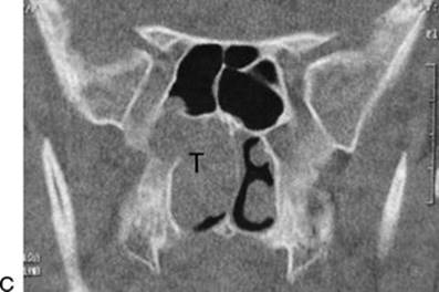
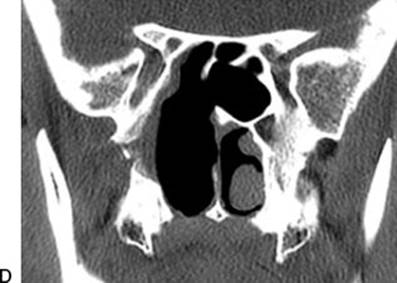
Figure 29.3 CT scan with contrast. A. Axial scan showing a heterogeneous tumor (T) enlarging the pterygopalatine fossa (Holman-Miller Sign) (arrow). B. Postoperative axial view. C. Coronal scan with tumor (T) protruding into the right sphenoid sinus. D.Postoperative coronal showing no residual tumor.
The diagnosis is made on clinical grounds of history, physical diagnosis, and imaging studies. Preoperative biopsy is unnecessary and not recommended due to the risk of catastrophic hemorrhage. Laboratory investigation including blood typing and preparation of fresh blood for transfusion is always indicated.
Because it is a highly vascular tumor, the embolization is indicated between 24 and 48 hours before surgery. Several materials may be used such as Gelfoam, Polyvinyl alcohol, or Onyx, a liquid embolic agent that allows deep penetration into the lesion, producing extensive tumor infarction. Embolization significantly reduces intraoperative blood loss and consequently minimizes the need for blood transfusion. Furthermore, endoscopic visualization is improved, providing more complete tumor removal. The maxillary and the ascending pharyngeal arteries are the most frequent vessels embolized (Fig. 29.4). Depending on the location, especially in advanced grades with intracranial and cavernous sinus involvement, branches from the internal carotid artery can provide high blood flow to the tumor. The embolization of these vessels can be dangerous, and in the majority of the cases is contraindicated.
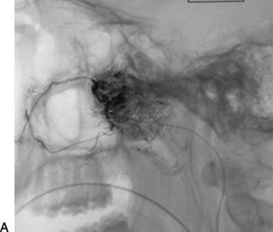

FIGURE 29.4 A. Angiography with the guide catheter inserted in the right maxillary artery showing a characteristic tumor blush. B.Postembolization with considerable reduction of tumor blood supply. The arrow indicates the occluded vessel.
SURGICAL TECHNIQUE
Microscopic Midface Degloving
Under general anesthesia, with the patient in the supine position the procedure begins with a bilateral incision in the gingival sulcus, as in a conventional Caldwell-Luc procedure. A complete transfixion incision of the membranous septum extended around the piriform aperture to the space between the superior and inferior lateral cartilages is made. The soft tissues of the nasal dorsum are then elevated in a subperichondrial and subperiosteal plane by using an elevator and Metzenbaum scissors. The remaining connections between the columella and the anterior nasal spine are dissected transnasally, joining the nasal cavity to the sublabial incision. The periosteum is then elevated, exposing the anterior maxillary wall, the ascending branch of the maxilla and the piriform fossa. The degloving approach is then completed by elevating the soft tissue of the upper lip, nasal dorsum, and superior maxillary region, thus exposing the bony structures of the middle third of the face (up to the infraorbital foramen leaving the infra orbital nerve intact and the infraorbital rim) (Fig. 29.5).

Figure 29.5 Midface degloving approach.
An ipsilateral wide resection of the anterior wall of the antrum is performed, leaving the infraorbital opening and its contents in place. The next step is the opening of the posterior wall of the maxillary sinus. Depending on the size of the tumor, the wall has the consistency of an eggshell, and, in the other cases, it may not be present.
The surgical microscope is then brought into the field in order to facilitate the ligation and section of the vascular pedicle of the tumor in the pterygomaxillary fossa. It is important to not touch the tumor until all the exposure is completed. The entire medial nasal wall is opened through a posterior and inferior detachment of the inferior turbinate that can be kept anterior until the end of the procedure or totally resected. The middle turbinate is displaced superiorly, increasing the visualization of the tumor.
An ethmoidectomy is done and the sphenoid sinus is opened, taking care to expose and resect the sphenoid rostrum, to allow visualization of the basisphenoid bone, which is one of the most important areas of the tumor’s origin.
After an entire exposure, using forceps, suction tubes, and bipolar electrocautery, the tumor is dissected free from the mucosa of the posterior wall of the nasopharynx, the mucosa of the posterior third of the nasal septum, the dura mater (if involved), and the basisphenoidal area of origin.
The tumor is removed, and a final look is done in order to avoid leaving some tumor remnants. It is important to drill the infiltrated surface of the basisphenoid bone with a diamond burr.
Finally, after hemostasis, the middle and inferior turbinates are sutured to the periosteum of the inferior orbital border (Fig. 29.6). The sublabial incision is sutured, and the surgical cavity is carefully packed. In Figure 29.7, is an example of a stage IVA angiofibroma resected by microscopic midfacial degloving.
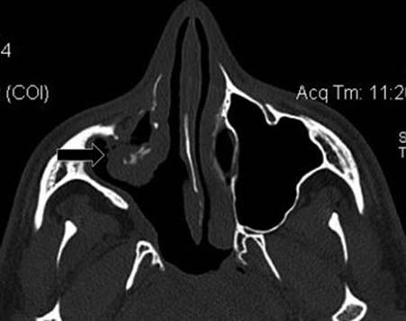
Figure 29.6 Postoperative axial CT view. Note the inferior right turbinate sutured to the orbital border (arrow).
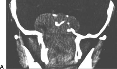
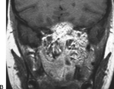
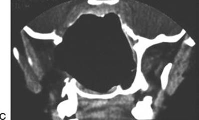
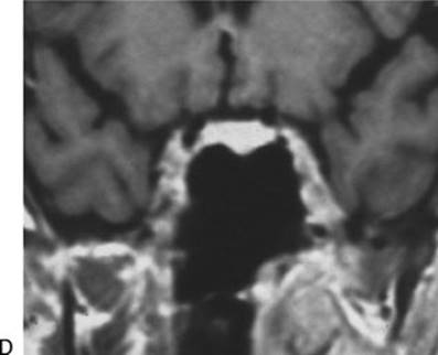
FIGURE 29.7 A and B. Coronal CT and MRI scans showing a stage IVA angiofibroma. C and D. Postoperative scans with complete resection using microscopic midface degloving approach.
Transnasal Endoscopic
The surgery is performed under hypotensive general anesthesia. The patient is placed in a supine position on the operating table, with the head elevated 30 degrees and with the neck slightly flexed and the head turned toward the surgeon. High concentration adrenaline-soaked cottonoids (1:1,000) are placed in the nasal cavity and over the tumor for 10 minutes before the surgical procedure begins.
The procedure begins with a wide middle meatus antrostomy to give maximal exposure of the posterior wall of the maxillary sinus. Then an anterior and posterior ethmoidectomy is performed. To avoid bleeding during the exposure and access, it is important not to inadvertantly touch the tumor. The bone of the posterior wall of the maxillary sinus is removed, exposing the pterygopalatine fossa, which is enlarged by the tumor, enabling the surgeon to control the internal maxillary artery which can be coagulated with bipolar forceps or clipped (Fig. 29.8A and B).
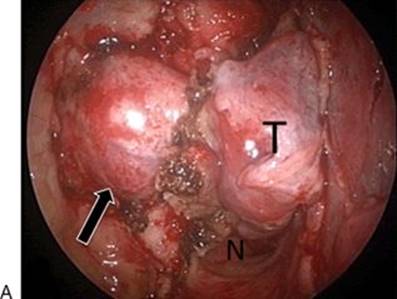
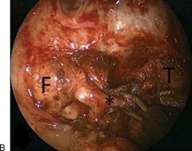
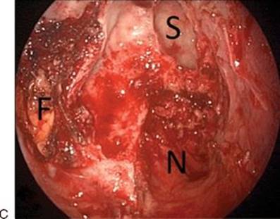
FIGURE 29.8 A. Complete tumor (T) exposure after a wide middle meatus antrostomy and removal of the posterior wall of maxillary sinus, remodeled by tumor (arrow). B. Bipolar coagulation of right maxillary artery (asterisk). C. Endoscopic view of the right nasal cavity after complete tumor removal. (N, nasopharynx; T, tumor; F, adipose tissue in the pterygopalatine and zygomatic fossa; S, sphenoid sinus.)
An anterior mucosal septal incision contralateral to the side of the tumor and another more posterior ipsilateral incision can be done allowing a binostril technique for four hands. At the end of the procedure, the incision is sutured to avoid postoperative septal perforation.
After the exposure, the tumor in the pterygomaxillary and infratemporal fossae is dissected using blunt instruments and bipolar cautery. To increase the mobilization of the tumor and free it from the pterygoid muscles and the infratemporal fossa adipose tissue, the base of the pterygoid bone must be drilled out.
The tumor is mobilized by separating it from the choana and posterior septum, then the nasopharyngeal portion is removed using bipolar cautery and surgical scissors, freeing it from the posterior pharyngeal wall and the prevertebral muscles (i.e., the longus capitis and longus colli muscles).
If there is invasion of the basisphenoid and sphenoid sinus, a wide sphenoidotomy is performed, and the floor of the sinus is drilled with a diamond burr, removing the tumor (Fig. 29.8C).
For larger tumors with lateral extension, the procedure may include a medial maxillectomy with removal of the inferior turbinate, entirely resecting the medial wall of the maxillary sinus to obtain adequate exposure of the posterior and posterolateral wall of the sinus or endoscopic-assisted approach (combined and open procedures) (Fig. 29.9).
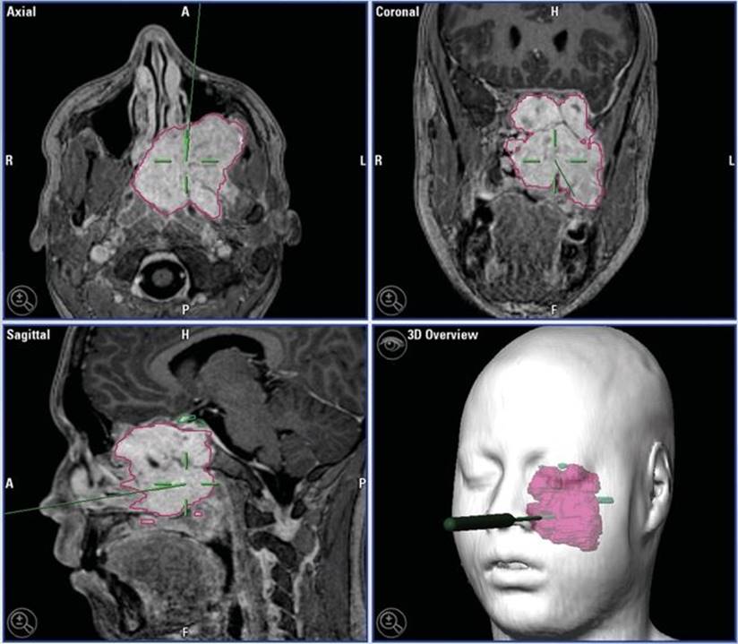
Figure 29.9 Intraoperative navigation of a stage IIIB angiofibroma using an endoscopic-assisted approach.
Finally, careful hemostasis is provided with hemostatic agents and bipolar cautery, and the entire surgical cavity is packed with rayon gauze soaked with antibiotic and supported by a Rapid Rhino pack.
POSTOPERATIVE MANAGEMENT
Care on the 1st postoperative day is commonly done in the intensive care unit. It is not mandatory, but due to the blood lost, it is safer. Patients are given systemic, broad-spectrum antibiotics for 1 week or more, depending on the duration of packing.
The inferior nasal packing (Rapid Rhino) is often removed in the hospital on the 3rd day. Patients are usually discharged within 3 to 5 days after surgery. The rayon gauze is removed after 7 days in the office. After removal of the pack, the patient receives saline nasal douches, and postoperative care is done every 2 weeks with nasal endoscopy in order to keep the nasal cavity without crusts in order to prevent scarring.
Frequent follow-up after surgery is necessary. MR scans with gadolinium or CT scans with contrast are performed after the re-epithelialization of the operative region and when the endoscopic examination does not show mucosal edema due to inflammation (Fig. 29.10). It avoids false-positive results in the radiologic study. Often the imaging is done in the 3rd month and then at 6 and 12 months.

Figure 29.10 Postoperative endoscopic view at 3 months of right nasal cavity after endoscopic transnasal tumor removal. (M, maxillary sinus; S, sphenoid sinus, and N, nasopharynx.)
COMPLICATIONS
Complications of surgical management of angiofibromas are related to two circumstances, embolization and surgical procedure. The first may have some important thromboembolic complications such as vasospasm that can impair the continuation of the examination or more rarely, vessel injury such as dissection or laceration. The formation of an embolus can occur and lead to deficits after the angiography, such as hemifacial and parotid swelling, necrosis of facial skin (lip, zygomatic area), and chemosis or, more important, decreased visual acuity due to central retinal artery occlusion.
Intraoperative hemorrhage is the most common complication during the surgical procedure for both open and endoscopic approaches. Branches from the internal maxillary artery and cavernous sinus are usually implicated. ICA damage during drilling of the pterygoid–infratemporal region can lead to catastrophic bleeding.
Nerve injury can occur during the external approach, especially to the second branch of the trigeminal nerve, causing infraorbital nerve dysesthesia. If the cavernous sinus is involved, during the tumor resection, cranial nerves III, IV, and VI can be injured causing a “frozen eye.” Finally, depending on the orbital involvement, an optic nerve or medial rectus muscle injury can occur, but this is transient in the majority of the cases.
In cases of dural defects due to the lesion or the procedure, the CSF leak needs to be repaired with multilayer-free grafts or pedicled flaps during the surgery.
RESULTS
The endoscopic endonasal approach to excise angiofibromas (stages I, II, and IIIA, B) has shown good results. Large angiofibromas can be treated by this approach but requires an experienced surgeon. Sometimes, the use of endoscope-assisted and external approaches with the microscope can achieve better results for stage IV tumors.
Recurrence rates are low and appear to be similar in endoscopic and open approaches.
Even with large angiofibromas, radiation therapy or chemotherapy is rarely used.
PEARLS
· The preoperative image evaluation includes CT and MR scans.
· Preoperative biopsy is unnecessary and not recommended due to the risk of severe bleeding.
· Preoperative embolization of the lesion, usually performed 24 to 48 hours before surgery, enables better results.
· The current concept of surgical cure does not require en bloc resection. A piecemeal resection seems to facilitate exposure of difficult anatomic locations.
· Follow-up includes periodic MR or CT scans in addition to routine endoscopic examinations.
PITFALLS
· Some tumor extensions can be hidden into the pterygoid and prevertebral muscles and fascia. Exploration of these regions is important to avoid residual tumor.
· In order to avoid leaving some residual tumor, it is important to drill the infiltrated surface of the basisphenoid bone with a diamond burr.
INSTRUMENTS TO HAVE AVAILABLE
· High definition video camera
· Surgical microscope
· Cutting forceps and endoscopic scissors
· Bipolar electrocautery
· Long-handled drill with cutting and diamond burrs
ACKNOWLEDGMENT
The author would like to thank the contribution of Leonardo Balsalobre, MD, MPH.
SUGGESTED READING
Andrews JC, Fisch U, Valavanis A, et al. The surgical management of extensive nasopharyngeal angiofibromas with the infratemporal fossa approach. Laryngoscope 1989;99:429–437.
Carrau RL, Snyderman CH, Kassam AB, et al. Endoscopic and endoscopic-assisted surgery for juvenile angiofibroma. Laryngoscope 2001;111(3):483–487.
Wormald PJ, Van Hasselt A. Endoscopic removal of juvenile angiofibromas. Otolaryngol Head Neck Surg 2003;129:684–691.
Lund VJ, Stammberger H, Nicolai P, et al. European position paper on endoscopic management of tumours of the nose, paranasal sinuses and skull base. Rhinol Suppl 2010(22):1–143.
Castelnuovo P, Pistochini A, Simoncello S, et al. Endoscopic surgery for juvenile nasopharyngeal angiofibroma. In: Stamm AC, ed. Transnasal Endoscopic Skull Base and Brain Surgery. New York: Thieme, 2011:301–309.