
Johnny B. Delashaw
INTRODUCTION
The major principle of intracranial skull base surgery is to provide access to the pathology at hand with minimal brain manipulation. When the lesion is extradural, the craniotomy should be tailored to the area of interest. Extradural brain retraction is usually well tolerated. However, intradural brain retraction is not and should be minimized. If the lesion is intradural, the surgeon should make every effort to carefully dissect the cerebrospinal cisterns and drain cerebral spinal fluid (CSF) to minimize brain retraction.
The modified orbitozygomatic (OZ) craniotomy is the cranial base approach used most frequently for surgical exposure of the anterior fossa, middle fossa, and orbit. The craniotomy incorporates the frontal bone, temporal bone, orbital rim, and a portion of the zygoma. This cranial bone flap usually is made in one piece unless the roof of the orbit is involved with the pathology. This versatile craniotomy provides access to vascular lesions and tumors of the basal frontal lobe, floor of the anterior cranial fossa, suprasellar and parasellar regions, anterior third ventricle, orbit, cavernous sinus, and floor of the middle fossa. Removal of the orbital rim and zygoma provides the surgeon with wider exposure and minimizes brain retraction when compared to the traditional pterional craniotomy.
HISTORY
The presentation of lesions that can be addressed with a modified OZ craniotomy is wide ranging and may include headache, vision loss, hydrocephalus, subarachnoid hemorrhage, upper cranial nerve palsies, proptosis, seizure, and even coma. The wide and varied regions for which this approach provides access make the possible presentations endless. A careful history should be taken focusing on visual complaints and upper cranial nerve symptoms.
PHYSICAL EXAMINATION
A complete neurologic examination should be carried out and tailored to the region of interest. Ophthalmologic evaluation for papilledema or oculomotor palsies is paramount. Trigeminal function should also be thoroughly investigated, especially in cases with involvement of Meckel’s cave or the cavernous sinus. Other signs should be sought based on the location of the specific lesion and are too numerous to detail here, given the wide range and flexibility provided by the modified OZ.
INDICATIONS
The OZ craniotomy is one of the most versatile approaches in skull base surgery. It can be used for any anterolateral pathology such as meningiomas of the sphenoid wing or lateral orbital tumors or for deep pathologies such as high-riding basilar aneurysms. It provides access to the suprasellar space and even the third ventricle for tumors such as craniopharyngiomas as well as the superior aspect of the clivus for some chordomas and any clinoidal process. The OZ approach may be ideal for anterior middle fossa and cavernous sinus pathologies such as meningiomas and schwannomas. Virtually any pathology in the anterior supratentorial space can be a potential indication for this modification.
CONTRAINDICATIONS
There are no absolute contraindications to an OZ craniotomy. Orbital manipulation should be avoided when possible in cases where there is preexisting contralateral loss of vision as the rare orbital complications become much more concerning in this setting.
PREOPERATIVE PLANNING
Magnetic resonance imaging is standard for planning any supratentorial tumor surgery. T2-weighted imaging can help to predict the consistency of the tumor, and T1-weighted imaging without and with contrast helps determine the differential diagnosis as well as vascularity of a tumor. Fine-cut computed tomography (CT) should be added for bone tumors or to determine the degree of bony involvement, hyperostosis, or erosion. MR or CT angiography can often provide adequate detail about arterial involvement or encasement, and computed tomographic angiography (CTA) can even provide useful details of venous involvement. Magnetic resonance angiography is routinely used for screening for unruptured aneurysms, and CTA can be used in subarachnoid hemorrhage. Digital subtraction angiography remains the gold standard for the detection of aneurysms and provides the greatest detail about venous drainage. Embolization can be performed to limit blood loss, but the modified OZ provides access to the accessible blood supply of most tumors.
The anesthesiologist and surgeon should attempt to maximize brain relaxation at the beginning of the procedure. Brain relaxation will prevent inadvertent dural tears when turning the bone flap and will improve exposure. The anesthesiologist should attempt to hyperventilate the patient to an end-tidal carbon dioxide level of 25. Intravenous infusion of 25 to 50 g of mannitol is also frequently performed to maximize brain relaxation. Frequently, the neurosurgeon will augment relaxation by insertion of a lumbar drain prior to scalp incision. The anesthesiologist can then slowly drain 30 to 40 mL of CSF at the time of the skin incision. If the surgical lesion is intradural and a wide Sylvian fissure dissection is planned, the lumbar drain is not inserted. Sylvian fissure dissection is easier when it is full of CSF.
SURGICAL TECHNIQUE
Positioning
After intubation and insertion of appropriate lines for venous access and monitoring of arterial blood pressure, the patient is positioned supine with the head in three-point fixation. The three-point fixation involves two pins inserted on the ipsilateral side of the planned craniotomy behind the ear in the parietal occipital bone. The third pin is placed in the contralateral frontal area. To avoid slippage in adults, the skull three-point fixation device is tightened until the pressure gauge measures 60 lbs/in2. The head is then turned 15 to 45 degrees toward the contralateral side depending upon the desired target. This three-point fixation device is then fastened to the operating table to prevent head movement during the surgical procedure. The head of the table is then elevated approximately 15 degrees above the level of the patient’s heart to facilitate venous drainage and reduce intracranial pressure.
Scalp Incision
The skin incision begins anterior to the tragus at the level of the zygoma. It extends cephalad in the hairline and crosses midline to the contralateral midpupillary line (Fig. 30.1A). It is important not to place the incision too far anterior to the auricle so as to avoid injury to the superficial temporal artery and frontalis branch of the facial nerve. This long incision allows the surgeon to turn a skin flap down to the orbital rim without tension. The scalp flap is dissected in the subgaleal plane leaving the pericranium intact. A vascularized pericranial graft based upon the supraorbital artery is then harvested separately down to the orbital rim. This pericranial flap can be used upon closure to repair skull base defects, such as the frontal sinus, at time of closure. As the pericranial flap is defined, the dissection continues over the orbital rim and exposes the periorbita. The supraorbital nerve is identified, and all attempts are made to preserve its integrity. If the nerve is in a notch, it is reflected down with the periorbita. If the nerve is in a foramen, then a drill with a side-cutting bit or osteotome is used to open the foramen and displace the nerve forward with the periorbita. The ipsilateral temporalis muscle is also dissected free from the skull in the subperiosteal plane and reflected forward with the scalp flap. A small cuff of temporalis fascia is left attached to the skull near the superior temporal line. The fascial cuff will be used at closure to reapproximate the temporalis muscle and secure it with suture. The orbital rim and the zygoma at the level of the frontozygomatic suture are dissected free.
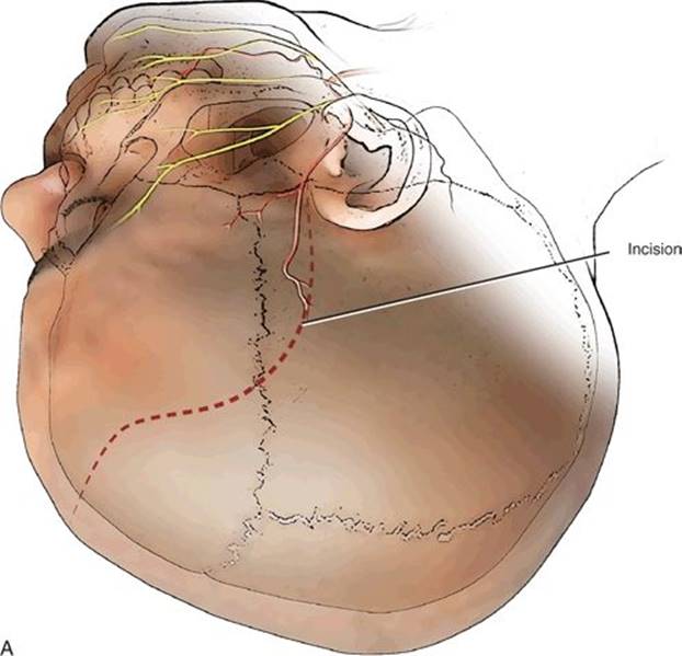


FIGURE 30.1 A. Planned ¾ coronal incision for a right modified orbitozygomatic craniotomy. B. Burr hole placement and cranial osteotomies for right modified orbitozygomatic craniotomy. C. An osteotome is used to weaken the attachment of the bone flap along the sphenoid wing and roof of the orbit.
Burr Holes
The McCarty burr hole is essential in performing the one-piece modified OZ craniotomy (Fig. 30.1B). The burr hole is placed over the frontosphenoidal suture 1 cm posterior to the frontozygomatic suture. This burr hole exposes the periorbita and dura of the frontal lobe. Two additional burr holes are made. One is placed in the posterior frontal region near the superior temporal line and the other is in the temporal squamosa area just above the root of the zygoma.
Creating the Bone Flap
Using a high-speed drill with a footplate attachment, the first cut is made from the temporal burr hole to the posterior frontal burr hole in the extradural plane. The cut is then extended forward to the level of the supraorbital notch or foramen. The second cut is made from the McCarty burr hole down to the sphenoid wing. The third cut is then made from the temporal burr hole forward and up to the sphenoid wing. A side-cutting bit on the high-speed drill is then used to cut the bone of the zygoma near the frontozygomatic suture down to the McCarty burr hole. This side-cutting bit drill is also used to weaken the orbital rim at the supraorbital notch or foramen. Using an osteotome, the attachment of the sphenoid wing is loosened (Fig. 30.1C). The osteotome is then used along the orbital rim to propagate a fracture line in the anterior roof of the orbit. The modified OZ flap is then cracked forward and separated from the dura and periorbita in one piece. The bone flap should be harvested easily, and the “crack” should not be forced. Upon removal of the bone flap, the anterior contents of the orbit and the frontal and temporal dura are exposed. A small rongeur can then be used to remove additional bone from the roof of the orbit and the sphenoid wing (Fig. 30.2A and B). The surgeon should remove this bone leaving the periorbita intact. Upon removal of the roof and wing, the surgeon will encounter the superior orbital fissure. Bone lateral to the superior orbital fissure down to the foramen rotundum can be removed easily with a rongeur. By removing bone over the superior orbital fissure, the temporal–orbital band is fully exposed.
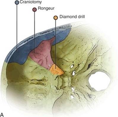

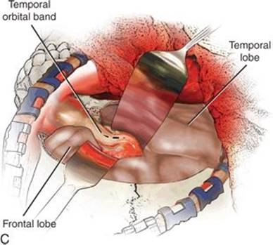
FIGURE 30.2 A. Extradural bone removal from craniotomy, rongeur, and diamond bit drill. B. Right orbitozygomatic exposure after removing the bone flap. C. Exposure of the temporal–orbital band (dotted line) after removing the roof of the orbit and opening widely the superior orbital fissure. D. Cutting the temporal–orbital band and peeling the outer layer of dura (lateral wall of the cavernous sinus) with the temporal lobe to expose the cavernous sinus and the cranial nerves.
Extradural Dissection
Extradural access to the optic foramen, anterior clinoid, and cavernous sinus is restricted by the temporal–orbital band (Fig. 30.2C). This temporal–orbital band is the dura along the superior temporal lobe that extends into the superior orbital fissure and is contiguous with the periorbita. The temporal–orbital band is comprised of two layers of dura. The outer layer is thick and continuous, and the inner layer is thin and not contiguous. Cutting the dural band with micro-scissors allows the surgeon to separate the layers of dura and reflect the temporal lobe posteriorly. As the temporal lobe is retracted, the wall of the cavernous sinus is peeled away with cranial nerves three, four, V1, and V2 exposed (Fig. 30.2D). In addition, the anterior clinoid and optic foramen are easily visualized.
Extradural Clinoidectomy and Optic Nerve Decompression
The optic canal is carefully thinned with a high-speed drill using a diamond burr. Copious irrigation is used during drilling. The bone is “eggshelled” over the foramen and then carefully removed with microcurettes. The clinoid can then be removed using a diamond drill bit. The center of the clinoid is drilled and the optic strut weakened until the bone can be easily removed with a curette.
Exposure
This craniotomy with extradural dissection provides a panoramic view of the orbit and access to the floor of the anterior and middle fossa and the cavernous sinus. If necessary, additional exposure can be obtained by opening the dura.
Bone Flap Reconstruction
The modified OZ bone flap can be placed back into position at time of closure. If the frontal sinus was violated, then a pericranial graft can be placed between its opening and the bone flap. The bone flap is secured with small cranial titanium plates. During this process, it is important to secure the orbital rim to the zygoma with a titanium “dog bone” plate. Reconstruction of the posterior roof of the orbit is not necessary.
POSTOPERATIVE MANAGEMENT
Patients should be kept overnight in an intensive care unit with frequent neurologic checks. The orbit should be checked for sign of muscle entrapment that, if noted early, can be corrected without long-term sequelae. Often the eyelid is so swollen that it must be opened by the examiner in order to check eye movements. When drains are used, they are kept until the edema has begun to resolve or their output has significantly decreased. The head of the bed should be kept elevated to facilitate cerebral venous drainage and minimize subcutaneous swelling. Intravenous steroids are administered briefly for neuroprotection.
COMPLICATIONS
In addition to the usual complications such as stroke or cranial neuropathy associated with any skull base approach, orbital entrapment is the most feared approach-related complication. Care should be taken to examine the orbit and periorbita following replacement of the orbital rim. If muscle entrapment has occurred postoperatively and is verified by the inability to move the eye in a given direction, fine-cut CT can confirm the location and source of entrapment. If identified early, the patient can be returned to the operating room for correction without long-term sequelae. Other orbital complications, such as ptosis, are usually transient.
Frontalis palsy can occur with any frontotemporal craniotomy, but zygoma dissection provides yet another opportunity for injury to or traction on this branch.
RESULTS
The results and efficacy of the OZ craniotomy for many pathologies, ranging from meningiomas to aneurysms, are well known and well reported. It remains a popular approach to the skull base and the gold standard for many anterior and anterolateral pathologies. When performed with proper knowledge and care, this modification to a frontotemporal craniotomy adds little morbidity and can provide profound improvement in access.
PEARLS
· Harvest the pericranial graft at the skin incision in case the frontal sinus is entered or the dura is difficult to close.
· A one-piece modified OZ craniotomy requires an osteotome to weaken the sphenoid wing and roof of the orbit to “crack” bone flap forward.
· Open the superior orbital fissure widely.
· Cut the temporal–orbital band to expose the anterior clinoid, optic foramen, and cavernous sinus extradurally.
PITFALLS
· A one-piece modified OZ craniotomy should not be attempted with hyperostosis of the roof of the orbit or sphenoid wing.
· Never force the cracking of the sphenoid wing or orbital roof.
INSTRUMENTS TO HAVE AVAILABLE
· Standard neurosurgical tray
· High-speed drill with side-cutting bit
· Osteotome
SUGGESTED READING
Schwartz MS, Anderson GJ, Horgan MA, et al. Quantification of increased exposure resulting from orbital rim and orbitozygomatic osteotomy via the frontotemporal transsylvian approach. J Neurosurg 1999;91:1020–1026.
Balasingam V, Noguchi A, McMenomey SO, et al. Fronto-temporo-orbito-zygomatic approach. Neurosurg Q 2005;15: 113–121.
Balasingam V, Noguchi A, McMenomey SO, et al. Modified osteoplastic orbitozygomatic craniotomy: technical note. J Neurosurg 2005;102:940–944.
Noguchi A, Balasingam V, Shiokawa Y, et al. Extradural anterior clinoidectomy. Technical note. J Neurosurg 2005;102: 945–950.