
Paul A. Gardner and Carl H. Snyderman
INTRODUCTION
The clivus is the anterior portion of the occipital bone and adjoins the petrous and sphenoid bones via synchondroses that gradually ossify throughout childhood and early adulthood. The clivus is typically divided into thirds based on neural foramina, as these determine the surgical approach (orbitozygomatic vs. transpetrous vs. far lateral). Given its midline location, endonasal approaches provide seamless access to the entire clivus with their lateral extension determined by neural foramina. Typically, the superior clivus is separated from the mid and lower clivus by Dorello’s canal, which transmits the abducens nerves from the intradural space, dorsal to the paraclival internal carotid artery (ICA), into the lateral cavernous sinus. From an endonasal viewpoint, this occurs at approximately the level of the midpoint of the paraclival ICA, halfway between foramen lacerum and the sella. A more practical division for endonasal approaches is to define the middle clivus as the bone between the floor of the sella and the floor of the sphenoid sinus while the inferior clivus extends from the floor of the sphenoid (at the level of the pterygoid “wedge”) to the foramen magnum.
Regardless of classification, the clivus makes up the pure midline of the inferior skull base, and as such, lesions that arise from it or its adjacent tissues, such as nasopharyngeal mucosa and dura, are ideally suited for an anterior approach such as the endoscopic endonasal approach (EEA). The limitations of this approach are the surrounding neurovascular structures and the surgeons’ experience, anatomical knowledge, and resources.
HISTORY
Headache and diplopia are the two most common symptoms caused by tumors arising from the clivus. Since the clivus is part of the occipital bone, tumors involving this bone typically present with purely occipital pain, though the headaches can be generalized or frontal. Bone tumors classically cause pain that is worse at night, even awakening the patient from sleep. Lesions such as meningiomas that involve or distend the dura or arachnoid can also cause headache of variable character. However, one must be careful about attributing headache to clival tumors, especially if small or otherwise asymptomatic. Lateral extension to Meckel’s cave or distension of the cisternal segment of the trigeminal nerve can cause retro-orbital pain or trigeminal neuralgia/neuropathy, respectively.
An abducens palsy with variable degrees of subjective or objective diplopia is a classic presentation for chordoma or chondrosarcoma. These tumors grow within the bone or interdural space, compressing or filling Dorello’s canal and thus causing nerve dysfunction. Intradural extension of clival tumors with brain stem compression can lead to gait dysfunction, swallowing difficulties, and even decreased mental status and hydrocephalus. Hearing loss and facial palsy can be signs of lateral extension to the petrous bone. Origination from or extension to the nasopharynx can lead to nasal airway obstruction, hyponasal speech, and epistaxis.
PHYSICAL EXAMINATION
A complete examination of the cranial nerves is critical in patients with clival tumors since these tumors can affect practically every cranial nerve depending on their size and extension. It is especially important to examine eye movement (CN III, IV, and VI) and palatal (CN IX and X) and tongue function (CN XII). Gait and swallowing function should be closely assessed as these can significantly affect patient recovery. Laryngoscopy to identify vocal cord paresis as well as a barium swallow evaluation should be performed in patients with a history of dysphagia or voice changes. Nasal endoscopy can identify masses with significant sinonasal involvement and occasionally provide for biopsy in appropriate settings such as sinonasal malignancy. This should be avoided in excessively vascular tumors or chordomas that have a potential for seeding. All patients should receive a complete examination of the head and neck to palpate for signs of cervical metastases or cervical extension.
INDICATIONS
Any tumor whose epicenter or origin is the clivus or associated structures can be approached endonasally. Given a midline origin, these tumors will displace surrounding neurovascular structures laterally, allowing direct access to the tumor through the paranasal sinuses without manipulation of these critical structures.
Chordomas are one of the best examples of a tumor with a midline origin that are ideally suited for an endonasal approach, the vast majority of which can be completely removed endonasally. Chondrosarcomas often involve the midline clivus but with their typical paramedian origin at the petroclival synchondrosis, each must be individually assessed and may require a combination with another approach (retrosigmoid, transpetrous, or orbitofrontal) for complete removal. Similarly, petroclival meningiomas have very variable epicenters. Purely or mostly clival meningiomas can be addressed with endoscopic endonasal surgery (EES) (Figs. 36.1 and 36.2), whereas purely or mostly petrous tumors cannot. The majority fall somewhere in between, and their relationship to associated cranial nerves (best visualized with FIESTA MRI sequences) determines the best approach (or combination of approaches).


FIGURE 36.1 A. T1-weighted postcontrast axial MRI. B. CT angiogram sagittal reconstruction demonstrating a primarily clival meningioma with midline origin and dural base. Following endoscopic endonasal resection, only a small, paramedian residual is left. C. Postcontrast axial. D. Sagittal T1-weighted MRI.


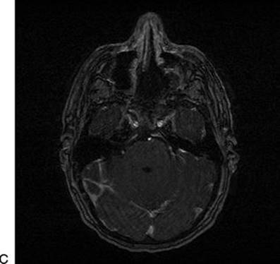
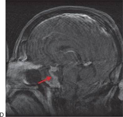

FIGURE 36.2 A. T1-weighted postcontrast sagittal. B. axial MRI demonstrating a primarily clival meningioma with midline origin and dural base. Following endoscopic endonasal resection, the clival location allows for complete resection demonstrated on (C) immediate postoperative postcontrast axial and (D) sagittal T1-weighted MRI. Note enhancing nasoseptal flap used for reconstruction (dashed arrow). E. T2-weighted, axial MRI showing the typical, hyperintense “bubbly” appearance of a clival chordoma (arrow).
Care should be taken to properly define the differential diagnosis of clival lesions. The diagnosis of benign fibro-osseous lesions such as fibrous dysplasia can usually be made radiographically, with their classic “ground glass” appearance, so that intervention can be avoided. Rare exceptions include progression with optic or cranial neuropathy, usually in children or adolescents and associated with cyst formation. Rarely, pituitary adenomas erode into and invade the clivus primarily, rather than extending into the suprasellar space. Extending a sellar exposure into the mid and even inferior clivus is required for complete removal of such tumors.
Nasopharyngeal cancers are usually advanced at the time of presentation and are treated primarily with radiation therapy. The primary role of surgery is biopsy for diagnosis and debulking of tumor to relieve symptoms prior to radiation therapy. Exceptions include small tumors that can be completely resected with adequate margins. For adenoid cystic carcinoma, the goal of surgery is maximal removal with minimal morbidity, followed by radiation therapy. It is not possible to achieve clear resection margins with adenoid cystic carcinoma of the skull base due to perineural spread, and the extent of surgery is limited by the surrounding neural and vascular structures. Surgical salvage of residual tumor following radiation therapy is of potential value for local control and should be considered based on posttreatment functional imaging with PET–CT or the results of biopsies.
CONTRAINDICATIONS
Tumors originating in the lateral skull base but extending medially will displace critical neurovascular structures medially, into the path of a midline approach such as an EEA. This remains the major contraindication to EES. Sinus infection is a transient contraindication to intradural (or potentially intradural) surgery that should be treated with antibiotics with or without surgical drainage as indicated before proceeding.
PREOPERATIVE PLANNING
Magnetic resonance imaging (MRI) and computed tomographic angiography (CTA) are complementary, and it is important to obtain both to help determine preoperative differential diagnosis. For example, MRI illustrates the typical heterogeneously hyperintense T2 “bubbles” of a chordoma or chondrosarcoma (Fig. 36.2), while CTA illustrates bony erosion, ICA stenosis, or occlusion and identifies benign fibro-osseous lesions such as fibrous dysplasia (that can appear very ominous on MRI). With meningiomas, MRI will show the dural tail while CTA illustrates the vascular relationships and degree of bony involvement or hyperostosis (Figs. 36.1 and 36.2).
SURGICAL TECHNIQUE (VIDEO 36.1)
All surgeries are performed by a team consisting of an otolaryngologist and a neurosurgeon performing a two-surgeon, three- or four-hand technique. This allows for appropriate care of both sinonasal and neural structures as well as critical dynamic endoscopy and bimanual microsurgical techniques. Additional benefits include improved problem solving (“copilot”) and efficiency.
Most patients are placed in three-pin head fixation with the head in a neutral or slightly extended position and rotated toward the side where the surgeons stand. This allows for precise positioning to optimize surgical ergonomics as well as prevent movement during critical portions of the operation. Registration of the navigation system is performed, and nasal decongestion with topical oxymetazoline (0.05%) is achieved. The external nose and nasal vestibule are prepped with an iodine solution, and perioperative antibiotic prophylaxis is provided with a third-generation cephalosporin.
Nasal endoscopy is performed with a 0-degree endoscope, and the turbinates are lateralized to provide greater access to the nasopharynx. The inferior portion of the middle turbinate is typically resected to provide room for endoscopy. If a dural defect is anticipated, a septal mucosal flap is elevated on the side contralateral to the bulk of the tumor and is placed into the maxillary sinus through a middle meatal antrostomy for storage during the surgery. The posterior septum is detached from the sphenoid rostrum, and the rostrum is resected to provide binarial access to the sphenoid sinus. The sphenoidotomy is maximized to provide full access from the sella to the floor of the sinus and lateral to both internal carotid arteries. Sphenoid septations are removed with bone rongeurs and drilling, and landmarks are identified with special attention to the course of the paraclival carotid arteries.
Fascia is elevated from the inferior margin of the sphenoidotomy, and the medial pterygoid plates are exposed bilaterally. The inferior portion of the clivus is exposed by resecting the nasopharyngeal mucosa and basopharyngeal fascia. This can be done either with a microdebrider or with monopolar electrocautery (needle tip or suction). Care should be taken to evaluate the course of the parapharyngeal ICA as it can become ectatic and loop medially at its most inferior (proximal) aspect. This is rarely an issue, but when it is, dissection of the fascia from the bone should occur in the subperiosteal plane, deep to the artery. Laterally, the supracondylar groove should be identified. This is a ridge of bone to which the fascia of the rectus capitis anterior muscle attaches; it reliably predicts the location of the hypoglossal canal.
Once the basopharyngeal fascia has been removed from the sphenoid floor to the foramen magnum, drilling of the entire clivus can be performed. The floor of the sphenoid represents the most prominent portion of the clivus and should be drilled to the depth of the clival recess. The initial width of this bone removal should be limited to the paraclival ICAs to prevent inadvertent injury. These are usually easily identifiable in a well-pneumatized sinus by their bony protuberances (Fig. 36.3). If they are not, their location can be verified by dissecting in the extradural space along the floor of the sella laterally until the downturn of the ICA as it enters from the paraclival segment is visualized.
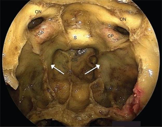
FIGURE 36.3 Endoscopic endonasal view of the clivus with a well-pneumatized clival recess (CR) allowing for easy identification of the paraclival internal carotid arteries (ICAs) (arrows). (S, sella; CP, carotid protuberance [parasellar]; ON, optic nerve.)
Once the floor of the sphenoid, the thickest portion of the clivus, has been drilled to the depth of the clival recess, the entire clivus is drilled to the depth of the inner cortex, which can be further thinned with the drill and removed with a Kerrison rongeur. The exposure should be extended as widely as possible to the paraclival ICA in the midclivus and to the medial eustachian tube in the lower clivus. The dura of the clivus has two layers (periosteal and meningeal) between which lies a very impressive venous plexus that must be managed before opening the inner layer. The bleeding can usually be packed off with flowable Gelfoam (e.g., Surgifoam, Floseal, Surgiflo) or other hemostatic materials. Careful stripping of the entire periosteal layer allows final packing of this plexus at its lateral margin and exposure of the inner, meningeal layer for more careful, controlled opening into the intradural space.
The vertebrobasilar system and abducens nerves are the key neurovascular structures associated with the mid and lower clivus and should be identified when possible with image guidance and electromyography, respectively (Fig. 36.4). The abducens nerve is at greatest risk with an endonasal approach and, because of its long course and association with multiple spaces, can have relationships with tumors that are difficult to predict. Chordomas typically displace the intradural segment posteriorly and laterally, but their tendency to invade the interdural space predisposes them to extend into Dorello’s canal. This can make identification of this portion of the nerve very challenging, and nerve involvement can make it impossible to preserve at times. Petroclival meningiomas can displace the abducens nerve in any direction, depending on their origin and growth pattern. Electromyographic stimulation of the overlying dura prior to opening into tumor can provide reassurance that the nerve is not interposed between the dura and tumor capsule and hopefully prevent nerve transection as part of the dural opening. If the origin of the nerve intradurally can be identified early, a “starting threshold” lowest limit of stimulation voltage or current can be established. If this is preserved throughout surgery, any palsy that may develop will be transient.
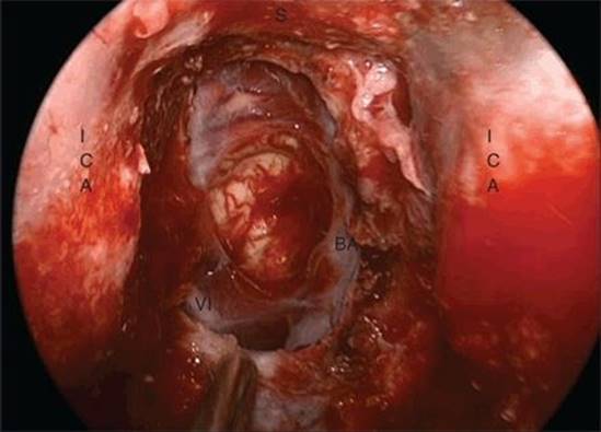
Figure 36.4 Intraoperative, endoscopic endonasal view following resection of the clival chordoma shown in Figure 36.2 with resultant wide dural defect and exposed basilar artery and abducens nerve (VI). (BA, basilar artery; ICA, internal carotid artery [paraclival]; S, sella.)
As with all intradural tumor resection, standard microsurgical dissection techniques should be employed. This is accomplished with extended endonasal instruments fashioned after open microsurgical dissectors and pistol-grip microscissors (see, Instruments section). The advantage of the EEA is that it can be used for tumors that directly abut or originate from the clivus. Therefore, these tumors are entered directly, and intradural tumors should be completely debulked internally prior to extracapsular dissection. Gentle countertraction with a teardrop-slotted suction will allow for blunt and sharp dissection of arachnoid bands or scar that are adherent to the tumor and neurovascular structures.
Reconstruction should be considered at the onset of the approach in order to preserve all reconstructive options. Clival defects without violation of the dura or significant exposure of the carotid arteries do not require reconstruction. In these cases, application of fibrin glue provides temporary protection of the surgical site and may promote healing. When repair of a dural defect or coverage of an exposed carotid artery is necessary, reconstruction with vascularized tissue is preferred. The dural defect is first repaired with an inlay collagen or fascial graft (e.g., allogenic dural substitute, cadaveric fascia, or autologous fascia lata) placed intradurally. Primary reconstructive options include a nasoseptal flap (see Chapter 42) or inferior turbinate flap (see Chapter 44). The nasoseptal flap is generally preferred due to the ease of dissection, minimal donor site morbidity, size, and wide range of rotation. It may be insufficient, however, for a deep or caudal clival defect. The coverage of the flap can be augmented by filling the clival defect with an autologous adipose tissue graft prior to placing the flap. Adipose tissue is also useful to bolster the reconstruction and prevents herniation of the brain stem into the defect. In this situation, care must be taken to ensure that the flap is in contact with normal mucosa or bone circumferentially to allow for healing. The rotation of the nasoseptal flap for a midclival or lower clival defect is more horizontal, and the width of the flap may be insufficient for the vertical dimension of the defect. This need is anticipated by extending the flap incisions to include the mucosa of the nasal floor, resulting in a wider flap. In addition, an onlay fascial graft can be placed to cover the entire defect, deep to the flap. An inferior turbinate flap is a useful option when a nasoseptal flap is not available due to prior surgery or tumor involvement of the vascular pedicle. The coverage area of an inferior turbinate flap can be augmented by including the mucosa of the nasal floor and even the ipsilateral nasal mucosa. When these flaps are not available, secondary options include an extracranial pericranial flap (see Chapter 46) or temporoparietal fascial flap (see Chapter 45).
POSTOPERATIVE MANAGEMENT
The usual postoperative precautions are taken following any intradural EEA. Transclival approaches are particularly at risk for catastrophic injury during passage of nasogastric tubes. If necessary, placement of a feeding tube should always be performed under direct endoscopic visualization to avoid disruption of the repair and passage of the tube intracranially. Signs to that effect should be placed at the patient’s bedside. Patients are cautioned to avoid nose blowing, bending over, or straining. Stool softeners are prescribed, and family members are advised to assist with any lifting. Sneezing is done with an open mouth to avoid the accompanying increase in intracranial pressure.
The role of lumbar drainage is not clear, but it probably plays a role in lowering postoperative cerebrospinal fluid (CSF) leak rates following intradural dissection. Reconstruction of large transclival defects is more difficult, and the reconstructive flap may provide limited coverage, increasing the risk of a CSF leak. A CT scan is obtained the evening of surgery to assess the degree of pneumocephalus (and ensure that there is no hemorrhage) prior to opening the lumbar drain. Elevation of the head of the bed, even during sleep, should be maintained for the first 2 weeks following intradural endonasal surgery.
When necessary, packing is left in place for 1 to 2 days following extradural surgery and 6 to 7 days following intradural dissection. Patients are maintained on a third- or fourth-generation IV cephalosporin (or equivalent if penicillin allergic) for 24 to 48 hours and then switched to an oral second-generation cephalosporin or equivalent until the packing is removed. Septal splints are maintained for 2 to 3 weeks postoperative if a septal flap has been employed. Nasal saline sprays are used liberally, and saline irrigations are instituted after several weeks. Patients are instructed to avoid activities that increase intracranial pressure for at 4 to 6 weeks if there has been a dural reconstruction. Gentle nasal debridement under endoscopic visualization is performed every few weeks for the first few months as needed.
Corticosteroids are generally reserved for patients who develop new cranial neuropathies that are thought to be transient and continued for approximately 48 hours, after which they are rapidly weaned to prevent complications of wound healing.
COMPLICATIONS
As emphasized above, the abducens nerve is at significant risk with a transclival approach. If the patient has a complete palsy, he or she will likely require temporary patching of the eye or prism placement in glasses. If the palsy is partial, patients are encouraged to tolerate this diplopia to speed adjustment or to alternate patching of the eyes. If the abducens nerve is sectioned or recovery is incomplete (after 6 months), referral to an oculoplastic surgeon should be made for consideration of correction.
Patients should be closely followed for signs of a CSF leak in the immediate postoperative period. With clival defects, CSF drainage can occur either from the nostrils or into the oropharynx, in which case the patient will complain of and should be questioned about salty drainage into the throat. This is typically worsened by the supine position.
Any vascular injury needs to be immediately evaluated with digital subtraction cerebral angiography to ensure that there is no pseudoaneurysm, thrombus, filling defect, or critical stenosis that would require anticoagulation, stenting, coiling, or arterial sacrifice.
Nasal morbidity is generally low. Patients notice diminished smell and taste function for several months following surgery. There is a small risk of epistaxis for several weeks from branches of the sphenopalatine artery. Nasal crusting is the greatest long-term morbidity but can be effectively managed with saline irrigations and periodic endoscopic debridement.
RESULTS
From April 2003 to September 2012, 84 patients with clival chordomas underwent EES at the UPMC Center for Cranial Base Surgery. Medical records and radiologic images were retrospectively analyzed and evaluated.
Eighty-four patients (59.5% male) with a median age of 44 years (range 4 to 88) underwent EES for primary (n = 46) or previously treated (n = 38) chordomas of the skull base. The overall rate of gross total resection (GTR) was 68% (n = 57). In the group of primary tumors, GTR was achieved in 36 cases (78%), near total resection (>95% of tumor removed) in 7 (15%), subtotal resection (>85% of tumor removed) in 2 (4%), and partial resection in 1. In the group of 38 previously treated chordomas, GTR was achieved in 21 (55%) patients, near total in 6 (16%), subtotal in 5 (13%), and partial in 6 (16%). Eighteen patients underwent staged surgeries, and in nine cases, EES was combined with craniotomy (11%). Fifty-five patients received adjuvant radiation therapy (proton beam in 43 cases). Surgical complications included CSF leakage in 16 cases (19%), new permanent cranial neuropathy in 5 (5.9%), and carotid injury in 3 (3.6%), all without neurologic sequelae. In a mean follow-up of 21 months (range 1 to 91), 28 patients developed recurrent disease (33%) and 20 among them underwent repeat EES. At the most recent follow-up, 49 patients (58%) are free of tumor, 26 (31%) have a stable residual or recurrent chordoma, and 9 (11%) died (eight due to disease progression). In total, the 84 patients underwent 143 surgical procedures.
PEARLS
· Petroclival meningiomas have extremely variable relationships to cranial nerves, and this should be evaluated and used as a major criterion for choice of approach to avoid manipulation of these structures.
· The paraclival ICA and vidian nerves provide the lateral boundaries for the midclival approach. Intraoperative navigation using CTA in combination with exposure of the sellar floor is helpful in identifying the paraclival ICA in a poorly pneumatized sinus.
· The abducens nerve is the nerve at greatest risk with an endonasal transclival approach. This can be minimized with gentle dissection technique and use of an electrical nerve stimulator.
· Inferior clival dural defects with a postoperative CSF leak can present as drainage down the back of the throat rather than from the nose.
PITFALLS
· Drilling of clival bone deep to the paraclival arteries can result in injury to the posterior surface of the arteries. Drilling from the contralateral nostril decreases the risk of injury to the artery from the shaft of the drill bit.
· The dura should be incised in the midline well above the vertebrobasilar junction to avoid injury to an abducens nerve displaced by tumor.
· Blind passage of nasogastric tubes postoperatively can result in intradural injury. This should be communicated to everyone involved in the care of the patient.
INSTRUMENTS TO HAVE AVAILABLE
· Zero- and 45-degree rod lens endoscopes (Storz)
· Extended tip micro-Doppler (for ICA identification)
· Standard sinus instruments
· Needle-tip and suction monopolar electrocautery tips
· High-speed electric drill with extended tip and extended drill bit (Stryker)
· Extended tip neurodissectors (KLS Martin)
· Pistol-grip Kurze microscissors (straight, curved left and right, and rotatable) (Storz)
· Pistol-grip bipolar cautery (side angle, “uptoe,” and fine, straight tip) (Storz)
ACKNOWLEDGMENT
Special thanks to Francisco Vaz Guimaraes Filho, MD, for his assistance with developing the figures for this chapter.
SUGGESTED READING
Madhok R, Prevedello D, Gardner P, et al. A direct corridor to the clivus: the expanded endonasal approach. A review of the transclival module in endoscopic skull base surgery. Skull Base 2008;18(Suppl 1):26.
de Notaris M, Cavallo LM, Prats-Galino A, et al. Endoscopic endonasal transclival approach and retrosigmoid approach to the clival and petroclival regions. Neurosurgery 2009;65(6 Suppl):42–50.
Barges-Coll J, Fernandez-Miranda JC, Prevedello DM, et al. Avoiding injury to the abducens nerve during expanded endonasal endoscopic surgery: anatomic and clinical case studies. Neurosurgery 2010;67(1):144–154.
Fraser JF, Nyquist GG, Moore N, et al. Endoscopic endonasal transclival resection of chordomas: operative technique, clinical outcome, and review of the literature. J Neurosurg 2010;112(5):1061–1069.
Koutourousiou M, Gardner PA, Tormenti MJ, et al. Endoscopic endonasal approach for resection of skull base chordomas: outcomes and learning curve. Neurosurgery 2012;71(3):614–625.
Gardner PA, Snyderman CH, Fernandez-Miranda JC. Sella and beyond: approaches to the clivus and posterior fossa. In Georgalas C, Fokkens WJ, eds. Rhinology. Stuttgart, AR: Thieme, 2013.