
Carl H. Snyderman and Paul A. Gardner
INTRODUCTION
Endonasal approaches to the ventral skull base include a midline corridor that extends from the frontal sinus to the superior aspect of the cervical spine. The transodontoid approach provides access to the region of the foramen magnum and includes the inferior aspect of the clivus and the superior cervical vertebrae (C1 and C2). The surgical field generally extends from the floor of the sphenoid sinus to the plane of the hard palate and is limited laterally by the parapharyngeal internal carotid arteries (ICAs) deep to the fossa of Rosenmuller. The inferior third of the clivus extends from the floor of the sphenoid sinus to the foramen magnum and is bounded superolaterally by the anterior genu of the petrous ICA. Inferolaterally, the inferior clivus is bounded by the occipital condyles and hypoglossal nerves. The inferior limit of the transodontoid approach is the body of C2 and is roughly defined by the nasopalatine line (NPL), a virtual line that is tangential to the inferior margin of the nasal bones and posterior edge of the hard palate (Fig. 37.1). The intersection of this line with the vertebrae establishes the inferior limit of endonasal access and corresponds to the lower dens or upper body of C2 in most patients. Posteriorly, the inferior clivus is bounded by the hypoglossal nerves, brainstem, and vertebral arteries.
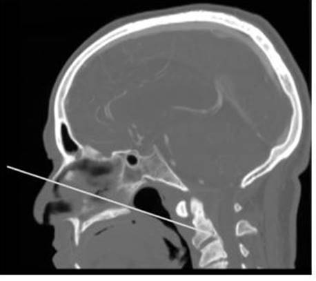
FIGURE 37.1 CT angiogram midsagittal reconstruction with a line drawn from the bony nasal bridge to the posterior hard palate and extended to the superior aspect of the spine. This nasopalatine line approximates the caudal extent of endonasal access.
The endonasal transodontoid approach avoids the morbidity of transoral/transpalatal approaches to the inferior clivus and superior cervical spine and is associated with a faster recovery (Table 37.1). Surgical advantages include improved visualization, greater access superiorly, and decreased bacterial contamination of the surgical field. Disadvantages include limited access to the inferior cervical spine and potentially greater risk of cerebrospinal fluid (CSF) leak following reconstruction of the dura. In our opinion, an endonasal approach is the preferred approach for lesions in the region of the foramen magnum that are bounded by the neural and vascular structures and for the resection of the odontoid process in the setting of basilar invagination.
TABLE 37.1 Comparison of Anterior Surgical Approaches to the Foramen Magnum
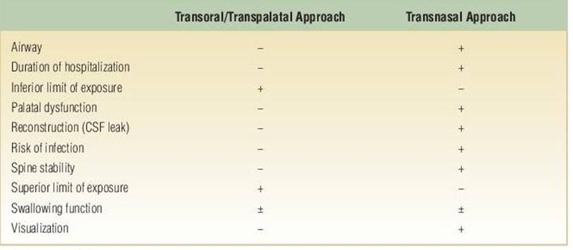
+, advantage; −, disadvantage.
HISTORY
Presenting symptoms will depend on the diagnosis, location, extent, and age of the patient. Patients may present with a serous middle ear effusion and conductive hearing loss due to eustachian tube dysfunction or obstruction. Involvement of cranial nerves can result in a wide variety of symptoms, including hypernasal speech and nasal reflux due to palatal dysfunction (CN IX/X); weak voice, dysphagia, and aspiration (CN X); and dysarthria (CN XII). Lower cranial nerve dysfunction can result from intradural compression or nerve involvement at their respective foramina. Coughing associated with meals or drinking should be questioned to check for active aspiration. A cervical mass (metastatic lymphadenopathy) is often the first symptom of nasopharyngeal cancer.
Bone disease with degenerative pannus or basilar invagination presents with myelopathy with or without dysphagia. Progressive loss of ambulation is a common complaint, and diagnosis is often delayed because of its insidious onset in elderly patients. Pain in the neck or occipital neuralgia can be indicative of active instability of the craniocervical junction.
PHYSICAL EXAMINATION
Physical examination includes a full examination of the head and neck including otoscopy, endoscopic visualization of the nasopharynx and hypopharynx, palpation of the neck, and assessment of cranial nerve function. Middle ear effusion may be the result of obstruction of the eustachian tubes. The type of hearing loss (conductive vs. sensorineural) can be confirmed with tuning fork testing (Weber and Rinne tests). The upper aerodigestive tract can be examined with a rigid nasal endoscope or flexible fiberoptic scope. The extent of mucosal lesions or masses should be noted. In particular, the fossa of Rosenmuller posterior to the eustachian tube should be carefully examined, as this is a common site for nasopharyngeal cancer. The neck should be palpated for metastatic cervical adenopathy. Assessment of cranial nerve function focuses on the lower cranial nerves: palatal dysfunction, vocal cord palsy, pooling of secretions in the hypopharynx with aspiration, and paresis of the tongue (deviation of tongue to the paralyzed side with protrusion). Range of mobility of the cervical spine can be grossly assessed with flexion, extension, and rotation. Signs of brainstem compression include weakness of the extremities and hyperreflexia. Gait assessment is important to determine if the gait is myelopathic or the result of another condition.
INDICATIONS
The primary principle of endonasal skull base surgery applies to the transodontoid approach: Avoid transgression or manipulation of major neural and vascular structures. If the lesion extends lateral to major vascular structures (carotid and vertebral arteries) or cranial nerves, an alternative approach or combination of approaches should be considered. For example, a large meningioma may require a combined approach (endonasal and retrosigmoid or far lateral) to access different areas of the tumor depending on its relationship to the neural and vascular structures.
Clinical indications for a transodontoid approach include neoplasms, inflammatory and degenerative disease with craniocervical compression, and trauma. The most common neoplasms include intracranial tumors (meningioma), neoplasms arising from bone (chordomas, chondrosarcoma), and malignancies of the soft tissues (nasopharyngeal carcinoma). Inflammatory or osteoarthritic degeneration or congenital abnormalities of the atlantoaxial joint and associated ligaments can result in chronic instability of the spine and resultant inflammatory pannus or progressive invagination with brainstem compression. Generally, only ventral bony compression needs to be addressed with an anterior approach, whereas soft tissue/pannus usually will resolve over time with posterior decompression and arthrodesis. In cases of severe pannus compression, we still perform an anterior approach to facilitate rapid decompression and avoid a posterior decompression, leaving the surface of the posterior C1 ring intact for fusion. Rarely, traumatic fractures or dislocations of C1 and C2 can be decompressed endonasally. This is more common in the delayed setting of an os odontoideum. The transodontoid approach is applicable to both adult and pediatric populations.
Neoplasms arising from the bone of the inferior clivus (chordoma, chondrosarcoma) are ideally suited for an endonasal approach. These tumors tend to have a midline or paramedian origin and displace neurovascular structures laterally. As a result, resecting the tumor through a midline approach minimizes or obviates any manipulation of these structures. Intracranial tumors such as meningiomas of the foramen can be approached endonasally if they are mainly midline and without significant involvement of the vertebral artery. Tumor relationship to the lower cranial nerves is key, with tumors that displace the nerves laterally being well suited for a midline, endonasal approach. CONTRAINDICATIONS
Active sinus infection is a contraindication for intracranial transnasal surgery but can usually be cleared rapidly with antibiotics with or without surgical drainage. Lesions that do not directly involve the occipitocervical joints in patients without preexisting instability should be approached with caution, taking care to avoid destabilizing the craniovertebral junction. Craniocervical meningiomas, which would require resection of the odontoid, should be considered instead for a posterolateral approach if it is more likely to maintain occipitocervical stability.
Rarely, especially in elderly patients, the parapharyngeal ICAs can become ectatic and loop medially to approach the midline behind the nasopharynx. This should be considered a relative contraindication to an anterior approach. Depending on the lesion and its location, though, dissection can still be performed superior and deep to the ICAs, mobilizing them in a cuff of soft tissue. PREOPERATIVE PLANNING
Any patient with signs of aspiration or subjective dysphagia should undergo a formal swallowing evaluation, including laryngeal endoscopy and an esophagram (barium swallow). If there is active aspiration preoperatively, the need for tracheostomy in the postoperative period should be discussed. Tracheostomy is not necessary for endonasal access to the craniocervical junction or superior spine, but it may be safest to prevent aspiration in a weakened postoperative state.
Magnetic resonance imaging (MRI) and computed tomography angiography (CTA) are complementary for both planning and intraoperative navigation. MRI demonstrates soft tissue involvement and neural compression as well as tumor characteristics that can help with the differential diagnosis. Craniocervical degenerative pannus can have a heterogeneous and atypical appearance but will appear more chronic on CT. CT can also show the degree of joint involvement, anomaly, or degeneration. CTA will demonstrate the degree of vertebral artery involvement and evaluate for an abnormal course of the parapharyngeal ICA, which can affect access. MRI should include both the skull base and cervical spine in the setting of pannus to determine if there is associated subaxial disease. Both modalities in addition to the clinical history and physical examination should be examined for evidence of acute or chronic sinus infection.
A midline sagittal reconstruction of the CTA should be evaluated for caudal extent of endonasal access. The simplest way to do this is to draw a line from the tip of the bony nasal bridge to the posterior hard palate and extend it to the spinal column (Fig. 37.1). This NPL is a rough approximation of the lowest point of access. The usual degree of access illustrated by postoperative imaging was 12.7 mm above that predicted by the NPL, likely due to the failure of the line to account for soft tissue limitations and the lack of need to always reach the most inferior point possible.
Patients with signs or symptoms of instability should undergo flexion/extension lateral cervical spine radiographs to check for evidence of atlantoaxial subluxation or subaxial mobility as this can be made worse by anterior decompression or impact the inferior extent of subsequent spinal fixation.
SURGICAL TECHNIQUE
The patient is positioned supine with the neck in a neutral position. When the head of the bed is elevated (reverse Trendelenburg position) to decrease bleeding, the angle of instrumentation to the upper cervical spine is optimal. Hyperextension can lower the spine relative to the plane of the hard palate and decrease inferior access. The head is rotated slightly toward the surgeons to provide a comfortable working position. The head position is fixed in pins with a Mayfield head holder, and registration of the navigation system is performed. Care should be taken during positioning to avoid excessive manipulation as these patients typically have significant cervicomedullary compression. Neuromonitoring includes measures of cortical brain function (somatosensory evoked potentials) with baseline potentials monitored prior to and throughout positioning to evaluate changes that may be related to positioning. Electromyography (EMG) of relevant cranial nerves is also performed. We typically monitor bilateral hypoglossal nerve EMG when performing an endonasal decompression of degenerative or congenital atlantoaxial disease since the anatomy is often abnormal and even a unilateral hypoglossal palsy in addition to preexisting dysphagia can be devastating for the patient’s quality of life. If brainstem compression or vascular involvement of the posterior circulation is present, brainstem function is also monitored with brainstem evoked response audiometry.
The nasal cavity is decongested with cottonoids soaked in 0.05% oxymetazoline. Antibiotic prophylaxis includes intravenous administration of a third- or fourth-generation cephalosporin or equivalent broad-spectrum coverage. The nasal aperture is prepped with Betadine solution, and the abdomen is prepped for a possible adipose tissue graft.
All operations are performed with a team consisting of an otolaryngologist/head and neck surgeon and a neurosurgeon. This combination allows for appropriate management of all relevant anatomy and pathology as well as providing for dynamic endoscopy and bimanual dissection.
The entire operation is usually performed with a 0 degree endoscope. The inferior and middle turbinates are lateralized to provide more room for instrumentation. If a large concha bullosa is present, the turbinate is partially resected. The surgical field for access to the foramen magnum and anterior arch of C1 extends from the floor of the sphenoid sinus down the wall of the nasopharynx to the plane of the soft palate and laterally from eustachian tube to eustachian tube (Fig. 37.2). Resection of a small amount of the posteroinferior nasal septum can improve initial visualization. This should be limited, however, as the field deepens rapidly allowing the endoscope to be placed posterior to the septum. This is the one exposure where a vascularized nasal septal flap does not have to be harvested at the beginning of the surgery because the entire surgical access is inferior to the vascular pedicle.
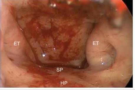
FIGURE 37.2 Endoscopic endonasal intraoperative view of the nasopharynx, which provides direct access to the craniocervical junction between the eustachian tubes (ET). Caudal access is limited by the hard palate (HP). The soft palate (SP) extends inferiorly from this and is not affected by an endonasal approach.
A suction monopolar electrocautery or microdebrider can be used to resect the soft tissue overlying the craniocervical junction (nasopharyngeal mucosa, basopharyngeal fascia, longus capitis, and rectus capitis anterior muscle attachments). Attempts to preserve the nasopharyngeal mucosa and underlying muscle for reconstruction are not successful due to restriction of the surgical field and the depth of the final surgical defect. For most anterior pathology of the craniovertebral junction, the lateral margin of the surgical field should be limited to the medial border of the eustachian tube (torus tubarius). The field can be widened by either retraction (with a suction tip) or resection of the torus tubarius. Care should be taken to evaluate the course of the parapharyngeal ICA since it can be directly subjacent to the eustachian tube. Radical resection of the medial eustachian tube is not necessary and may affect palatal function.
As the longus capitis and rectus capitis muscles are detached, their point of attachment should be identified. This supracondylar groove directly overlies and predicts the location of the hypoglossal canal in the depth and, as such, represents an important landmark if the condyle is to be exposed. Monopolar electrocautery is useful to completely remove fascial and muscle attachments from the foramen magnum and anterior arch of C1. Care should be taken if there is any gap between the foramen magnum and C1, though this is rare. The anterior tubercle of C1 should be identified since it serves as a valuable midline landmark.
An extended, downward curved drill with a thin, flexible drill bit allows extension of the caudal exposure. Additionally, reduction of the posterior maxillary crest (bony attachment of the nasal septum to the hard palate) to the plane of the hard palate will improve caudal access as this is the “highest” point of the hard palate. Care should be taken not to drill completely through the hard palate and violate the oral mucosa as this would create an oronasal fistula.
The anterior arch of C1 can be removed with a high-speed drill with a cutting or coarse diamond burr. For resection of the odontoid, the width of resection should be enough to expose the dens in its entirety (Fig. 37.3). Inferior access should be maximized at every step of the exposure. Once the dens is exposed, any soft tissue overlying its tip should be cauterized or resected with through-cutting sinus instruments. In cases of basilar invagination, the foramen magnum may need to be drilled to expose the invaginated odontoid peg. The dens should be drilled and removed from the tip down, as disconnection from the body of C2 at the neck would result in a floating tip with numerous dense ligamentous attachments that are difficult to detach from free-floating bone. Careful dissection of the inner cortex of the dens from the ligaments, especially at the tip, can be performed with extended tip dissectors (see instruments section below) or simply a Kerrison rongeur, with care taken to avoid further compression of the immediately subjacent neural tissues.
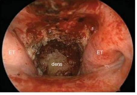
FIGURE 37.3 Endoscopic endonasal view following removal of the anterior arch of C1 to expose the dens/odontoid process. (ET, eustachian tubes.)
In cases of purely bony anterior compression, decompression to the tectorial membrane is adequate (Fig. 37.4). If the tectorial membrane is pulsatile and widely decompressed, it does not need to be removed. Even if there is extensive pannus, it can usually be debulked without fully exposing the underlying dura, since this dura is often thinned, friable, or eroded. If intradural resection of the tumor is intended, the tectorial membrane should be stripped to expose the dura completely. Intradural structures associated with the ventral craniocervical junction include the vertebral and anterior spinal arteries, hypoglossal nerves, and C1 nerve rootlets. With the exception of the anterior spinal artery, all of these are structures that originate laterally and are usually displaced by the pathology away from the surgeon. As always, intradural dissection should be performed with strict adherence to microsurgical, bimanual technique.
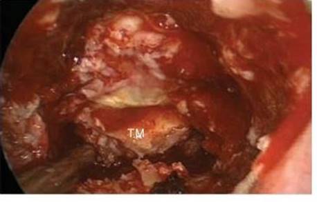
FIGURE 37.4 Endoscopic endonasal view following resection of the odontoid and cervicomedullary decompression, leaving only the tectorial membrane (TM) overlying the dura.
Intraoperative imaging is important in order to ensure adequate decompression. At a minimum, AP and lateral radiographs with instillation of radiopaque dye into the defect should be performed. We are fortunate to have intraoperative CT, which is an ideal way to confirm complete decompression (Fig. 37.5).
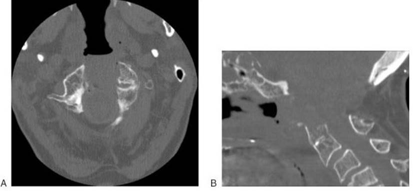
FIGURE 37.5 Intraoperative CT (A) axial and (B) sagittal reconstruction confirming adequate width and craniocaudal extent of decompression.
In cases of purely extradural pathology without inadvertent dural breach, simple coverage of the surgical site with fibrin glue is adequate for repair. Secondary scar formation and remucosalization will occur naturally and effectively. If there is a small dural tear, reconstruction with an adipose tissue graft filling the soft tissue defect is usually effective. The defect tends to be deep and narrow, ideal for filling with adipose tissue, and is at low risk for a postoperative CSF leak.
If there is a wide dural opening, reconstruction with vascularized tissue is recommended. The nasal septal flap, pedicled on the posterior nasal branch of the sphenopalatine artery, provides a robust flap, but it may not reach a deep, caudal defect such as can be created with intradural endonasal surgery. Often, the deep defect created after removal of the lower clivus, anterior arch of C1, and/or dens needs to be filled in with an adipose tissue graft and the flap placed over this with its edges in alignment with the nasopharyngeal mucosa. An onlay allo- or autograft (such as Alloderm, pericardium, or fascia lata) is often placed deep to the adipose tissue to provide dural reconstruction. Another vascularized option for craniocervical defects is an inferior turbinate flap.
Silastic splints are sutured to the nasal septum. Packing is not necessary unless there has been repair of a dural defect.
POSTOPERATIVE MANAGEMENT
Many patients with craniovertebral disease, especially degenerative or rheumatoid, will require a posterior arthrodesis, regardless of the anterior approach. This can be performed before or after an anterior decompression. When indicated, we prefer to perform the endonasal decompression first, since this provides rapid neural decompression, which may increase the safety of the posterior approach (as the cervicomedullary junction is no longer at risk from the anterior vector). It can also provide more bone surface for fusion since the posterior arch of C1 can often be left intact if there is adequate anterior decompression. Patients may undergo arthrodesis during the same anesthetic or the following day, depending on the length of surgery and associated patient factors.
If patients require fixation, they are left in either a hard cervical collar or halo vest for 2 to 4 weeks. A halo vest is not required with modern arthrodesis techniques, but may aid healing of the occipitocervical incision by avoiding all pressure on this area.
Regardless of other treatment, patients with intradural approaches should have typical endonasal postoperative precautions: avoidance of nose blowing with strict orders to avoid positive pressure ventilation and nasal instrumentation unless under direct endoscopic visualization. The head of bed is elevated to 30 degrees at all times for 2 weeks. Nasal splints are removed at 1 week if a septal flap has not been used and at 3 weeks if employed. Saline nasal sprays are instituted immediately postoperative, and saline irrigations can be started at 2 to 3 weeks. Periodic nasal endoscopy with debridement of nasal crusts is performed until healing is complete.
Patients with craniocervical disease should be evaluated pre- and postoperatively for signs of dysphagia, vocal cord paresis, or other lower cranial neuropathies, since this can result in aspiration and resultant lung injury. Most patients should only be fed once this has been assessed. However, one of the main advantages of an endonasal approach is that patients can resume a transoral diet immediately if safe and appropriate.
Patients who do not have preexisting or clear postoperative instability should be followed for sign or symptoms of progression. Regular cervical spine films with flexion/extension views should be obtained during the first 2 postoperative years or at any time when the patient develops new onset of pain in the neck, headache, or radiculopathy.
COMPLICATIONS
CSF leak remains a concern for any patient undergoing intradural endonasal surgery. Large dural defects in the craniocervical junction can prove a challenge for reconstruction, but tend to heal well. Injury to the lower cranial nerves can be subtle and go unnoticed until patients become quite sick (aspiration and associated complications) and thus should be carefully evaluated. Patients who have intraoperative vascular injuries, though rare with this approach, should undergo immediate angiography to determine the extent and consequences of the injury as well as need for further treatment such as endovascular coiling or sacrifice of the artery.
RESULTS
We conducted a retrospective review of all patients who underwent a purely endoscopic endonasal odontoid resection for decompression of the cervicomedullary junction from 2004 to 2010. Patient outcomes were assessed using the Neck Disability Index and NURICK cervical myelopathy scale. Special attention was also paid to complications and postoperative swallowing function.
Twenty-nine patients underwent complete endoscopic endonasal resection of the odontoid. The most common pathology (n = 23) treated was spondylitic pannus. Other pathologies included chondrosarcoma, meningioma, type 2 odontoid fracture, os odontoideum, and metastatic carcinoma. The mean patient age was 67.9 years. Twenty-seven of the twenty-nine patients underwent a posterior fusion.
All patients had either improvement or stabilization of their neurologic status (none worsened). NURICK and NDI data were available for 15 patients who had a mean follow-up of 27.6 months (range 3 to 57). The mean NURICK score postoperatively was 1.0, and no patient received a score of 5. The mean NDI score was 9.2 (range 0 to 32). Nine of twelve patients had an NDI score of less than 15.
Two patients had preoperative swallowing dysfunction, which required a preemptive gastrostomy. Postoperatively, it appeared there was no change in the need for supplementation. Five patients (17%) required placement of a gastrostomy tube for postoperative dysphagia. In two of these patients, the tube was removed within 6 weeks postoperatively as the patient resumed normal swallowing function. The remaining 23 patients (73%) were able to continue oral feedings with minimal or no problems. No patient was found to have developed permanent velopharyngeal dysfunction postoperatively although one patient did have velopharyngeal insufficiency preoperatively related to a prior inadequate transoral approach for odontoid resection. This patient did not experience worsening velopharyngeal insufficiency following the endonasal approach.
Prior to leaving the OR, all patients underwent either instillation of radiopaque dye under fluoroscopy or intraoperative CT to assess the adequacy of decompression. All patients underwent a postoperative CT scan to assess the adequacy of their decompression. No patient had evidence of residual compression, and no patient required a repeat anterior approach for further decompression.
Two patients required intraoperative repair of CSF leaks at the time of the initial surgery. In both patients, the dura was approximated with U-clips and augmented with Duragen and an autologous adipose tissue graft. Both patients were treated with lumbar drainage for 5 days and suffered no sequelae.
Respiratory complications were the most frequently encountered nonneurologic complication. Four patients experienced respiratory failure secondary to pneumonia. These four patients required a temporary tracheostomy. One additional patient required a tracheostomy for respiratory failure secondary to chronic pulmonary disease and deconditioning; this patient was eventually decannulated. No patient had a tracheostomy performed as part of the surgical procedure. Two patients had aspiration pneumonia treated with antibiotics. One patient had a deep venous thrombosis without evidence of pulmonary embolism and was placed on anticoagulation. One patient died 8 days following surgery from cardiac arrest.
PEARLS
· The NPL drawn on a midsagittal CT approximates the most inferior point of endonasal access.
· Reduction of the posterior maxillary crest with a drill until it is flush with the rest of the hard palate provides a significant increase in caudal access.
· In general, the approach should be chosen based on the relationship of the pathology to associated nerves and vessels. For anterior or midline pathologies that displace neurovascular structures posterolaterally, an endonasal approach is ideal. However, potential destabilization must be taken into account when addressing the craniovertebral junction, as this can have equal impact on patient outcome and quality of life.
PITFALLS
· Tortuous parapharyngeal ICAs may approach the midline of the nasopharynx and be at risk for injury.
· Failure to drill the tip of the dens before detaching it from the body of C2 makes it difficult to detach the ligaments.
· Care should be taken not to drill through the entire hard palate since an oral nasal fistula may occur if the underlying mucosa is violated.
INSTRUMENTS TO HAVE AVAILABLE
· A full set of standard sinus instruments (Storz).
· Extended tip dissectors (KLS Martin).
· An extended, downward curved drill with thin, flexible drill bit allows extension of the caudal access. Cutting and coarse diamond bits are ideal.
· Fine and angled tip, pistol-grip bipolar electrocautery forceps (Storz) are critical for extra- and intradural hemostasis.
SUGGESTED READING
Nayak JV, Gardner PA, Vescan AD, et al. Experience with the expanded endonasal approach for resection of the odontoid process in rheumatoid disease. Am J Rhinol 2007;21(5):601–606.
De Almeida JR, Zanation AM, Snyderman CH, et al. Defining the nasopalatine line: the limit for endonasal surgery of the spine. Laryngoscope 2009;119(2):239–244.
Gardner P, Kassam A, Spiro R, et al. Endoscopic endonasal approach to the odontoid. In: Mummaneni P, Kanter A, Wang M, et al., eds. Cervical spine surgery: current trends and challenges. St. Louis, MO: Quality Medical Publishing, 2009.
Morera VA, Fernandez-Miranda JC, Prevedello DM, et al. “Far-medial” expanded endonasal approach to the inferior third of the clivus: the transcondylar and transjugular tubercle approaches. Neurosurgery 2010;66(6 Suppl Operative):211–219, discussion 219–220.
Gardner PA, Tormenti MJ, Kassam AB, et al. Endoscopic endonasal approach to the odontoid and craniocervical junction. In: Kassam AB, Gardner PA, eds. Endoscopic approaches to the skull base. Progress in neurological surgery. Vol. 26. Basel, Switzerland: Karger, 2012:152–167.
Thirumala PD, Kodavatiganti HS, Habeych M, et al. Value of multimodality monitoring with brainstem auditory evoked potentials and somatosensory evoked potentials and in endoscopic endonasal surgery. Neurol Res 2013;35(6):622–630.