
Chandranath Sen
INTRODUCTION
Tumors involving the clivus are usually difficult to access because of their deep, midline location anterior to the brainstem and anterior to the cranial nerves. The position of the petrous portion of the temporal bone and the configuration of the tentorium are some of the limiting factors to the use of traditional approaches such as the retrosigmoid approach or a transsylvian approach, where a surgeon ends up working along a very long and narrow path leading to the tumor. The working area is very restricted, creating a less than satisfactory situation. The combined approach is designed to overcome some of these limitations. Partial or complete drilling of the temporal bone brings the surgeon closer to the target pathology, while cutting the tentorium enlarges the working space by allowing the surgeon to use both the supra- and infratentorial compartments, which are combined into one.
HISTORY
Since these tumors are usually slow growing, most patients will have very few complaints even with large tumors. Headache, gait, and balance problems are the most common symptoms.
PHYSICAL EXAMINATION
Neurologic examination may reveal ataxia, weakness of the upper or lower extremities. Compression of cranial nerves can produce hearing loss, diplopia, facial numbness or pain, and occasionally, facial weakness.
INDICATIONS
This approach is primarily useful for extra-axial tumors anterior and anterolateral to the brainstem. It can also be used for some intra-axial processes such as cavernous angiomas in the brainstem that come close to the anterolateral surface of the brain. Examples include intradural tumors such as meningiomas, schwannomas, and epidermoid tumors; extradural tumors such as chordomas and chondrosarcomas that may also have intradural extensions are also candidates. Some of the smaller tumors that are confined to the upper clivus can also be removed by the anterior transpetrosal approach (described in Chapter 32).
CONTRAINDICATIONS
There are no contraindications to using this approach.
PREOPERATIVE PLANNING
A high-quality MRI scan done before and after administration of gadolinium is essential. Meningiomas and schwannomas enhance vividly with gadolinium. A trigeminal schwannoma can occur anywhere along the course of the nerve, posterior fossa, middle fossa, and infratemporal fossa, or in a dumbbell-shaped configuration straddling the compartments. Those that occupy the posterior fossa and middle fossa are eligible for this approach. Meningiomas show vivid and uniform enhancement and have a broad dural base at the clivus and petrous bone (Fig. 38.1A and B). Schwannomas are more rounded in appearance. They may contain areas of cystic change. In most instances, there is an arachnoid plane separating the brainstem, the blood vessels, and the cranial nerves from the tumor. Infrequently, the tumor may violate the arachnoid and invade the pia causing edema of the brainstem. T2-weighted images and FLAIR sequences will demonstrate edema of the brainstem when present (Fig. 38.2A and B). A high T2 signal within the tumor may indicate that the tumor is soft in consistency. Hyperintensity or diffusion-weighted images are diagnostic of an epidermoid tumor. A CT scan is helpful if additional anatomical information about bone is needed with respect to the temporal bone. Evaluation of the surrounding vascular anatomy can be performed using MRI (Fig. 38.3A and B), an MR arteriogram and venogram, or a digital subtraction angiographic study. The MR arteriogram is adequate for assessing large vessels and venous sinuses, but a more detailed assessment of the arterial and venous system and flow characteristics is best obtained by an angiogram (Fig. 38.4A–C). If there is significant blood supply from the external carotid branches or the intracavernous internal carotid artery, embolization may be helpful for surgery. The venous anatomy and drainage is carefully evaluated with respect to the temporal draining veins and the size and dominance of the sigmoid sinus, in case the sigmoid sinus is ligated or gets injured during the operation.
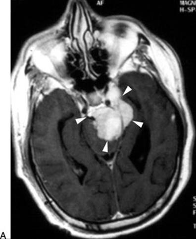
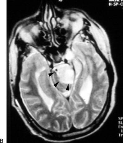
FIGURE 38.1 A. Axial MRI T1 with gadolinium showing the well-defined tumor left of the midline with extension into the left Meckel’s cave (arrowheads). B. Axial T2 shows tumor and brainstem border (arrowheads). There is no edema within the brainstem. The ventricles are enlarged in this case.
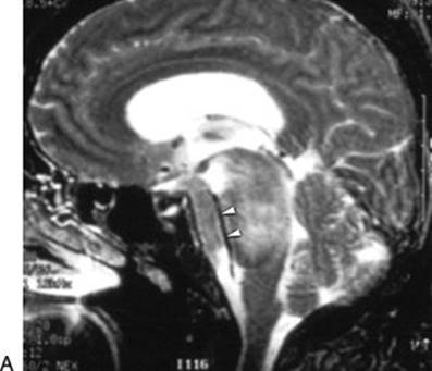

FIGURE 38.2 A. Sagittal T2 MRI shows encasement of the basilar artery within the tumor (arrowheads). B. There is extensive high signal within the brainstem indicating invasion of the pia by the tumor producing edema (asterisk); arrowheads indicate the tumor–brainstem interface; T, tumor.
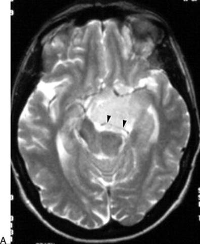
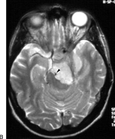
FIGURE 38.3 Axial T2 MRI showing that the tumor surrounds the posterior cerebral artery (arrowheads) (A) and the basilar artery (arrowhead) (B).
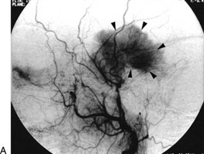
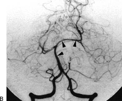
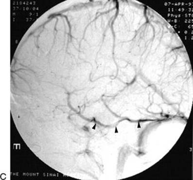
FIGURE 38.4 A. Cerebral arteriogram, external carotid injection showing the blood supply to the tumor from the middle meningeal artery (arrowheads). B. The basilar artery and its branches are displaced by the tumor (arrowheads).Figure 38.4(Continued) C. The venous phase shows a large temporal draining vein (arrowheads) coursing along the inferior surface of the temporal lobe to drain into the tentorium and then into the sigmoid–transverse junction. This venous channel must be preserved while the tentorium is being incised.
SURGICAL TECHNIQUE
Anesthesia, Intraoperative Neurophysiologic Monitoring, and Positioning
General intravenous anesthesia is used during the tumor phase of the operation because of the need for intraoperative neurophysiologic monitoring. Intra-arterial and venous lines are place. Continuous neurophysiologic monitoring of cranial nerves VII and VIII, somatosensory and motor evoked potentials, and transcranial facial nerve evoked potentials are very helpful during these operations. Lumbar spinal drains and external ventricular drains are usually not necessary. Broad-spectrum antibiotics and anticonvulsant prophylaxis are administered at the beginning of the operation.
The patient is positioned supine with a soft roll under the ipsilateral shoulder, and the head is turned to the contralateral direction about 60 degrees and stabilized in a three-point headrest (Fig. 38.5). The extent of the head rotation is for the surgeon to be able to visualize and access both the base of the tumor along the petrous ridge and the surface of the tumor pressing into the brainstem. The head is kept elevated relative to the chest and body to facilitate venous drainage.
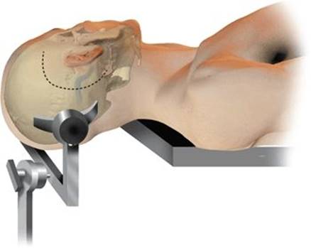
Figure 38.5 The patient is positioned with the head turned away from the side of the tumor about 60 degrees. The surgeon sits behind the patient. The incision is “C” shaped such that the posterior limb is about 1 inch posterior to the tip of the mastoid.
Scalp Incision and Soft Tissue Flaps
A large “C”-shaped flap is used located above, behind, and below the ear and centered at the ear (Fig. 38.5). The posterior limit of the incision should extend about an inch posterior to the mastoid tip. The scalp flap is elevated up to the external ear canal, which has to be carefully identified and separated from the bony ear canal. The muscle flaps are elevated superiorly and inferiorly, by making an incision horizontally separating the temporalis muscle from the nuchal muscles. The posterior temporal line, which is the posterior extension of the zygomatic arch, is identified, which separates the floor of the middle cranial fossa from the mastoid.
Craniotomy and Bone Work
The opening of the bone can be done in two ways: One way is to perform a craniotomy first and then a retrolabyrinthine mastoidectomy, and the other way is to perform the mastoidectomy first and then use that as the starting point for the craniotomy. In order to perform the craniotomy first, one burr hole is made above the sigmoid–transverse junction (Fig. 38.6A), at the junction of the squamosal suture with the posterior temporal line. The second burr hole is made below the sigmoid–transverse junction at the asterion (Fig. 38.6A and B). Since there is a deep groove housing it, the sinus can be readily injured here. Therefore, a bony trench is drilled joining the two burr holes. Once the dura has been separated from the bone, a combined temporal and posterior fossa craniotomy is made with the high-speed craniotome following the dotted line in Figure 38.6A. Traversing the posterior part of the transverse sinus with the saw is safe because there is no bony groove. The retrolabyrinthine mastoidectomy is now carried out unroofing the sigmoid sinus down to the jugular bulb and the superior petrosal sinus.
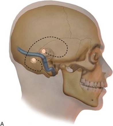

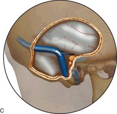
FIGURE 38.6 A–C. Sequence of the bony opening starting with the craniotomy. The burr holes A and B are made above and below the transverse/sigmoid junction as described in the text. In this sequence, the mastoidectomy is performed later in the bone removal.
In the other option, the mastoidectomy is performed first, skeletonizing the labyrinth and exposing the sigmoid sinus entirely from the sigmoid–transverse junction down to the jugular bulb. The subtemporal and presigmoid dura are exposed so that the superior petrosal sinus and the endolymphatic sac are fully unroofed. It is very important that the unroofing of the sigmoid sinus be carried out all the way down to the jugular bulb and the thin ridge of bone between the two structures be drilled down carefully. This reduces the venous bleeding from the area of the jugular bulb that can occur when the sigmoid sinus is retracted posteriorly during the tumor resection phase. The labyrinth must be fully skeletonized so that the maximal exposure of the dura between the sigmoid sinus and the posterior semicircular canal can be achieved. The petrous bone can be drilled down further anteriorly by performing a partial or complete labyrinthectomy and even by drilling down the cochlea to provide a larger exposure while minimizing brain retraction. A combined temporal and posterior fossa craniotomy is started at the mastoidectomy defect. The anterior extent of the temporal craniotomy should extend to above the external ear canal. The inferior extent of the posterior fossa craniotomy should be almost down to the foramen magnum. It must provide enough temporal exposure to allow for placement of a temporal lobe retractor without limiting the retractor by the bone edge of the craniotomy. Posteriorly, the craniotomy must extend far enough behind the sigmoid sinus to allow the sinus to be retracted posteriorly and also to allow the surgeon to perform a retrosigmoid exposure should it be needed in addition to the presigmoid exposure (Fig. 38.7).
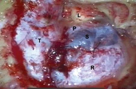
Figure 38.7 Intraoperative view of a right-sided exposure. The vertex is to the left, and the ear is superior. Dural exposure after completion of the bone work. (T, temporal lobe dura; R, retrosigmoid dura; S, sigmoid sinus; P, presigmoid dura; L, labyrinth.)
Dural Exposure and Opening
The brain must be relaxed prior to opening the dura. The exposure can be markedly compromised if the brain is tight. Administration of mannitol, hyperventilation, and elevation of the head are the usual maneuvers to facilitate relaxation of the brain. If hydrocephalus is present, an external ventricular drain can be placed prior to positioning for the craniotomy. Drainage of cerebrospinal fluid (CSF) can also be performed by making a limited opening in the posterior fossa dura, elevating the cerebellum and opening the cisterna magna or the lateral cerebellomedullary cisterns.
The temporal dura is opened horizontally along the lower part of the temporal lobe starting at the anteriormost limit of the temporal craniotomy and extending posteriorly past the sigmoid–transverse junction (Figs. 38.8A and 38.9). A “T”-shaped extension is made in the inferior direction toward the superior petrosal sinus. The temporal lobe is gently elevated to look for temporal bridging veins entering the venous sinuses below. These veins have to be preserved while cutting the superior petrosal sinus and the tentorium below. This dural incision has to be made anterior to these bridging veins, such that the veins will continue to drain posteriorly into the venous sinuses (Fig. 38.8B). The posterior fossa dura is opened vertically, in front of the sigmoid sinus and behind the opening of the endolymphatic sac. Inferiorly, it is taken down to the jugular bulb and superiorly, up to the superior petrosal sinus. The superior petrosal sinus is coagulated with the bipolar cautery, staying anterior to the temporal bridging veins, and then divided with microscissors (Fig. 38.10).
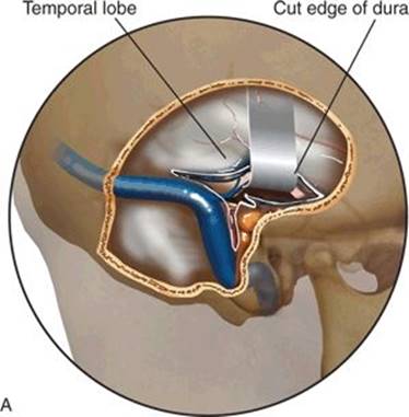
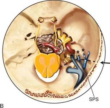
FIGURE 38.8 A. Dural opening: The temporal dura is opened in a direction parallel to the floor. The presigmoid dura is opened vertically from the jugular bulb up to the superior petrosal sinus. The temporal lobe is elevated to locate temporal draining veins. The superior petrosal sinus is coagulated, and tentorial incision is made anterior to these veins as shown in (B), in an axial view. The location of the tumor is shown relative to the direction of the approach and the tentorial incision. (SPS, superior petrosal sinus; **, temporal draining veins.)
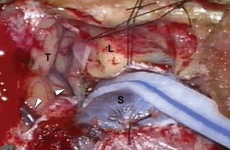
Figure 38.9 Intraoperative photo of the case in Figure 38.7. The temporal and presigmoid dura has been opened. Two separate temporal veins are noted to be draining posteriorly (arrowheads). (T, temporal lobe; L, labyrinth; S, sigmoid sinus.)
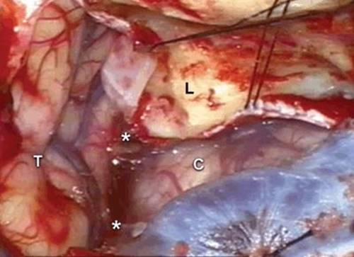
Figure 38.10 Intraoperative photo of the case in Figure 38.9. The tentorium has been divided. The two asterisk indicate the cut ends of the superior petrosal sinus. (T, temporal lobe; C, cerebellum.)
Prior to extending this incision into the tentorium, a Cottonoid patty is passed along the bottom of the temporal lobe without disrupting the draining veins and another one is over the cerebellum to protect these respective surfaces. The tentorium can be quite vascular, especially in the case of meningiomas, and needs to be coagulated prior to cutting. The incision is made parallel to the petrous ridge and slightly behind it all the way to the incisura. Prior to cutting down to the incisura, the trochlear nerve is visualized separated from the tentorium by a layer of arachnoid at a point posterior to its entry into the incisura. The anterior edges of the dura adjacent to the labyrinth are tented up with sutures (Fig. 38.9).
A retractor is placed to gently elevate the temporal lobe, which is being protected by a Cottonoid. This retractor must be placed such that the posterior leaf of the cut edge of the tentorium is lifted up with the temporal lobe and its draining veins all together such that there is no stretch or avulsion of these veins. The tumor can now be visualized anterior to the brainstem, nestled medial to the cranial nerves (Fig. 38.8B).
Tumor Removal
Careful dissection following the arachnoid planes is very important in order to separate the tumor from the cranial nerves, the vascular structures, and the brainstem. The cerebellum is carefully retracted posteriorly after dissecting it free from the seventh and eighth cranial nerves, which are usually displaced laterally and inferiorly by the tumor. The safest area to begin debulking the tumor is under the temporal lobe and above the seventh and eighth cranial nerves, where the trigeminal nerve root will be located, usually displaced laterally and inferiorly by the tumor. Thorough intracapsular debulking of the tumor is essential before any significant dissection, and separation of the tumor capsule can be carried out. An ultrasonic device with a fine tip is useful. Debulking is carried out by working in between the cranial nerves starting at the top and identifying the trigeminal nerve. The temporal lobe may need to be retracted upward while working on the upper pole of the tumor. However, continuous retraction of the temporal lobe should be avoided, and the retractor should be moved away while working on the other parts of the tumor. While debulking the tumor, it is preferable to stay toward the base of the tumor, which simultaneously devascularizes the body of the tumor since the blood supply is coming from anteriorly at the base. The 6th nerve is at risk of injury at the base of the tumor and needs to be carefully sought out and preserved. As the tumor is progressively debulked, the capsule gets softer and can be gradually delivered and cut away or removed with the ultrasonic aspirator. Pulling on a tense capsule prior to adequate debulking is dangerous since it can avulse vessels and damage cranial nerves.
The inferiormost limit of exposure by the presigmoid approach is up to the top of the 9th and 10th cranial nerves. If the lower pole of the tumor reaches below the level of these nerves, removing or delivering this may be difficult. In such instances, the retrosigmoid dura is opened to provide access to the lower pole of the tumor. Like all lateral approaches, the presigmoid as well as the retrosigmoid approach requires the surgeon to work in between the cranial nerves. It is therefore very important to be as gentle as possible with these nerves. Intraoperative neurophysiologic monitoring of these cranial nerves may be helpful to minimize the trauma.
Because of the curvature of the clivus and petrous bone, the attachment of the tumor at the ipsilateral part of the clivus and petrous bone is usually difficult to visualize from the lateral perspective of the presigmoid approach. The overhang of the labyrinth and petrous apex can be reduced by performing a partial labyrinthectomy in which the superior and posterior semicircular canals are drilled down and occluded with bone wax. If the patient has poor or no hearing, the entire labyrinth can be drilled away, uncovering the internal auditory canal over 270 degrees circumference. If this additional bone drilling is not an option, opening the retrosigmoid dura and looking along the cerebellum provides an excellent visualization of the ipsilateral clivus and petrous apex dura.
The degree of tumor removal is dictated by factors such as its size, consistency, adherence to important structures, and presence of an arachnoid plane. If the preoperative MRI shows a high T2 signal within the brainstem adjacent to the tumor, it indicates that there is invasion of the pia by the tumor and some tumor will have to be left against the brainstem in order to avoid permanent damage to the brainstem and perforator vessels (Fig. 38.2A and B).
Closure
The dura is closed primarily or with a dural graft to get as much of a watertight closure as possible. The mastoid is occluded with bone wax, and special attention is paid to the mastoid antrum. A small chip of bone and bone wax are used to close this off so that CSF is not able to enter it. The mastoid defect is filled with a free adipose tissue graft usually harvested from the abdomen. Fibrin glue is used to seal this in place. The craniotomy bone flap is replaced and fixed with titanium miniplates. A titanium mesh cranioplasty is performed to cover the mastoidectomy defect. The two muscle flaps are sutured back into position on to the titanium mesh, and the skin and subcutaneous tissues are closed. A subcutaneous drain tube is left in place, brought out through a separate stab wound and attached to a closed, gravity drainage system since CSF tends to build up and can stress the suture line. The drain is usually left in for 2 to 3 days.
POSTOPERATIVE MANAGEMENT
The patients are monitored in the neurosurgical intensive care unit. Temporal lobe edema and contusion are closely monitored as reflected by the patient’s level of consciousness. A noncontrast CT scan is performed the next day before the patient is transferred out of the unit and mobilized out of bed. Seizure prophylaxis is also maintained postoperatively. Sequential compression boots are maintained as deep vein thrombosis prophylaxis, and subcutaneous heparin or enoxaparin is given 24 hours after surgery.
COMPLICATIONS
Complications can be divided into the following:
1.Cranial nerve problems
2.Temporal lobe problems
3.Brainstem problems
4.Arterial injury
5.CSF leaks
1.Cranial nerve problems: The third through the tenth cranial nerves on the ipsilateral side are encountered during this approach. The tumors are usually located medial to these nerves, and hence, the surgeon has to work in between these nerves to debulk and remove the tumor. To make matters more complex, the nerves are often stretched posteriorly or even surrounded by the tumor, making them attenuated and compromised to begin with and therefore more vulnerable to manipulation. Temporary impairment of their function is often seen although they tend to improve with time. If a patient experiences diplopia or ophthalmoplegia, wearing an eye patch over the eye is sufficient until the function returns. In some cases, oculoplastic procedures may be needed if the function does not improve in 6 to 9 months. Conductive hearing loss can result from effusion in the middle ear space from the mastoidectomy and is self-limiting. Nerve deafness is irreversible. The incidence of facial palsy can be minimized by using facial nerve monitoring with direct stimulation as well as transcranial stimulation. Protection of the eye is important if a patient has complete facial paralysis. Placement of a gold weight and lubrication of the eye need to be rigorously implemented, and the patient has to be educated regarding these. A combined trigeminal nerve and facial nerve deficit is a serious problem for the eye. A tarsorrhaphy may be needed in order to avoid repeated corneal ulcers and eventual blindness. Lower cranial nerve palsies will result in dysphagia and hoarseness. A modified Barium swallow test is performed early in the postoperative period if significant manipulation of the lower cranial nerves has been performed. Since postoperative nutrition is very important in the recovery of the patient, gastrostomy tube placement is considered early to minimize the chance of malnutrition and aspiration pneumonia. In most instances, this is only a temporary measure and the tube can be removed in a few months when the patient resumes oral intake.
2.Temporal lobe problems: The majority of problems with the temporal lobe are caused by retraction and injury to the veins draining to the temporal lobe that travel from the undersurface of the temporal lobe to the tentorium. Prevention is the main strategy, since, once the problems have started, the neurologic consequences are serious and can be irreversible, especially on the side of the dominant temporal lobe. There can be more than one vein in this area, and all of them need to be preserved. The tactics described above need to be diligently implemented. The surgeon should retract sparingly and intermittently, by working in different portions of the tumor instead of working in one part of the tumor for a long period of time needing prolonged retraction.
3.Brainstem problems: It is very important to review the preoperative T2-weighted and FLAIR MR images prior to the operation. High signal within the brainstem adjacent to the tumor indicates that the tumor has violated the arachnoid and the pia producing edema in the brainstem. The dissection plane is absent in these situations, and also the perforating arteries to the brainstem are often adherent to the tumor or even supply the tumor. This situation presents a risk of direct injury to the brainstem while peeling the tumor capsule or perforator injury resulting in a stroke. In these situations, it is advisable to leave a portion of tumor on the brainstem.
4.Arterial injury: The basilar and vertebral arteries and their branches are often intimately involved with these tumors. If there is complete encasement of the vessel, there is a high risk of injury and the surgeon has to be extremely cautious in such cases. The risk increases in firm tumors and vascular tumors because visualization of the structures and planes becomes more difficult in such cases. Blood supply to the tumor from the vertebrobasilar system and its branches seen on an angiogram is a warning sign and demands meticulous technique and judgment as to aggressiveness of resection.
5.CSF leak: During closure, the mastoid antrum leading to the middle ear space needs to be closed thoroughly and completely. Watertight dural closure is not possible in most instances, and a dural substitute is placed over the dural repair as well as over the drilled surface of the mastoid. A sealant is used to hold these in place, and an adipose tissue graft is laid on top of this.
RESULTS
This approach allows excellent access to the tumors described above. The resectability of these tumors depends on several factors. The consistency of the tumor plays an important role; the softer the tumor, the easier it is to remove it without undue manipulation of the nerves or the brainstem. If the tumor invades important blood vessels or even surrounds them, complete removal may not be possible due to the risk of causing a brainstem infarct. If the plane between the tumor and the brainstem is not defined by a distinct arachnoid layer, the surgeon will be obligated to leave some tumor behind on the brainstem to avoid injury. Tumors that are very vascular are a particular challenge because the blood supply comes from its attachment at the clival dura. The surgeon does not gain access to the blood supply till quite late in the procedure, and therefore devascularization of the tumor early on is not usually possible unless some preoperative embolization has been carried out.
PEARLS
· Careful analysis of the preoperative imaging studies is critical in defining whether the approach is suitable for the lesion to be treated.
· The patient and the family need to be thoroughly educated regarding the potential risks and how they will be managed. The recovery process can be quite stormy, and it is important that a good understanding and relationship be established with the patient and family prior to the surgery to avoid their dissatisfaction.
· The patient must be positioned carefully so that the head is not turned too sharply compromising the venous return. This can produce a “full brain” that will severely limit the exposure and can even prevent the surgeon from proceeding with the operation. If a full brain is encountered after opening the dura, the surgeon may elect to place a ventricular drain in the lateral ventricle through the temporal lobe, elevate the head, or even “un-turn” the head to some degree to achieve relaxation.
· The temporal bone should be drilled down to the jugular bulb, and there should be no bone left on the sigmoid sinus. This is important in order to be able to mobilize the sigmoid sinus adequately to work in front of it.
PITFALLS
· If careful arachnoid plane dissection is not followed, damage to the cranial nerves, the blood vessels, and the brainstem will likely occur with many adverse consequences.
· Retraction of the sigmoid sinus for any prolonged period should be avoided to prevent thrombosis of this structure.
INSTRUMENTS TO HAVE AVAILABLE
· Surgical drill
· Reciprocating saw
· Temporal lobe retractor
· Bipolar electrocautery
· Ultrasonic aspirator
· Titanium mesh
SUGGESTED READING
Abdel Aziz KM, Sanan A, van Loveren HR, et al. Petroclival meningiomas: predictive parameters for transpetrosal approaches. Neurosurgery 2000;47:139–150.
Cho CW, Al-Mefty O. Combined petrosal approach to petroclival meningiomas. Neurosurgery 2002;51:708–718.
Little KM, Friedman AH, Sampson JH, et al. Surgical management of petroclival meningiomas: defining resection goals based on risk of neurological morbidity and tumor resection rates in 137 patients. Neurosurgery 2005;56:546–559.
DelaCruz A, Teufert KB. Transcochlear approach to cerebellopontine angle and clivus lesions: indications, results and complications. Otol Neurotol 2009;30:373–380.