
William T. Couldwell
INTRODUCTION
Several pathologies can be addressed with a far lateral approach to the lower clivus and upper cervical spine. The most common lesions for which surgeons would choose this approach are skull base tumors, such as meningiomas of the foramen magnum and lower clivus, and vascular lesions, such as aneurysms of the vertebral artery or posteroinferior cerebellar artery (PICA). Less common lesions include clival chordomas and tumors of the lower cranial nerve sheath, in addition to metastases from various sites and occasionally an extensive glomus tumor. The approach is chosen to provide a lateral approach to enable the surgeon to remove tumors and treat vascular lesions situated lateral and anterior to the upper cervical spine and medulla (Fig. 39.1).
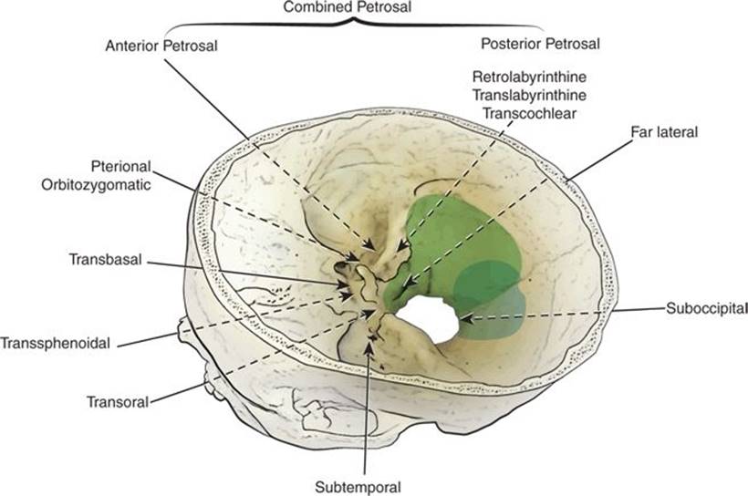
Figure 39.1 Illustration of various approaches to the cranial base. The far lateral transcondylar transtubercular approach (dark gray shading) provides a posterolateral trajectory to the craniovertebral junction to access lesions in the inferior clivus and ventral foramen magnum. The midline suboccipital approach (light gray shading), which provides direct access to the posterior foramen magnum, is limited in accessing ventral or ventrolateral lesions of the foramen magnum.
HISTORY
The most common symptoms of a lesion of the lower clivus and upper cervical spine are headache and pain in the neck. If the lower clivus and condyles are involved, as in cases with an extensive chordoma, pain may be associated with movement of the head. Schwannomas of the lower cranial nerves may have symptoms referable to the involved nerve (e.g., weakness of the tongue, hoarseness, or difficulty swallowing). If the lesion is large enough, there may be compression of the cervicomedullary junction, with associated long tract signs such as weakness or numbness, which is usually asymmetric. In such cases, upper motor neuron weakness, with spasticity, gait difficulties, and occasionally sensory loss, may be present.
Obviously, in cases of aneurysmal subarachnoid hemorrhage (SAH), the presentation will involve acute severe headache and impairment of the neurologic sensorium. Severe meningismus will accompany the SAH with neck rigidity. There is nothing specific to the presentation of a ruptured aneurysm of the posterior circulation in most cases, but a local hematoma may produce specific cranial nerve deficits, such as hearing loss or swallowing difficulty.
PHYSICAL EXAMINATION
During a thorough neurologic evaluation, particular attention should be paid to the cranial nerve examination. Even in the absence of symptomatology, subtle cranial nerve deficits may be noted. A diminished gag reflex may be the result of cranial nerve IX and X deficits that may not have reached the magnitude for the patient to note difficulty with swallowing. Long tract signs may manifest as upper motor weakness and spasticity with increased tone and associated reflexes in the affected arm or leg. Similarly, there may be associated sensory deficits of various modalities. Gait evaluation may reveal ataxia with a positive Romberg sign. In addition to the signs and symptoms of SAH, I have also seen presentation of dissecting aneurysms, common in the vertebral artery and PICA, presenting with specific ischemic syndromes, such as the lateral medullary syndrome of Wallenberg.
INDICATIONS
Classic indications for a far lateral approach include pathology anterior to the lower brainstem and upper cervical spine. This includes meningioma (at the foramen magnum or upper anterior spine in location), chordoma of the lower clivus or upper cervical spine, lower cranial nerve schwannoma, or vascular lesion (most commonly aneurysms) involving the vertebral artery or PICA.
CONTRAINDICATIONS
Contraindications for this approach are few, but include congenital anomalies of the upper cervical spine, which might predispose to instability and perhaps an isolated vertebral artery when the contralateral vertebral artery is compromised. This latter condition could increase the chances of devastating posterior circulation ischemia with injury to a sole vertebral artery. Relative contraindications to the approach are paralysis of the contralateral vocal cord or other dysfunction of the lower cranial nerves, which would predispose to a life-threatening bilateral vocal cord paralysis or swallowing difficulties with additional cranial nerve injury on the operated side.
PREOPERATIVE PLANNING
In most instances, magnetic resonance imaging with contrast enhancement is the best imaging study to characterize the nature of the tumor in the upper cervical or clival region. The signal characteristics of specific tumors are characteristic in this location. A meningioma usually will be isointense with the brain on T1-weighted imaging and will avidly enhance with the administration of gadolinium contrast. Chordomas may involve the bone of the clivus or upper cervical spine and are usually erosive. They do enhance with contrast administration, but more specifically are of high signal intensity on T2-weighted imaging, which is a useful tool to differentiate from other tumors such as metastasis. Any involvement or encasement of major vessels, often the vertebral artery or PICA at its origin, should be noted.
In cases of aneurysms, computed tomography (CT) will more accurately demonstrate subarachnoid blood and the predominant location (cervicomedullary cistern or 4th ventricle) may indicate a vertebrobasilar or PICA location. In these cases, a CT angiogram (CTA) study is indicated, which may demonstrate the aneurysm. In some cases, with extensive bone and beam-hardening artifacts in the base of the skull, a conventional digital subtraction angiogram will be necessary to demonstrate the vascular lesion, particularly in cases with dissecting aneurysms.
The rationale for choosing a far lateral approach to the cervicomedullary junction is to provide a more lateral trajectory to the region to avoid or reduce retraction on the brainstem and cerebellum (Fig. 39.1). The region is replete with important cranial nerves and the vertebral and basilar arteries and their branches and perforating vessels. During preoperative planning, imaging is carefully studied, noting the size and location of the tumor and its relationship to the brainstem and vasculature. It is common to encounter tumors in this location that completely encase one vertebral artery in its intradural course. As such, the potential for injury during resection must be considered during planning, and, in such cases, I like to document bilateral vertebral arteries that have a normal course and anastomose at the basilar junction. A common variant is a small vertebral artery on one side that terminally ends in PICA; in such cases, a balloon occlusion test of the involved vertebral artery should be performed, and planning will include repair or revascularization by bypass if the vessel cannot be sacrificed safely.
An important consideration when resecting tumors of this region is the stabilization of the craniocervical junction. I have had several cases in which extensive chordomas and glomus tumors in this location have resulted in bone erosion such that after their removal the craniocervical junction is destabilized by the tumor erosion and additional drilling of the bone. This needs to be anticipated, and I have performed both simultaneous and staged craniocervical stabilization procedures with spine specialists in these cases.
With vascular lesions, especially aneurysms of the vertebral artery or PICA, surgical planning will include any options for endovascular treatment of these lesions. If an open surgical approach is deemed the optimal treatment, planning for treatment will include anticipating options such as vessel sacrifice if tolerated or clip–wrap and bypass techniques.
SURGICAL TECHNIQUE
The location of the tumor, its area of attachment, and involvement of any vascular structures are noted and included in the decision-making process. The extent of drilling of the skull base and condyle is not uniform and is tailored to the particular tumor or vascular lesion. In general, more bone drilling is used for extensive chordomas in this location than for other tumors. The amount of the condyle removed is planned based on the trajectory needed. In most cases of vertebral artery or PICA aneurysms, extensive resection of the condyle or drilling of the jugular tubercle is not necessary.
It has been estimated by some authors that the posteromedial one-third of the occipital condyle may be resected without inducing instability. This issue should be considered on opening, and it should be determined prior to drilling whether bone removal in combination with tumor erosion will result in destabilization of the unilateral condyle region. I have had experience with such cases in which, in addition to the condyle itself, the bone superior to the condyle was involved with tumor such that the condyle was disconnected from the remainder of the skull base, which produced the same destabilizing result.
Far Lateral Transcondylar Approach
The patient is placed in the lateral position with the head in Mayfield three-point fixation (Fig. 39.2), with extensive padding between the legs and in the axilla. The upper arm is supported by an airplane rest and padded accordingly. An important feature of positioning is that the upper arm is directed inferiorly to pull the ipsilateral shoulder as low as possible. This, in combination with lateral flexion of the neck, provides more operative room in the lateral suboccipital–cervical region. A Foley catheter and arterial line are placed prior to positioning.

FIGURE 39.2 A> . Diagram showing the patient placed in the lateral position. The head is held in three-point pin fixation with the neck slightly flexed, the vertex angled slightly down, and the face slightly rotated ventrally, so that the ipsilateral external auditory meatus and the mastoid bone are at the highest point. B. Retroauricular curvilinear skin incision (dotted line).
Several options have been described for skin incision for a far lateral exposure. I use one of two options depending upon the extent of exposure noted and whether the need for the occipital artery for revascularization is anticipated. The first is a curved incision extending from the retromastoid region (approximately 2 to 3 cm behind the mastoid) to the lateral neck down to approximately C4 in most cases (Fig. 39.2). This approach provides rapid exposure to the region of interest with the least soft tissue disruption and postoperative pain. Alternatively, if more exposure is needed to the midline for placement of stabilization hardware or in cases involving harvesting of the occipital artery, I use a larger incision extending to the midline and then laterally in a hockey-stick fashion producing a flap. The occipital artery may then be dissected as necessary in preparation for bypass.
The skin flap is elevated in two layers. First, the incised skin and galea are elevated to expose the pericranium above the superficial fascia of the neck, which may be harvested as a fascial graft for later watertight dural closure. The pericranium and the superficial fascia are then elevated to expose the underlying musculature.
During the dissection, three layers of muscle are identified. The superficial layer, including the trapezius and sternocleidomastoid muscles, and the middle layer, consisting of the splenius capitis, longissimus capitis, and semispinalis capitis muscles, are incised and reflected as a single layer posteriorly. This exposes the suboccipital triangle (Fig. 39.3), which is opened by detaching the insertions of the superior and inferior oblique muscles from the transverse process of C1 and reflecting them posteriorly. The rectus capitis major is detached from the inferior nuchal line and reflected posteriorly, after which the C1 lamina and vertebral artery will become more apparent. Further exposure of the laminae of C2 or C3 may be performed if more inferior exposure is needed.
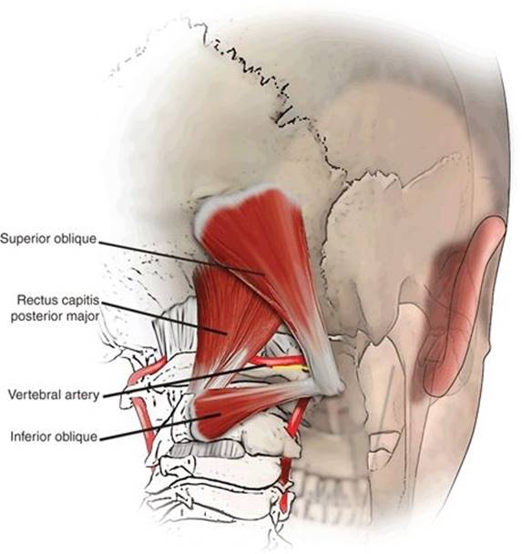
Figure 39.3 Illustration showing the suboccipital triangle, which is bound medially by the rectus capitis posterior major (RCM) muscle, inferiorly by the inferior oblique (IO) muscle, and superolaterally by the superior oblique (SO) muscle and serves as an anatomic landmark for identifying the dorsal ramus of the C1 nerve root and the V3 horizontal segment of the vertebral artery (VA).
The extradural course of the vertebral artery from the foramen transversarium of C2 to the occiput is identified to facilitate exposure. The ventral ramus of the C2 nerve root, found between the laminae of C1 and C2, can be traced laterally until it crosses dorsal to the vertical segment of the vertebral artery, coursing between the foramen transversarium of C2 and C1 (Fig. 39.4). Small muscular branches and the posterior meningeal artery arising from the horizontal segment of the vertebral artery can be safely coagulated. In some cases, the posterior spinal artery and the PICA arise extradurally; care must be taken to avoid injury to these structures. Subperiosteal dissection of the vertebral artery from the vertebral groove reduces bleeding from the venous plexus by leaving the periosteal sheath around the artery intact. The atlanto-occipital membrane is sharply divided to expose the underlying dura. Vertebral artery transposition can be performed by opening the foramen transversarium of C1 with a high-speed diamond drill and mobilizing the artery inferomedially away from the atlanto-occipital joint. Although transposition is not necessary in most cases for the standard far lateral transcondylar approach, this maneuver is important for gaining a direct lateral (extreme lateral) trajectory to resect the lateral mass of C1, the lateral aspect of the occipital condyle, the odontoid process, and the inferior clivus, as described in the extreme lateral approaches for resection of extradural lesions of the craniovertebral junction. Subsequent occipitocervical stabilization, which is necessary if the atlanto-occipital joint is resected, can be performed unilaterally with the same surgical exposure immediately after resection.

Figure 39.4 Diagram of the course of the vertebral artery. The ventral ramus of the C2 nerve root, found between the laminae of C1 and C2, can be traced laterally until it crosses dorsal to the vertical segment of the vertebral artery, coursing between the foramen transversarium of C2 and C1. As the vertebral artery exits the foramen transversarium of C1, it is encased in a venous plexus and courses posteriorly behind the lateral mass of C1 in the vertebral groove and turns medially to pierce the atlanto-occipital membrane and dura.
I use a craniotome and rongeurs initially to create a lateral suboccipital craniectomy or craniotomy extending toward the midline medially, to the inferior nuchal line superiorly, to the posterior rim of the foramen magnum inferiorly, and up to the occipital condyle laterally (Fig. 39.5). For more superior access to the cerebellopontine angle, I extend the craniectomy up to the transverse–sigmoid junction. Then, the sigmoid sinus and jugular bulb are exposed by using rongeurs and a high-speed drill. The posterior condylar emissary vein will be encountered as it travels from the jugular bulb and exits the condylar fossa via the condylar canal to join the extradural venous plexus. Hemostasis can be achieved by packing the vessel with Surgicel. An ipsilateral hemilaminectomy of C1 extends the exposure of the dura inferiorly, and removal of the hemilamina of C2 and C3 provides additional inferior exposure for lower-lying lesions.
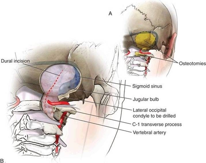
FIGURE 39.5 A. Illustration of retrosigmoid lateral suboccipital craniectomy (dotted line), extending toward the midline medially, to the inferior nuchal line superiorly, to the posterior rim of the foramen magnum inferiorly, and up to the occipital condyle laterally. B. The craniectomy can be extended up to the transverse–sigmoid junction to provide more superior access to the cerebellopontine angle, as shown. The dural incision (dotted line) is made in a curvilinear fashion several millimeters posterior to the sigmoid sinus and extends inferiorly toward the C2 lamina, staying posterior to the vertebral artery where it pierces the dura. A relaxing “T” incision is made just superior to the entry of the vertebral artery, leaving a cuff of dura around the vertebral artery for later watertight closure.
Extradural reduction of the occipital condyle is one of the key maneuvers in maximizing exposure to the ventral aspect of the craniovertebral junction while avoiding brainstem retraction (Fig. 39.6). Partial resection of the condyle increases the angle of exposure, the working space at the level of the foramen magnum, and the visualization of the ventral and ventrolateral aspect of the craniovertebral junction and the contralateral aspect of the inferior clivus. Although the degree of removal of the occipital condyle necessarily varies widely, from no resection to complete resection, my experience indicates that removal of the posterior and medial one-third of the condyle usually is adequate if more ventral exposure is needed. It is important to study preoperative CT images of the cranial base, because not all patients require resection of the condyle. Resection of the condyle may not be necessary to widen the surgical corridor (Fig. 39.7) if the patient has small occipital condyles and a large foramen magnum or if the tumor has eroded the condyle and displaced the brainstem medially. The craniovertebral junction becomes more unstable when more than 50% of the condyle is resected or destroyed by the lesion; in these patients, occipitocervical stabilization may be necessary. The posteromedial aspect of the occipital condyle is removed with a high-speed diamond drill, taking care to protect the vertebral artery (Fig. 39.8). Once the cortical layer of bone is removed, the soft cancellous bone is encountered. Further drilling will expose another cortical layer of bone that covers the hypoglossal canal. Identification of the medial aspect of the hypoglossal canal usually indicates that approximately one-third of the posterior condyle has been removed. Because the hypoglossal canal is directed anteriorly and laterally at a 45-degree angle with the sagittal plane, further skeletonization of the canal to its lateral extent usually results in removal of the lateral aspect of approximately the posterior two-thirds of the condyle. Removal of bone is next directed superiorly toward the inferior margin of the jugular bulb.

Figure 39.6 Illustration showing reduction of the occipital condyle (OC) and jugular tubercle (JT), which increases the angle of exposure and visualization of the ventral foramen magnum past the midline of the clivus (shaded triangle).

FIGURE 39.7 A. Diagram demonstrating a ventral foramen magnum tumor compressing the brainstem. A far lateral transcondylar transtubercular approach (shaded in gray) provides excellent exposure for a lesion in this region. B. A very large tumor of the clivus compressing the ventral and lateral aspects of the brainstem that has eroded the occipital condyle. A far lateral approach without transcondylar or transtubercular resection would be sufficient in removing this tumor because the tumor has created a large surgical window by displacing the brainstem medially. Postoperative occipitocervical stabilization should be strongly considered because the tumor has eroded a significant portion of the occipital condyle.

FIGURE 39.8 A. Illustration showing inferior view of the base of the cranium and the anatomic relationship of the jugular tubercle, hypoglossal canal, and occipital condyle. The jugular tubercle is situated superior to the hypoglossal canal. The occipital condyle is inferior to the hypoglossal canal. B. Extradural reduction of the posterior third of the occipital condyle and the superomedial aspect of the jugular tubercle is the key maneuver for gaining access to the ventral foramen magnum.
Extradural reduction of the jugular tubercle is the key step in maximizing intradural exposure across the anterior surface of the brainstem and midclivus (Fig. 39.8). This maneuver also may make it easier to visualize some vertebrobasilar junction aneurysms and vertebral artery–PICA aneurysms. Failure to reduce a prominent jugular tubercle adequately may result in an obstructed view of the basal cisterns and clivus anterior to the lower cranial nerves. Reduction of the jugular tubercle should focus on the superomedial aspect portion, which is the major area of obstruction. Where they cross over the deep aspect of the jugular tubercle into the jugular foramen, cranial nerves IX, X, and XI may be at risk of damage by direct trauma, stretching of the dura mater, and heat generated by the drill. To minimize these risks, the center of the tubercle is cored out with a high-speed diamond drill and copious irrigation, leaving an eggshell-thin layer of bone covering the dura mater that can be elevated with a microdissector.
Intradural exposure is necessary for accessing intradural lesions, such as meningiomas, schwannomas, aneurysms, and vascular malformations (Figs. 39.9 and 39.10). I make a curvilinear incision of the dura several millimeters posterior to the sigmoid sinus, extending inferiorly toward the C2 lamina and staying posterior to the vertebral artery where it pierces the dura mater (Fig. 39.5). I prefer to extend the dural opening anteriorly in a “T” fashion, just superior to the vertebral artery, to enable increased exposure. The incision can be extended up to the junction of the transverse–sigmoid sinus if more exposure of the cerebellopontine angle is needed. A dural cuff is preserved around the vertebral artery for later watertight closure. The anterior leaflet of the dura mater is reflected laterally and is held with tacking sutures for maximal exposure. Adequate reduction of the occipital condyle and jugular tubercle should provide a straight surgical trajectory to the craniovertebral junction parallel to the intracranial course of the vertebral artery. Structures of the inferior aspect of the cerebellopontine angle and the cerebellomedullary angle are visualized. Sharp arachnoid dissection is performed, and cranial nerves V through XII, the basilar artery, the vertebral artery, the vertebrobasilar junction, the PICA, and the anteroinferior cerebellar artery can be visualized (Fig. 39.10).

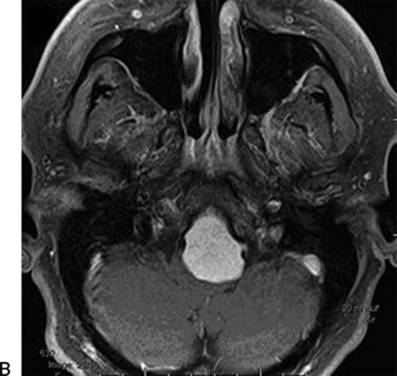
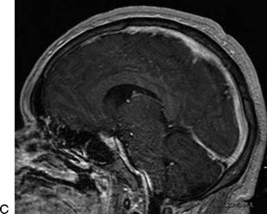
Figure 39.9 Preoperative MRI scans. Sagittal FLAIR (A) and axial T1 with contrast (B) views demonstrate a ventrally based foramen magnum meningioma compressing the cervicomedullary junction in a 36-year-old man who presented with progressive myelopathy. Gross total resection was achieved by use of a far lateral approach. C. Postoperative sagittal MRI scan.

Figure 39.10 Diagram illustrating intradural exposure of the far lateral approach. (AICA, anteroinferior cerebellar artery; PICA, posteroinferior cerebellar artery.)
Closure
A primary watertight closure of the dura should be performed. If necessary, an autologous pericranium or fascial graft can be harvested from the neck wound and supplemented with autologous adipose tissue and fibrin glue or fascia lata or abdominal fascia may be used. The exposed mastoid air cells are closed with bone wax. The muscle layers are reapproximated carefully to avoid postoperative leakage of cerebrospinal fluid (CSF). Temporary CSF diversion with a lumbar drain can be used to promote sealing of the wound and reduce the risk of a pseudomeningocele developing.
In Video 39.1, a 56-year-old man has suffered a severe subarachnoid hemorrhage from a ruptured proximal fusiform aneurysm of the left posteroinferior cerebellar artery. The plan was to either repair the aneurysm after inspection or perform distal bypass and trap the aneurysm. Shown in the video is the harvesting of the occipital artery from the flap in preparation for the bypass if needed. Ultimately, the aneurysm was clip–wrapped with muslin gauze to prevent rebleeding.
POSTOPERATIVE MANAGEMENT
The patient is monitored in the intensive care unit postoperatively. Depending on the nature of the lesion, the major risks are damage to the lower cranial nerves and vertebral artery. Careful airway management is essential, and evaluation of vocal cord and swallowing function is performed in the intensive care unit to determine if there is a risk for aspiration.
COMPLICATIONS
The most common complication noted is development of CSF pseudomeningocele from lack of an adequate dural closure. To prevent this complication, I use an adipose tissue graft extradurally to eliminate dead space and bolster the dural closure. The abdomen or lateral thigh may be used for donor sites. The most serious complications relate to vascular injury to the vertebral artery or lower cranial nerve palsy (swallowing, airway protection, or hypoglossal nerve weakness). Rarely, overresection of the occipital condyle–C1 joint may lead to instability of the neck.
RESULTS
The approach is ideal for the resection of a meningioma of the foramen magnum (Fig. 39.9). The surgeon has an excellent trajectory anterior to the brainstem and upper cervical spine. Similarly, this is the approach of choice for aneurysms of the vertebral artery or proximal aspect of the PICA. The wound heals well, with a cosmetically acceptable scar whether a curvilinear or larger incision is used. The patient may complain of stiffness of the neck early postoperatively, but, in the absence of instability or fixation, should be reassured that full mobility of the neck will resume with time (usually a few weeks). If the smaller curvilinear incision is used, the postoperative pain is minimized as there is relatively little posterior cervical muscular disruption.
PEARLS
· Prior to surgery, a rigorous evaluation includes magnetic resonance imaging, with evaluation of vascular involvement by computed tomographic angiography or digital subtraction angiography if necessary.
· Preoperative counseling of the patient should include a thorough discussion of the possibility of lower cranial nerve dysfunction.
· Meticulous bimanual microsurgical dissection is the best option for safe surgical removal. Blunt dissection is to be avoided, and careful attention is paid to all perforating vessels to the brainstem and to careful preservation of all lower cranial nerves.
PITFALLS
· Attention must be paid to minimize intraoperative manipulation of lower cranial nerves since unilateral lower cranial nerve palsies may be significantly morbid and bilateral palsies are potentially fatal.
· CSF leak or pseudomeningocele development is a risk if closure of the dura is not performed adequately. Dural closure is challenging in this region and use of dural grafts is usually necessary.
INSTRUMENTS TO HAVE AVAILABLE
· Needle-tip monopolar electrocautery
· Bipolar cautery
· High-speed cutting (3 to 4 mm) and diamond (3 to 4 mm) drills
· Microscope
· Suction–irrigator or surgical assistant to provide irrigation while drilling
· Microsurgical dissectors
ACKNOWLEDGMENTS
The author would like acknowledge the superb editorial help of Kristin Kraus. Parts of the intraoperative description were adapted from Liu JK, Rao G, Schmidt MH, Couldwell WT. Far lateral transcondylar transtubercular approach to lesions of the ventral foramen magnum and craniovertebral junction. Contemp Neurosurg 2007;29(10).
SUGGESTED READING
Bertalanffy H, Seeger W. The dorsolateral, suboccipital transcondylar approach to the anterior portion of the craniocervical junction. Neurosurgery 1991;29:815–821.
Liu JK, Couldwell WT. Far-lateral transcondylar approach: surgical technique and its application in neurenteric cysts of the cervicomedullary junction. Neurosurg Focus 2005;19(2):E9.
Liu JK, Rao G, Schmidt MH, Couldwell WT. Far lateral transcondylar transtubercular approach to lesions of the ventral foramen magnum and craniovertebral junction. Contemp Neurosurg 2007;29(10):1–8.
Garber ST, Couldwell WT. Resection of an inferior pontine cavernous malformation using a far-lateral transcondylar approach. Acta Neurochir 2011;153:2461–2464.