
Cerebrospinal fluid (CSF) rhinorrhea is a condition that in most patients is not diagnosed early unless it is precipitated by trauma, either penetrating or blunt. CSF leaks encountered during sinus surgery could be the result of progression of the disease process, which may be inflammatory or neoplastic in origin. Benign or malignant tumors may produce spontaneous CSF leaks due to local growth that may penetrate the dura or the arachnoid membrane. CSF leaks can be divided into four categories in order to plan a repair of the dural defect: spontaneous leaks, leaks resulting from penetrating trauma, leaks resulting from blunt trauma, and surgically planned leaks.
HISTORY
It is important to elicit a careful history in the diagnosis and management of CSF leaks. A history of clear nasal discharge following nasal trauma is useful in assessing the etiology of the condition. Spontaneous clear nasal discharge is usually an indication of a leak from one of the sites of nonfusion of the skull base, or erosion from a lesion at the same region. Blunt, nonpenetrating trauma produces leaks by an imploding pattern of injury. This contrasts with surgical trauma following endoscopic surgery in the skull base region where there is a clear site of penetration.
PHYSICAL EXAMINATION
Physical examination in patients with CSF leaks is strictly limited to an endoscopic examination of the nasal cavity under topical vasoconstriction. Endoscopic examination with a 0-degree and a 30-degree rigid endoscope is valuable in searching for the anatomic location of the defect and the surrounding pathology (Fig. 40.1). The nasal discharge should be collected in a clear tube and analyzed for beta-2 transferrin enzyme, which is the most accurate laboratory test for diagnosing CSF leak. This enzyme is contained in the CSF, vitreous humor, and cochlea. Collection of a minimal amount of 0.4 mL is adequate to make the diagnosis biochemically.

Figure 40.1 Zero-degree nasal endoscopic view of the left middle meatus revealing a CSF leak from an encephalocele in the roof of the ethmoid sinus.
INDICATIONS
All dural defects associated with CSF leaks should be surgically closed in order to prevent meningitis or cerebritis, which otherwise will ensue.
CONTRAINDICATIONS
There are relatively few contraindications for this surgical procedure. If a CSF leak is seen in the face of an infection, it may be wise to treat the inflammation or infection prior to the surgery. This is especially seen in patients with spontaneous leaks or when there is evidence of blunt trauma to the head. It is preferable to have stable mucosa that has better potential to heal well postoperatively.
PREOPERATIVE PLANNING
In the preoperative planning of the reconstruction of defects in the skull base, radiologic examinations in the form of a CT scan and MRI scan are absolutely necessary. A CT scan with a triplanar view for image guidance will help in analyzing the defect and also help in the surgical planning. These are usually nonoverlapping axial scans of the head with sagittal and coronal reconstructions (Fig. 40.2). The second imaging modality that should be obtained in all of these cases is an MRI scan with contrast. This helps to identify the extent of brain and meningeal involvement in these defects. They also help to identify important neurovascular structures near the site of the defect. When planning a surgical repair of these defects, the choice of nonvascular material for closure of small defects is usually an autologous homograft. The best choices are a free adipose tissue graft and fascia lata graft, which have the highest potential to heal well. The use of the lumbar drain should be considered in select cases where the potential to obtain a satisfactory result is improved by decreasing intracranial pressure for a short period of time. This is usually the case in spontaneous leaks where the patient’s body mass index is high and associated with high intracranial pressure.


Figure 40.2 Coronal (A) and sagittal (B) CT scan of the sinuses revealing a CSF leak from an encephalocele (arrows) of the left lateral lamellar area and the roof of the ethmoid sinus.
In my experience, the use of intrathecal fluorescein has been a valuable adjunct in identifying the site of the CSF leak in the skull base. Occasionally, there may be multiple leaks that may be overlooked when there is one particular site that is large. This is usually seen in congenital, small skull base bone defects. Fluorescein is injected after a lumbar puncture at the beginning of the surgical procedure. Patients are usually pretreated with diphenhydramine and dexamethasone upon arrival to the operating room, prior to induction of anesthesia. The dosage of fluorescein is usually 0.25 mL of 10% fluorescein diluted in 10 mL of CSF. The use of blue light with or without an amber blocking filter is also helpful in certain cases (Fig. 40.3). It should be noted that this use of fluorescein is off-label. I usually favor a team approach with a neurosurgeon for the closure of these defects, as it is helpful in the entire management of the patient and improves the outcome.


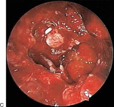
FIGURE 40.3 A. Endoscopic view of the roof of the sphenoid sinus with CSF leak seen with fluorescein staining (arrow) with and without blue light. B. Blue light filter view of CSF leak with fluorescein staining. C.Endoscopic view after complete closure with no evidence of leak.
SURGICAL TECHNIQUE
The patient is turned in lateral position, and a lumbar puncture is performed. If a lumbar drain is planned, it should be placed at this time. Now 10 mL of CSF is withdrawn and diluted with 0.25 mL of 10% fluorescein and reinjected into the intrathecal space. The patient is placed in the supine position. Head pins are then placed, and the image guidance system is calibrated for accuracy. CT scan image guidance is used in most cases. Dual MRI scan image guidance can also be used simultaneously.
Once the patient is prepared for surgery, cottonoids soaked in 4 mL of 4% cocaine are inserted into the intranasal cavity for vasoconstriction. This is helpful in diagnostic intraoperative endoscopic examination. Once the defect is localized, a regional vascular strip injection for ethmoidectomy, sphenoidotomy, or frontal recess surgery is carried out based on the location of the defect. It is ideal to have a clear view of the site of the defect prior to instrumentation. The bone defects in the skull base are usually well identified with a 30-degree or a 45-degree rigid endoscope, and the edges of the defect are freshened up. Small defects require careful attention during this process, as aggressive instrumentation may enlarge the defect, requiring different forms of closure. Small skull base defects are usually easily identifiable. For low-pressure, low-volume leaks, a free adipose tissue graft, harvested from the anterior abdominal wall, can be placed using an underlay technique for adequate closure. Once closed, endoscopic visualization with blue light may be used to verify a watertight closure. Subsequently, Duraseal and Floseal can be applied in that area to augment the closure. Defects in the roof of the sphenoid, the ethmoid, or the nasal cavity medial to the vertical lamella of the middle turbinate are relatively easy to close as the plane of closure is horizontal, and positioning the graft is convenient and uncomplicated. Small leaks in the lateral lamella of the frontal recess or the lateral wall of the sphenoid sinus require more careful planning, as the plane of closure is oblique and requires careful attention when freshening up the defect prior to closure. Finally, a Telfa sponge is applied to support the graft.
If the defect is large enough to warrant a fascia lata graft (2 cm × 2.5 cm), a gasket seal closure should be considered. This helps to improve the rate of successful closure in small defects that are more challenging. The edges of the bone are first freshened up, and fascia lata is harvested from the lateral aspect of the thigh. The graft is fashioned to be larger than the defect and then inset into the defect. A Medpor prosthesis is then countersunk into the defect to ensure complete closure at the edges of the defect (Fig. 40.4). Duraseal is usually applied thereafter, followed by Floseal, which is a useful adjunct in these closures. Once the procedure is completed, the patient is extubated without coughing to ensure that the graft is not displaced. Antibiotics are used in the high-risk category of patients: patients with resolving infections, those with comorbid conditions, and patients with poor wound healing conditions.
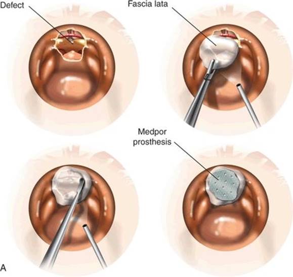

FIGURE 40.4 A. Schematic view of the various steps of gasket seal closure. B.Schematic coronal view of the gasket seal demonstrating closure of the edges of the leak and with obliteration of the dead space with adipose tissue when there is an indication for it.
POSTOPERATIVE MANAGEMENT
Postoperative management of these patients should be carefully planned and carried out. I usually see patients 1 week postoperatively to examine the wound healing process. Careful endoscopic debridement is useful in removing the epithelial debris and crusts to enhance wound healing. I have my patients use saline spray mixed with an antibiotic to ensure rapid healing. Patients are then seen at 3-week intervals until the wound is well epithelialized as confirmed by nasal endoscopy (Fig. 40.5). Patients are advised to refrain from physical activity for a period of 3 weeks after surgery.
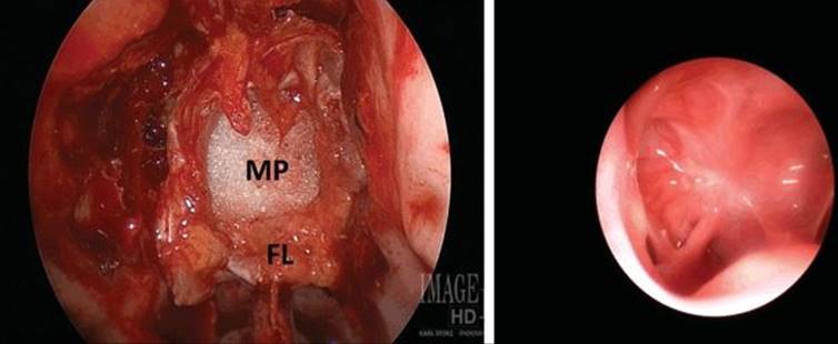
Figure 40.5 Endoscopic view of the gasket seal closure intraoperatively (FL, fascia lata; MP, Medpor prosthesis) and a healed gasket seal of the roof of the ethmoid sinus.
COMPLICATIONS
Postoperative complications are relatively uncommon following endoscopic closure of small defects. Recurrent leaks due to poor wound healing or displacement of the graft in the postoperative period are the most common complications. Meningitis, cerebritis, brain abscess from an infected graft, unexplained headaches, and seizures are very unusual complications that one may encounter in the closure of small defects. The position of the Medpor prosthesis is usually not exposed after wound healing is complete. Occasionally, granulation may be seen at the surgical site, and gentle debridement promotes successful healing.
RESULTS
The wound healing and the choice of the graft material with the technique of closures usually have a major role in the success of the closure. The closure rate is usually in the high 90% range and improved further by placement of a gasket seal.
PEARLS
· An accurate radiologic, biochemical, and endoscopic diagnosis should be made prior to surgery.
· The etiology and the mechanism of the leak should be evaluated prior to surgery.
· The choice of graft material must be planned prior to surgery.
· Consider short-term lumbar drainage (48 hours) in spontaneous, small high-pressure leaks.
· Use of intrathecal fluorescein is a valuable adjunct with image guidance for identifying the site of leak for closure.
· A team approach with a neurosurgeon leads to improvement of the results of CSF leak closure.
· Endoscopic postoperative surveillance of the surgical site is absolutely necessary until the wound heals well.
· It is important to have longer instruments than the conventional sinus instruments to access these areas in the skull base.
PITFALLS
· Poor preoperative evaluation with poor planning always leads to poor results.
· Surgical closure in the presence of an infection at the surgical site area leads to a higher incidence of graft rejection.
· Compromising on the choice of instruments to use in endoscopic skull base surgery leads to poor outcome including leaks.
INSTRUMENTS TO HAVE AVAILABLE
· Standard endoscopic sinus surgery instruments
· Endoscopic skull base surgery instruments
SUGGESTED READING
Hegazy HM, Carrau RL, Snyderman CH, et al. Transnasal endoscopic repair of cerebrospinal fluid rhinorrhea: a meta-analysis. Laryngoscope 2000;110:1166–1172.
Tabaee A, Placantonakis DG, Schwartz TH, et al. Intrathecal fluorescein in endoscopic skull base surgery. Otolaryngol Head Neck Surg 2007;137(2):316–320.
Leng LZ, Brown S, Anand VK, et al. “Gasket-seal” watertight closure in minimal-access endoscopic cranial base surgery. Neurosurgery 2008;62(5 Suppl 2):ONSE342–ONSE343.