
Allan Vescan
INTRODUCTION
The nasoseptal flap (NSF), based on the posterior septal branch of the sphenopalatine artery, is a popular flap used to repair defects in the skull base. It is versatile and reliable and can be used to repair defects of the anterior, middle, clival, and parasellar skull base. Along with multilayered reconstruction, it has reduced the incidence of postoperative cerebrospinal fluid (CSF) leaks to that of open approaches (3% to 5%).
The flap was first described in 2006 as a novel reconstruction technique by Hadad et al. and was named the Hadad-Bassagasteguy flap (HBF). More commonly, the flap is described as the nasal-septal flap (NSF). The NSF has been described in the reconstruction of a variety of skull base defects, and its use has been reported in patients as young as 14 years of age. In my personal experience, it can be used in younger patients with favorable anatomy.
HISTORY
A full rhinologic history should be obtained, including a history of recurrent sinus infections, chronic sinusitis, previous septoplasty or other sinonasal surgery, trauma, and granulomatous diseases. Patients should be asked about their use of topical nasal sprays and of cocaine abuse. A previous septoplasty is not a contraindication to a NSF but will make raising the flap more challenging.
PHYSICAL EXAMINATION
Office-based endoscopy with rigid or flexible endoscopy must be performed preoperatively. The septal flap is usually harvested from the right side of the nasal septum; however, it can also be harvested from the left side if the approach dictates sacrifice of the right pterygopalatine fossa contents. Septal deviation, previous septoplasty, septal spurs, or perforations should be considered when planning for the harvest of the NSF.
INDICATIONS
· CSF leak repair
· Reconstruction of anterior fossa, middle fossa, clival (posterior fossa), and parasellar skull base defects
· Vascularized coverage of internal carotid artery
CONTRAINDICATIONS
· Previous posterior septectomy
· Large septal perforation
· Previous wide sphenoidotomy with sacrifice of posterior septal artery
· Transpterygoid approaches requiring sacrifice of the sphenopalatine artery
· Tumor involvement of the septum or pterygopalatine fossa
· Embolization of terminal vasculature of internal maxillary artery
PREOPERATIVE PLANNING
· Examine the CT scan to look for a septal deviation and for evidence of previous septal surgery.
· If a revision case, review previous operative notes to understand what septal surgery was performed.
· Determine the length and width of flap based on the extent of planned skull base resection.
· Determine the side of maximal tumor dissection (transpterygoid approach).
SURGICAL TECHNIQUE (VIDEO 42.1)
General anesthesia is induced and the patient placed in the supine position. The nasal mucosa is decongested with one-inch neuropledgets soaked in 1:1,000 topical adrenaline. The right side is usually selected for flap harvest, although the left is an appropriate option. The septum is infiltrated with 1% lidocaine with 1:100,000 epinephrine in the submucoperichondrial and submucoperiosteal plane. The inferior turbinate is out fractured, and the middle turbinate is usually resected.
Exposure should then develop to allow for the planned inferior and superior incisions and includes resection of the inferior two-thirds of the superior turbinate and a posterior ethmoidectomy (Figs. 42.1 and 42.2). To aid visualization of the surgical field, insert a flexible suction catheter in the other nasal cavity to suction the plume from electrocautery. The inferior incision, performed with a needle-tip monopolar cautery, starts at the lateral aspect of the roof of the choana and is brought medially onto the vomer and then anteroinferiorly onto the nasal floor (Fig. 42.3). This incision is then continued anteriorly to the squamociliary junction, at the nasal vestibule.

Figure 42.1 Initial view of posterior nasal cavity. (C, roof of choana; SF, anterior wall of the sphenoid sinus; S, septum; NP, nasopharynx; IT, inferior turbinate.)
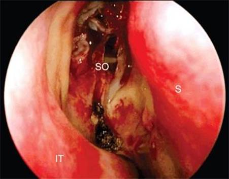
Figure 42.2 Endoscopic view after middle turbinectomy and posterior ethmoidectomy. (S, septum; IT, inferior turbinate; SO, sphenoid ostium.)
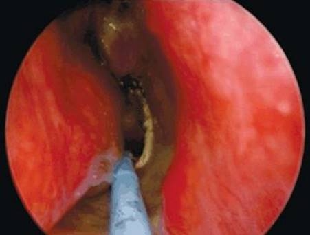
Figure 42.3 Inferior incision. Note how it is brought from the roof of the choana, onto the vomer, and then anteroinferiorly onto the floor of the nose.
The superior incision starts at the ostium of the sphenoid sinus and is then brought anterosuperiorly in a sharp curve to 1 cm below the skull base (Fig. 42.4). This is then brought anteriorly to the same vertical plane as the anterior limit of the inferior incision, and a vertical incision connects the two (Fig. 42.5).

Figure 42.4 Superior incision beginning at the ostium of the sphenoid sinus, curving sharply superiorly onto the septum.
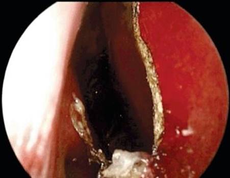
Figure 42.5 Anterior vertical incision connecting the two incisions.
The flap is then elevated in the submucoperichondrial and submucoperiosteal plane, back onto the face of the sphenoid, and as far as possible laterally (Fig. 42.6). This can be done with a combination of a Cottle elevator and endoscopic scissors. The flap is tucked into the nasopharynx until the tumor is removed, and it is needed for reconstruction (Fig. 42.7). The location of the pedicle must be kept in mind during the surgery so that it is not injured, especially during drilling of the floor of the sphenoid sinus. Alternatively, if a clival resection is planned, a wide antrostomy is performed and the flap is stored in the maxillary sinus to avoid obstruction of the surgical field throughout the procedure.

Figure 42.6 Flap elevated in the submucoperichondrial/submucoperiosteal plane. The free end is reflected posteriorly to aid visualization and to provide tension. (SC, septal cartilage; HBF, Hadad-Bassagasteguy flap.)

Figure 42.7 Flap elevated laterally off the anterior wall of the sphenoid sinus and tucked into nasopharynx. (V, vomer.)
As part of the repair of the defect, the flap is brought up from the nasopharynx, usually with a suction in the left hand and neurosurgery pituitary forceps in the right. The flap is smoothed out in its normal rotation so that the mucosal side is facing externally and the pedicle not kinked. Ensure that mucosa is not folded on itself to help prevent late mucocele formation (Fig. 42.8).

Figure 42.8 The nasoseptal flap is placed as an overlay, ensuring close approximation with underlying tissue.
In addition to the NSF, a multilayered reconstruction can help ensure a CSF leak–free postoperative outcome. This is usually done with collagen matrix (Duragen®) as a dural underlay to reconstitute the arachnoid plane, followed by the NSF as an extradural, extracranial bony overlay. The edges of the flap are covered with Surgicel, and the whole area is matted with fibrin glue. Saline-soaked Gelfoam follows, and a Foley catheter is gently inflated and buttressed against the repair. Doyle Silastic splints are sutured across the septum.
In general, because the flap is harvested before the actual size of the defect is known, the surgeon should plan a generous-sized flap to overcompensate. If, however, a larger defect is anticipated, such as in a resection of the anterior skull base, the flap can be made larger by extending the inferior incision into the inferior meatus and the superior incision to the cribriform plate. Conversely, anticipated small defects can be covered with a smaller flap, with the incisions on the septum, and with the anterior incision further posteriorly.
POSTOPERATIVE MANAGEMENT
General principles of management of a CSF leak are recommended. The Foley catheter is removed 3 to 5 days later, and the Doyle splints are removed at 3 weeks postoperatively. The patient is instructed to use gentle saline irrigations on a daily basis. The patient is seen at 4 to 6 weeks postoperatively and regularly thereafter for gentle debridement without disrupting the flap.
COMPLICATIONS
Complications of the NSF are generally mild and well tolerated. Complications related to elevation of the flap include a septal perforation, epistaxis, nasal crusting, and loss of olfaction. In patients with prior septal surgery (septoplasty, transseptal approach to skull base), the dissection of the flap is more difficult and the contralateral mucosa is prone to injury. The contralateral mucosa should also be protected from instrumentation during surgery. Nasal crusting is a consequence of the loss of normal nasal mucosa and exposed cartilage. This can be minimized by covering the flap donor site with a free mucosal graft from a resected middle turbinate. Epistaxis is usually a consequence of bleeding from the flap pedicle or a branch of the sphenopalatine artery. Bipolar cautery should be used sparingly since excessive cauterization may injure the vascular pedicle of the flap.
The most significant complication is a postoperative CSF leak. Potential risk factors are myriad and include patient factors, technical issues, and perioperative care. Inadequate coverage of the defect requires supplementation with nonvascularized tissue (fascia or adipose tissue grafts). Factors contributing to failure with CSF leak include inexperience, high intracranial pressure, large defects, and lack of a multilayer closure supported by a Foley catheter. Flap necrosis due to injury to the vascular pedicle is extremely rare. The best management for a postoperative CSF leak is usually early surgical intervention with endoscopic repair supplemented with a lumbar spinal drain.
RESULTS
There are increasing numbers of studies reporting on the successful use of the NSF for a variety of indications. A recent retrospective review of 32 patients reported a CSF leak rate of less than 2.5%. Another study reported no leaks in 14 patients when used to repair traumatic CSF leaks. In a meta-analysis of the skull base literature, reconstruction with vascularized tissue has been shown to be superior to nonvascularized tissue for the repair of large dural defects and results in fewer postoperative CSF leaks.
Quality of life studies demonstrate that sinonasal outcomes (SNOT-22) are temporarily decreased following reconstruction with a NSF, but these differences disappear after several months. Temporary adverse effects of the flap include nasal congestion, crusting, and decreased olfaction. A permanent decrease in olfaction has been associated with the use of NSFs, but the mechanism is unclear.
PEARLS
· Obtain proper exposure (including middle turbinate resection and posterior ethmoidectomy) to help plan the incisions.
· Use a guarded needle-tip monopolar cautery. Bend the tip 45 degrees.
· The inferior incision should always be on bone. If it is brought out inferior to the vomer into soft tissue, elevation of this area will be difficult.
· Ensure all areas of the incision are through the mucosa before attempting elevation of the flap.
· When elevating the flap, flip the free edge posteriorly to provide tension and expose the remaining area to be separated.
· Be careful to not injure the pedicle when drilling the floor of the sphenoid sinus; protect the pedicle with a suction.
· Store the septal flap in the maxillary sinus for transclival approaches.
· Modifications of the flap are possible to meet the anticipated size of the defect and location requirements.
PITFALLS
· Previous septoplasty can make it very challenging to harvest the flap.
· Exercise caution with the superior incision as this can lead to olfactory loss.
INSTRUMENTS TO HAVE AVAILABLE
· Guarded needle-tip monopolar cautery
· Cottle elevator
· Endoscopic scissors
· Flexible tracheal suction catheter
ACKNOWLEDGMENTS
The author would like to thank Dr. Kristian Macdonald FRCSC for his effort in the creation of this chapter and capturing video and still images of NSF harvest and inset.
SUGGESTED READING
Snyderman CH, Janecka IP, Sekhar LN, et al. Anterior cranial base reconstruction: role of galeal and pericranial flaps. Laryngoscope 1990;100:607–614.
Neligan PC, Mulholland S, Irish J, et al. Flap selection in cranial base reconstruction. Plast Reconstr Surg 1996;98: 1159–1166; discussion 1167–1158.
Hadad G, Bassagasteguy L, Carrau RL, et al. A novel reconstructive technique after endoscopic expanded endonasal approaches: vascular pedicle nasoseptal flap. Laryngoscope 2006;116:1882–1886.
Kassam AB, Thomas A, Carrau RL, et al. Endoscopic reconstruction of the cranial base using a pedicled nasoseptal flap. Neurosurgery 2008;63:ONS44–ONS52; discussion ONS52-43.
Shah RN, Surowitz JB, Patel MR, et al. Endoscopic pedicled nasoseptal flap reconstruction for pediatric skull base defects. Laryngoscope 2009;119:1067–1075.
Eloy JA, Choudhry OJ, Friedel ME, et al. Endoscopic nasoseptal flap repair of skull base defects: is addition of a dural sealant necessary? Otolaryngol Head Neck Surg 2012;147(1):161–166.