
Robert E. Merritt
 INDICATIONS/CONTRAINDICATIONS
Video-assisted thoracic surgery (VATS) left upper lobectomy is a minimally invasive technique that is indicated for resection of stage I lung cancers located in the left upper lobe. Induction therapy, chest wall invasion, and endobronchial tumor location are relative contraindications for VATS lobectomy. The VATS left upper lobectomy is contraindicated in patients who have had a previous pleurodesis procedure or in patients with an FEV1 predicted of 40% or less. The reported benefits for VATS lobectomy include less postoperative pain, shorter hospital length of stay, and a faster recovery.
The author believes that patients with an FEV 1 that is less than 40% predicted should be considered for a VATS sublobar resection or stereotactic radiosurgery as opposed to VATS lobectomy. The risk for pulmonary complications is higher when the preoperative FEV1 is less than 40% regardless of the technique (VATS or open thoracotomy). An FEV1 of less than 40% predicted is not a contraindication for lobectomy; however, the perioperative risk will be higher for pulmonary complications.
 PREOPERATIVE PLANNING
Preoperative staging and a complete medical assessment should be completed for all patients undergoing VATS lobectomy. A complete history and physical examination should uncover medical comorbidities that may increase the perioperative risk for lobectomy. A chest CT scan and an integrated PET CT scan should be obtained to evaluate patients for lymph node metastasis or distant metastatic disease. Lymph nodes that are larger than 1 cm in diameter or have a maximum SUV value above 2 should undergo further staging with either cervical mediastinoscopy or endobronchial ultrasound (EBUS). Patients with metastatic carcinoma involving the N2 mediastinal lymph nodes should be referred for induction chemoradiotherapy.
Prior to VATS left upper lobectomy, a complete cardiopulmonary evaluation is necessary to identify patients who are at risk for perioperative morbidity and mortality. Complete pulmonary function tests with spirometry and diffusion capacity should be obtained. Patients with an FEV1 less than 50% predicted or a diffusing capacity less than 50% predicted should be referred for quantitative perfusion scanning and exercise oxygen consumption testing. Patients with a maximum consumption (VO2max) of less than 10 mL/kg/min are at high risk for lobectomy and should be considered for referral for stereotactic radiosurgery.
 SURGERY
VATS Left Upper Lobectomy
The multiple variations of pulmonary artery anatomy that can be encountered during a left upper lobectomy make this particular VATS procedure the most challenging lobectomy. The VATS left upper lobectomy should be performed with extreme caution. The left main pulmonary artery may give off as many as seven branches to the left upper lobe.
Positioning
I routinely perform a fiberoptic flexible bronchoscopy before every lobectomy procedure to assess the airway anatomy and detect aberrant anatomy that may alter the operative plan. After the fiberoptic flexible bronchoscopy is completed with a single-lumen endotracheal tube, the patients are reintubated with a double-lumen endotracheal tube for selective lung ventilation. A right-sided double-lumen tube is preferable for a VATS left upper lobectomy because the subcarinal space is easier to dissect without a rigid endotracheal tube in left mainstem bronchus. Patients are then positioned in the right lateral decubitus position for left VATS upper lobectomy. All pressure points should be padded with gel pads to prevent nerve compression and skin necrosis. A padded bean bag is used to hold the patient in position. The operating room table is flexed with the patient’s anterior-superior iliac crest below the break in the bed. The flexion opens up the rib spaces to allow placement of VATS ports. The video monitors are placed at the head of the table and the surgeon stands in front of the patient.
Incisions
Three standard VATS incisions are used for left upper lobectomy (Fig. 26.1). The first incision is placed in the eighth intercostal space posterior axillary line. A 5-mm trocar is placed through the incision and 5-mm thoracoscope inserted in the left chest. The left hemithorax is inspected for evidence of pleural dissemination, such as a pleural effusion or tumor implants on the chest wall. The 5-mm thoracoscope is lower profile and is less likely to place torque on the intercostal nerve. A 10-mm incision is placed 3 to 4 fingerbreadths below the tip of scapula. A 4-cm utility incision is placed on the anterior border of the latissimus dorsi muscle. The particular interspace for the utility incision is based on the location of the left superior pulmonary vein. The index finger can be placed through the access incision for direct palpation of lung nodules. A 10-mm incision is also placed anteriorly at the level of the inframammary crease. I use low profile lung clamps and vascular clamps, which easily pass through the VATS incisions without placing pressure on the intercostal nerve.
Lymph Node Dissection
The lymph node evaluation is an essential component of VATS lobectomy. Patients who have metastatic disease involving interlobar or mediastinal lymph nodes will typically be referred for adjuvant chemotherapy. The author prefers to perform the lymph node dissection prior to completion of the lobectomy. The clearance of the mediastinal and interlobar lymph nodes will often provide better exposure to the arterial and vein branches. The 9L lymph nodes are located in the inferior pulmonary ligament, which is incised with bovie electrocautery and the nodes can be collected with a ring clamp. Next the mediastinal pleura over the posterior hilum is incised up to the AP window. The subcarinal lymph nodes can be assessed by retracting the pericardium and the left mainstem bronchus anteriorly and dissecting the space anterior to the thoracic aorta. Placement of a right-sided double-lumen tube will facilitate this maneuver.

Figure 26.1 Incision 1 is the anterior 10-mm incision. Incision 2 is the 10-mm camera port. Incision 3 is the 3- to 4-cm utility incision. Incision 4 is the 10-mm posterior incision. Rib spreading is not utilized during VATS lobectomy.
The level 5 lymph nodes are located in the AP window between the aortic arch and the left main pulmonary artery. The level 5 lymph nodes can be grasped with a ring clamp and dissected with a harmonic scalpel or Bovie. Injury to the recurrent nerve should be avoided in this location. The level 6 para-aortic lymph nodes can be dissected along the aortic arch. The interlobar lymph nodes (11L) are located along the left upper lobe bronchus and between branches of the pulmonary artery. The interlobar lymph nodes should be removed during dissection of the pulmonary artery branches and the left upper lobe bronchus. In general, lymph nodes should be removed intact as opposed to removing them in pieces. Surgicel can be utilized to improve hemostasis after dissection of the lymph node stations.
Dissection of the Superior Pulmonary Vein
The left upper lobe is retracted posteriorly with a lung clamp, thus exposing the anterior hilum. The superior pulmonary vein is located anterior to the left pulmonary artery and branches into the apicoposterior branch and the lingular branch (Fig. 26.2). The mediastinal pleura is incised with a harmonic ultrasonic scalpel. The left phrenic nerve should be identified and avoided during this step. The left superior vein can be dissected with an endo-Kittner until the vessel is free of all of the areolar tissue. A right-angle clamp is then passed behind the vein and a heavy silk suture is passed behind the superior vein. A linear endo-GIA stapler with a vascular reload is placed through the posterior VATS incision and is passed behind the superior vein and the structure is divided (Fig. 26.3). The left main pulmonary artery lies posterior to the superior pulmonary vein, so care must be taken to avoid injury to the pulmonary artery when dissecting the vein posteriorly.
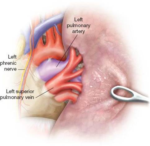
Figure 26.2 The left superior pulmonary vein dissection. The left main pulmonary artery courses behind the vein. The left phrenic nerve is located medially to the pulmonary artery and the superior vein.
Dissection of the Anterior Trunk Branch of the Left Pulmonary Artery
The anterior trunk branch of the left pulmonary artery should be readily visualized after division of the superior pulmonary vein. The interlobar lymph nodes should be removed along the upper lobe bronchus and anterior trunk. A thoracoscopic right-angle vascular clamp is then passed behind the anterior trunk branch and gently spread to clear space for the passage of the endoscopic stapler (Fig. 26.4). Excessive torque should be avoided on the pulmonary artery to avoid avulsion of the branches and subsequent hemorrhage. Once the anterior trunk branch is completely dissected, a linear endo-GIA stapler is passed behind the vessel, which is then divided.

Figure 26.3 The division of the left superior pulmonary vein with a linear endoscopic stapler.
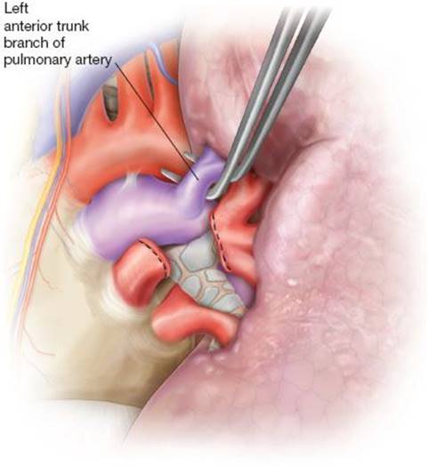
Figure 26.4 The left anterior trunk branch of the pulmonary artery being dissected with a right-angle clamp.
Completion of the Anterior Major Fissure and Division of the Lingular Artery
The left upper lobe is then retracted superiorly and the lower is retracted inferiorly. Once the main pulmonary artery is visualized; the overlying pleura can be divided with the endoscopic stapler to complete the incomplete anterior fissure. A vascular clamp can be used to dissect the overlying lung tissue and identify the tract of the stapler passage. The underlying pulmonary artery should be in view at all times during the dissection of the anterior portion of the fissure. The lingular branch of the left pulmonary artery usually has two branches. The vessel can be dissected with a right-angle clamp and divided with an endoscopic stapler that is passed through the inferior VATS incision.
Division of the Left Upper Lobe Bronchus
The left upper lobe bronchus is located anterior to the left main pulmonary artery. The passage of an endoscopic stapler between these structures is partially blind maneuver and result in an injury to the artery resulting in significant hemorrhage. The left upper lobe bronchus must be carefully dissected from the pulmonary artery with blunt dissection. A right-angle clamp can be passed through the utility incision and passed between the two structures to create a tunnel for passage of the endoscopic stapler (Fig. 26.5). The left lower lobe is routinely reinflated to ensure that the left mainstem and lower bronchi are not within the jaws of the endoscopic stapler. The bronchus is then divided with the endoscopic stapler with a 4.8-mm reload cartridge.
Division of the Posterior Major Fissure and the Posterior Ascending Branch of the Pulmonary Artery
The final step of VATS upper lobectomy includes the division of smaller remaining branches of the pulmonary artery. These branches may be dissected individually and divided with a linear endoscopic stapler or clips. The remaining posterior major fissure is divided with a linear endoscopic stapler (Fig. 26.6). The lobectomy specimen is then placed in a specimen bag and removed through the access incision. I typically place a single 28-French chest tube for drainage of fluid and air.
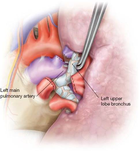
Figure 26.5 The dissection of the left upper lobe bronchus with a right-angle clamp. The left main pulmonary artery lies posterior to the bronchus.

Figure 26.6 The division of the anterior portion of the major fissure with an endoscopic stapler. The upper lobe is retracted cephalad and the lower lobe is retracted inferiorly.
TABLE 26.1 Published Contemporary VATS Lobectomy Series

 POSTOPERATIVE MANAGEMENT
The postoperative management for patients undergoing left VATS upper lobectomy is relatively straightforward. Our patients are admitted to a monitored surgical floor. Routine ambulation and incentive spirometer usage is encouraged. I prefer to set the pleuravac on 10 cm of suction for 24 hours. The chest tube is typically removed when the pleural fluid drainage is less than a 250 mL/24 hours and there is no evidence of an alveolar air leak. Most patients will have an epidural catheter placed for continuous infusion of local anesthetic such as Marcaine. The average length of stay is 3 to 4 days after VATS lobectomy.
 COMPLICATIONS
The possible complications after VATS lobectomy included pneumonia, prolonged air leak (>5 days), atrial fibrillation, hemorrhage, lobar lung collapse/mucous plugging, acute myocardial infarction, pulmonary embolism, and acute respiratory failure. The perioperative mortality rate for VATS lobectomy is less than 1% at our institution.
 RESULTS
The clinical outcomes for VATS lobectomy have been excellent (Table 26.1). The reported perioperative mortality rates have ranged from 0.3% to 2.7%. The length of hospital stay has been consistently less than 5 days. The reported complication rates are relatively low compared to open lobectomy series. The lymph node dissection achieved by VATS lobectomy has been comparable to open lobectomy in a number of reports. Currently, there are limited randomized clinical trials comparing VATS lobectomy to open lobectomy.
 CONCLUSIONS
 Left VATS upper lobectomy is indicated for stage I lung carcinoma involving the left upper lobe. Induction chemoradiotherapy and chest wall invasion are relative contraindications.
The pulmonary artery anatomy for the left upper lobe is variable and there may be multiple branches arising from the main pulmonary artery.
The left upper lobe bronchus lies anterior to the main pulmonary artery, which may be injured during the dissection of the bronchus.
The complication rates for VATS lobectomy are relatively low and the length of hospital stay is usually less than 5 days.
There are no large randomized clinical trials comparing VATS lobectomy to open lobectomy.
Recommended References and Readings
D’Amico TA, Niland J, Mamet R, et al. Efficacy of mediastinal lymph node dissection during lobectomy for lung cancer by thoracoscopy and thoracotomy. Ann Thorac Surg. 2011;92:226–232.
Flores RM, Park BJ, Dycoco J, et al. Lobectomy by video-assisted thoracic surgery (VATS) versus thoracotomy for lung cancer. J Thorac Cardiovasc Surg. 2009;138:11–18.
Mahtabifard A. Left upper lobectomy. In: McKenna RJ Jr, Mahtabifard A, Swanson SJ, eds. Altas of Minimally Invasive Thoracic Surgery (VATS). Philadelphia, PA: Elsevier; 2011:103–118.
McKenna RJ Jr, Houck W, Fuller CB. Video-assisted thoracic surgery lobectomy: Experience with 1,100 cases. Ann Thorac Surg. 2006; 81:421–426.
Onaitis MW, Petersen RP, Balderson SS, et al. Thoracoscopic lobectomy is a safe and versatile procedure: Experience with 500 consecutive patients. Ann Surg. 2006;244:420–425.
Paul S, Altorki NK, Sheng S, et al. Thoracoscopic lobectomy is associated with lower morbidity than open lobectomy: A propensity-matched analysis from the STS database. J Thorac Cardiovasc Surg.2010;139:366–378.
Swanson SJ, Herndon JE, D‘Amico TA, et al. Video-assisted thoracic surgery lobectomy: Report of CALGB 39802–a prospective, multi-institution feasibility study. J Clin Oncol. 2007;25:4993–4997.