Wendy I. Sligl
Stephen D. Shafran
Infections of the central nervous system (CNS) are often rapidly progressive and can be fatal if left undiagnosed and/or treatment is delayed. Prompt diagnosis and treatment are, therefore, crucial to decreasing morbidity and mortality. Patients with CNS infections commonly require intensive care unit (ICU) support, particularly for airway protection and mechanical ventilation in the presence of an altered mental status. Similarly, patients with undiagnosed CNS infections may be admitted to the ICU, offering intensivists the opportunity to make challenging diagnoses and alter patient outcomes with early and effective therapy.
Identifying the presence or absence of focal neurologic findings is the most important distinction to be made in patients with suspected neurologic infections. This distinction helps to focus the differential diagnosis and identifies patients in whom lumbar puncture may be contraindicated—at least until neuroimaging is completed. The major neurologic infections encountered in the critically ill include acute bacterial meningitis, encephalitis, brain abscess, subdural empyema, epidural abscess, and suppurative intracranial thrombophlebitis. Neurologic findings may also be the result of primary nonneurologic syndromes such as bacterial endocarditis and are covered in other chapters. Neurologic infections in advanced HIV/AIDS are also covered separately elsewhere.
The central nervous system is normally protected by various host defenses, the most important of which is the blood–brain barrier. Once micro-organisms gain entry, however, they are able to proliferate rapidly due to the low concentration of immunoglobulins and leukocytes in the CNS. Central nervous system infections can be caused by viral, bacterial, mycobacterial, fungal, or parasitic agents. Patient age, underlying host factors, and epidemiologic exposures including travel, animal or vector exposures, and contacts with infectious cases are important risk factors for acquiring specific types of infections or pathogens. Prompt physical examination to identify patients in need of urgent interventions—including endotracheal intubation—should be performed, followed by lumbar puncture and/or imaging studies. New techniques in the areas of molecular diagnostics and neuroimaging have revolutionized the approach to the diagnosis and management of patients with central nervous system infections. New therapeutic options, as well as improvements in intensive care support, have also enhanced outcomes in these patients.
The remainder of this chapter will specifically address the epidemiology, clinical presentation, diagnosis, management, and prevention of neurologic infections in the critically ill.
Meningitis
Key Points
· Untreated acute bacterial meningitis is universally fatal; early recognition, rapid diagnostic testing, and emergent administration of antimicrobials are crucial.
· The classic triad of fever, nuchal rigidity, and change in mental status occurs in less than 66% of patients with bacterial meningitis; however, the absence of all three of these findings effectively excludes the diagnosis with 99% to 100% sensitivity.
· Lumbar puncture should be performed urgently in all patients with suspected meningitis.
· Neuroimaging with computed tomography (CT) or magnetic resonance imaging (MRI) to rule out mass lesions should precede lumbar puncture in those with an abnormal level of consciousness, focal neurologic deficits, papilledema, a history of CNS disease, immune compromise, or seizure within 1 week of presentation.
· Empiric antimicrobial agents should be administered as soon as possible after blood cultures are collected if neuroimaging is to be performed prior to lumbar puncture or immediately following cerebrospinal fluid (CSF) collection.
· The specific microbiology and choice of empiric therapy in bacterial meningitis depend on patient risk factors, especially age, underlying host immune status, and history of preceding infections or neurosurgical procedures.
· Dexamethasone has been shown to decrease mortality in adults with Streptococcus pneumoniae meningitis and children with Haemophilus influenzae, and should therefore be administered before or concomitant with the first dose of antimicrobial in all cases pending Gram stain and culture results.
· Neurologic complications of bacterial meningitis include seizure, cerebral edema, cerebral infarction, cranial nerve involvement, venous sinus thrombosis, brain abscess, subdural empyema, and coma. Intracranial pressure monitoring and/or other surgical interventions may be required.
· Chemoprophylaxis and/or immunoprophylaxis are available for Neisseria meningitidis, Haemophilus influenzae, and infections in specific circumstances.
· In suspected meningococcal or Haemophilus influenzae meningitis, droplet isolation should be strictly enforced until 24 hours of effective antimicrobial therapy has been completed or an alternate diagnosis is reached. Isolation in other cases of meningitis, including pneumococcal meningitis, is not required.
· Aseptic meningitis refers to inflammation of the meninges not attributed to bacterial infection. CSF analysis usually reveals a normal glucose, elevated protein, elevated white blood cell count with lymphocytic predominance, and negative Gram stain and bacterial cultures.
· Viral, mycobacterial, syphilitic, fungal, amoebic, and parameningeal infections should be considered in the differential diagnosis of aseptic meningitis.
Meningitis, or inflammation of the meninges, may be caused by a wide variety of micro-organisms (Table 108.1). Infectious agents gain entry to the CSF via hematogenous, transdural, or transparenchymal routes. It is important to consider noninfectious syndromes in the differential diagnosis of meningitis. Such examples include meningeal carcinomatosis, vasculitic syndromes, or drug effect (e.g., nonsteroidal anti-inflammatories, antimicrobials, immunosuppressants, anticonvulsants). Identification of such noninfectious conditions is essential, as their therapies differ from those used in the treatment of infectious syndromes; specifically, high dose corticosteroid therapy may be indicated in some of these cases. Aseptic meningitis refers to inflammation of the meninges not attributed to bacterial infection. Critically ill patients, however, present much more commonly with bacterial meningitis by virtue of the more rapid and fulminant presentation of bacterial as opposed to aseptic meningitis.
|
Table 108.1 Causes of Acute Meningitis |
||||||||||||||||||||||||
|
||||||||||||||||||||||||
|
Table 108.2 Predisposing Host Factors to Specific Etiologic Agents of Meningitis |
||||||||||||||||||||
|
||||||||||||||||||||
Bacterial Meningitis
Acute bacterial meningitis accounts for approximately 1.2 million cases annually worldwide (1). Because untreated bacterial meningitis is universally fatal, early recognition, rapid diagnostic testing, and emergent administration of antimicrobial and adjunctive agents are crucial. The most common meningeal pathogens include Streptococcus pneumoniae and Neisseria meningitidis, although specific etiologic agents and their frequencies vary with underlying host factors such as age, immune status, and route of acquisition. The case fatality rate for adults with bacterial meningitis is approximately 25%, with transient or permanent neurologic sequelae in 21% to 28% of survivors (2,3).
Bacterial meningitis develops as a result of several mechanisms (4). Certain micro-organisms that colonize the nasopharynx may invade local tissues and subsequently spread to the bloodstream and CNS. Bacteremia and subsequent CNS invasion may also develop from localized sources such as pneumonia or urinary tract infection. Last, direct entry from contiguous infection (such as via the sinuses or mastoids), trauma, neurosurgery, or prosthetic devices such as CSF shunts or cochlear implants also occurs. Host factors including functional or anatomic asplenia, complement deficiency, and congenital or acquired immunodeficiency predispose to bacterial meningitis (Table 108.2). Other risk factors for the development of meningitis include recent exposure to a patient with acute bacterial meningitis, recent travel to areas with endemic meningococcal disease, injection drug use, recent neurotrauma or CSF leak, and otorrhea.
The median duration of symptoms prior to admission in bacterial meningitis is impressively short, averaging approximately 24 hours (5). The classic triad of fever, nuchal rigidity, and change in mental status occurs in less than 66% of cases; however, almost all patients have at least one of these findings (6). It bears reiterating that the absence of any of these findings effectively excludes the diagnosis with 99% to 100% sensitivity.
Nuchal rigidity can be detected with passive or active flexion of the neck. Tests, such as the Kernig and Brudzinski signs, are well-described physical examination techniques but are neither sensitive nor specific (7). In addition to severe headache, patients often note photophobia, and seizures, focal neurologic deficits, and papilledema may be seen on physical examination. Some patients may not manifest the classic signs and symptoms of bacterial meningitis, particularly neonates and those with underlying immunosuppressive conditions including diabetes, chronic organ failure, neutropenia, chronic corticosteroid use, transplantation, and HIV infection.
Certain micro-organisms may present with specific physical findings. Meningococcal meningitis may present with characteristic skin manifestations consisting of diffuse petechiae and purpura on the distal extremities. Skin findings occur in approximately one fourth of bacterial meningitis cases, over 90% of which are due to Neisseria meningitidis infection (8).
As a result of the widespread use of conjugate vaccine for H. influenzae type b in infants, Streptococcus pneumoniae has become the most frequently observed cause of bacterial meningitis, accounting for 47% of total cases (9). S. pneumoniae serotypes causing bacteremic disease are also those commonly responsible for meningitis. Focal infection is common with contiguous or distant sites, including sinusitis, mastoiditis, pneumonia, otitis media, and endocarditis. The major risk factors for pneumococcal meningitis include asplenia, hypogammaglobulinemia, alcoholism, chronic renal or hepatic disease, malignancy, diabetes mellitus, basal skull fracture with CSF leak, and the presence of a cochlear implant. Mortality rate range from 19% to 30% (9,10,11,12,13).
Neisseria meningitidis commonly causes meningitis in children and young adults. Serogroups B, C, and Y are responsible for most endemic disease in North America, accounting for 32%, 35%, and 26% of cases, respectively (14). Epidemic disease is most commonly caused by serogroup C, with fewer outbreaks due to serogroup A. In 2000, epidemic W-135 was associated with the Hajj pilgrimage to Mecca in Saudi Arabia (15). Subsequently, meningococcal vaccination has become legally required prior to undertaking this activity. Risk factors for invasive meningococcal disease include nasopharyngeal carriage, terminal complement deficiency, and properdin deficiency (16,17). Although a characteristic rapidly evolving petechial or purpuric rash strongly suggests N. meningitidis, a similar rash may be seen in splenectomized patients with overwhelming S. pneumoniae or H. influenzae type b infection.
Haemophilus influenzae previously accounted for a large proportion of cases of bacterial meningitis; however, widespread vaccination against H. influenzae type b has now markedly decreased its incidence (Table 108.3). Isolation of H. influenzae type b in adults suggests the presence of an underlying condition such as sinusitis, otitis media, pneumonia, diabetes mellitus, alcoholism, CSF leak, asplenia, or immune deficiency.
|
Table 108.3 Incidence by Cause of Bacterial Meningitis in the United States, 1995 |
||||||||||||||
|
||||||||||||||
Listeria monocytogenes meningitis carries a mortality rate of 15% to 29% (9,12). It occurs in neonates, adults older than 50 years of age, and in those with risk factors including alcoholism, malignancy, pregnancy, and immune suppression secondary to corticosteroid therapy or transplantation. It is interesting that this infection is seen infrequently in HIV-infected patients for unknown reasons. Pregnant women may be asymptomatic carriers and transmit infection to their infants. L. monocytogenes commonly makes up part of the fecal flora of farm animals and can be isolated from soil, water, or contaminated vegetables. Outbreaks have been associated with unpasteurized dairy products such as milk and cheeses, vegetables, and processed meats (18,19,20).
Aerobic Gram-negative bacilli can cause meningitis in specific groups of patients. Predisposing risk factors include neurosurgical procedures, neonatal status, advanced age, immune suppression, Gram-negative bacteremia, and disseminated Strongyloides stercoralis infection with hyperinfection syndrome. Escherichia coli is a common cause of meningitis in neonates.
Staphylococcus aureus or Staphylococcus epidermidis can both cause meningitis but are, however, less common than the previously described micro-organisms. Both staphylococcal species exist as part of the normal skin flora, predominantly causing infections following neurosurgery or neurotrauma, or when prosthetic material is present, particularly external ventricular drains or ventriculoperitoneal shunts. Some patients with staphylococcal bacterial meningitis have underlying infective endocarditis, paraspinal or epidural infection, sinusitis, osteomyelitis, or pneumonia.
Other less common causes of bacterial meningitis include enterococci, viridans group streptococci, beta-hemolytic streptococci, diphtheroids and Propionibacterium acnes—generally only in the setting of prosthetic material—and anaerobic species.
Viral Meningitis
Viruses are the most commonly isolated pathogens in aseptic meningitis. The nonpolio enteroviruses, especially Coxsackie viruses A and B, and echoviruses are common, accounting for 85% to 95% of all cases of aseptic meningitis with an identified pathogen (21). Enteroviruses occur worldwide, are transmitted by fecal-oral or respiratory droplet spread, and exhibit summer and fall seasonality in temperate climates. Infants, children, and young adults are commonly affected. Clinical manifestations depend on host age and immune status but generally include abrupt onset of severe headache, fever, nausea, vomiting, photophobia, nuchal rigidity, and malaise. Rash and upper respiratory symptoms are common. Only rarely is illness severe enough to require critical care.
Arboviruses more commonly cause encephalitis but have also been isolated in cases of aseptic meningitis. Arboviruses endemic to North America include the flaviviruses—such as St. Louis encephalitis virus, Colorado tick fever, Japanese encephalitis virus, and West Nile virus—and California encephalitis viruses. Arboviruses occur predominantly in the summer and early fall when vector exposure is most likely. St. Louis encephalitis virus is mosquito borne and causes epidemic disease in the Mississippi River area. Japanese encephalitis virus less commonly causes meningitis, is endemic in Asia, and requires prolonged stays in rural settings for acquisition so is uncommon even in returned travelers.
West Nile virus (WNV) came to widespread attention in 1999 when the first North American cases were identified. Since then, the virus has spread extensively across North America and should be considered in all patients with meningitis, particularly in late summer or early fall. WNV infection is asymptomatic in 80% of cases. The remaining patients present with West Nile nonneurologic syndrome (approximately 20%; formerly named West Nile fever) or West Nile neurologic syndrome (WNNS; less than 1%). West Nile nonneurologic syndrome is a self-limited febrile illness characterized by fever, headache, malaise, myalgias, and often a rash (50%). WNNS may present as encephalitis, meningitis, or flaccid paralysis. Meningitis, however, is the least common presentation of WNNS.
Lymphocytic choriomeningitis (LCM) virus is a zoonotic disease, transmitted by contact with infected rodents—such as house mice, rats, hamsters—or their feces. Though now rare, LCM virus was one of the first viruses to be associated with aseptic meningitis (4). Infection is more common in the winter months. Presenting manifestations include an influenza-like syndrome and meningismus, with occasional rash, orchitis, arthritis, myopericarditis, and transient alopecia.
Six of the eight recognized human herpesviruses can cause meningitis. Herpes simplex viruses (HSV) are most commonly associated with aseptic meningitis during primary genital infection, affecting 36% of women and 13% of men with primary genital herpes (22). HSV-2 infection is responsible for most infections; nonetheless, HSV-1 genital infection and concomitant meningitis can also occur. Meningitis is much less likely in the setting of genital herpes recurrences. Headache, photophobia, and meningismus are common presenting symptoms. Genital lesions are present in 85% of patients with primary HSV-2 meningitis and generally precede meningeal symptoms by several days.
Herpes zoster aseptic meningitis, with or without typical skin lesions, has also been reported, particularly in older patients. Cytomegalovirus (CMV), Epstein-Barr virus (EBV), and human herpes virus 6 (HHV-6) are all capable of causing aseptic meningitis but occur very rarely, predominantly in immune-suppressed populations.
HIV-associated aseptic meningitis can occur with primary infection in approximately 5% to 10% of patients (22). Cranial neuropathies may be present along with headache, fever, and meningismus. Symptoms are usually self-limited.
Mumps, now rare as a result of universal vaccination programs, was once a relatively common cause of aseptic meningitis. The clinical manifestations include fever, vomiting, headache, and parotitis in approximately 50% of patients. Meningismus, lethargy, and abdominal pain may also be present.
Other Less Common Infectious Causes
Spirochetal meningitis may be caused by Treponema pallidum or Borrelia burgdorferi. T. pallidum, the etiologic agent of syphilis, is acquired by sexual contact, placental transfer, or direct contact with active lesions; these include condyloma lata, mucous patches, or the rash of secondary syphilis. Syphilitic meningitis usually occurs during primary or secondary infection, complicating 0.3% to 2.4% of untreated infections during the first 2 years (4). B. burgdorferi is transmitted by the Ixodes tick and causes Lyme disease. It is the most common vector-borne disease in the United States. Meningitis can occur during the first stage of disease, concurrently with erythema migrans at the tick bite site. Dissemination of the micro-organism in the second stage of disease, 2 to 10 weeks following exposure, may also result in aseptic meningitis. Late or chronic disease may include subacute encephalopathy but not meningitis.
Mycobacterium tuberculosis may cause a subacute or chronic form of meningitis. Infection of the meninges results from rupture of a tuberculous focus into the subarachnoid space. In very young patients, concomitant disseminated systemic infection is common. Epidemiologic risk factors include a known prior history of tuberculosis (TB) exposure, residence in an endemic area, contact with an active case, incarceration, homelessness, and HIV infection. Tuberculin skin testing is negative in over half of patients with tuberculous meningitis (23,24). A negative skin test, therefore, cannot be used to exclude tuberculous meningitis, as is also the case with other active tuberculous infections. Newer tests, such as the QuantiFERON-TB Gold test, may be available in some centers (25).
Fungal meningitis, although uncommon, should be considered, particularly given the high mortality associated with untreated infection. Cryptococcus neoformans predominantly affects immunocompromised hosts but can also infect the immunocompetent. The encapsulated yeast is distributed worldwide but prefers wet forested regions with decaying wood and is found in particularly high concentrations in pigeon guano. Risk factors for cryptococcal infection include HIV/AIDS, prolonged corticosteroid therapy, immunosuppression post-transplantation, malignancy, and sarcoidosis. Clinical presentation is typically indolent, occurring over 1 to 2 weeks, and is characterized by fever, malaise, and headache. Meningismus, photophobia, and vomiting occur in less than 33% of patients. Cryptococcus gattii, a serotype usually restricted to tropical climates, emerged on Vancouver Island, British Columbia (BC), Canada in 1999 and has since been responsible for numerous cases of CNS infection in predominantly immunocompetent hosts in BC and the U.S. Pacific Northwest.
Coccidioides immitis, a dimorphic fungus, is found in soil in the dry desert regions of the southwest United States, Mexico, and Central and South America. Infection results after inhalation of arthroconidia, usually following a dust storm or during building construction. Infection is usually confined to the respiratory system in those with competent immune systems. However, extrapulmonary dissemination to the meninges can occur in patients with immune compromise or during pregnancy. Patients present with headache, vomiting, and altered level of consciousness. Risk factors for the development of disease include travel to or residence in an endemic region and immune deficiency. Coccidioidal meningitis is universally fatal if untreated.
Less common fungal causes of meningitis include Blastomyces dermatitidis, Histoplasma capsulatum, Sporothrix schenckii, and rarely, Candida species. B. dermatitis, H. capsulatum, and S. schenckii are all dimorphic fungi with similar presentations to coccidioidal meningitis. Primary infection occurs via inhalation, and disseminated infection occurs predominantly in immune compromised populations. B. dermatitidis and H. capsulatum are endemic in the Mississippi and Ohio River Valleys. S. schenckii has been reported worldwide, with most cases in the tropical regions of the Americas.
Candida exists only in yeast form and is part of the normal flora of skin and gastrointestinal tract. CNS involvement is most commonly due to candidemia with subsequent meningeal seeding. Predisposing risk factors for candidemia include the use of broad-spectrum antibiotics, the presence of indwelling devices such as vascular or urinary catheters, parenteral nutrition, intensive care unit admission, prolonged hospital stay, and immune compromise. Specific risk factors for Candida CNS infection include ventricular shunts, trauma, neurosurgery, or lumbar puncture (26,27). C. albicans is the most commonly isolated species; however, nonalbicans species are becoming more prevalent, particularly in ICU populations (28,29,30).
Meningitis caused by protozoa or helminths is extremely rare. The free-living amoebas Acanthamoeba, Balamuthia, and Naegleria fowleri are associated with fresh water exposure. They are usually acquired by individuals diving into contaminated lakes or swimming pools. CNS invasion occurs via penetration of the nasal mucosa and cribriform plate. N. fowleri can cause a primary amoebic meningoencephalitis. Acanthamoeba and Balamuthia rarely cause meningitis; they commonly present as encephalitis.
Angiostrongylus cantonensis, the rat lungworm, is the classic infectious cause of eosinophilic meningitis (>10% eosinophils in the CSF) (Table 108.4). Humans are incidental hosts and develop neurologic symptoms as a result of larval migration through the CNS. A. cantonensis is endemic in Southeast Asia and the Pacific Islands and is acquired by ingesting raw mollusks such as snails or slugs. Gnathostoma spinigerum, acquired by ingestion of raw and undercooked fish and poultry, is not primarily neurotropic like A. cantonensis but may also cause eosinophilic meningitis as a result of migration of larvae up the nerve tracts to the CNS. Gnathostomiasis is endemic in Asia, especially Thailand and Japan, and more recently in Mexico. Baylisascaris procyonis, a roundworm infection of raccoons, rarely causes human eosinophilic meningoencephalitis following accidental ingestion of ova from raccoon feces in contaminated water, soil, or foods.
Diagnosis
Lumbar puncture (LP) should be performed emergently in all patients suspected of having bacterial meningitis unless contraindicated, although it is commonly unnecessarily delayed while neuroimaging is performed to exclude mass lesions. Complications associated with lumbar puncture are uncommon; however, the incidence of life-threatening brain herniation has been reported to range from less than 1% to 6% (31,32). A recent study evaluating the clinical features at baseline associated with abnormal findings on CT scan, and thus, increased risk of brain herniation, identified: age greater than or equal to 60 years; a history of CNS disease such as a mass lesion, stroke, and focal infection; immune compromise such as HIV or immunosuppressive therapy; a history of seizure less than or equal to 1 week before presentation; and specific abnormal neurologic findings (33). Based on these findings, guidelines for which adult patients should undergo CT prior to LP have been recommended (Table 108.5) (34).
|
Table 108.4 Cerebrospinal Fluid Tests in Suspected CNS Infection |
||||||||||||||||
|
||||||||||||||||
Nosocomial meningitis is rare in nonneurosurgical patients; nevertheless, lumbar punctures are often performed in hospitalized patients with unexplained fever and/or decreased level of consciousness. The yield of performing an LP in the nonneurosurgical population is extremely low and of questionable utility (35).
|
Table 108.5 Indications for Imaging Prior to Lumbar Puncture in Adults with Suspected Bacterial Meningitis |
||
|
CSF analysis is extremely important in the diagnosis of meningitis. Basic laboratory analyses, including cell count and differential, protein, glucose, Gram stain, and bacterial cultures, are most useful in distinguishing between viral, bacterial, tuberculous, and fungal infection (Table 108.3).
Bacterial Meningitis
Bacterial meningitis usually presents with an elevated systemic white blood cell (WBC) count and left shift (immature forms such as bands and myeloids). Leukopenia is occasionally present in severe infection. Thrombocytopenia may be the result of sepsis, disseminated intravascular coagulation, or meningococcemia alone. Renal and hepatic dysfunction may occur as part of multiorgan failure in severe disease. Blood cultures are often positive and should always be drawn prior to the administration of antimicrobials, particularly if the LP cannot be performed immediately. Approximately 66% of patients with bacterial meningitis have positive blood cultures (8).
CSF analysis in bacterial meningitis classically reveals a neutrophilic pleocytosis with hundreds to thousands of cells and greater than 80% neutrophils. In fact, a low CSF WBC count is usually a marker of poor prognosis in this setting. The CSF glucose concentration is usually low and should always be compared with a simultaneous serum glucose measurement. An abnormal CSF-to–serum glucose ratio (less than 0.5) is common in bacterial meningitis—and it is often much lower than 0.5. Acute illness in diabetics may increase serum glucose levels markedly, making the CSF-to–serum glucose ratio inaccurate. In the postoperative neurosurgical patient, elevated CSF lactate concentrations (greater than or equal to 4.0 mMol/L) have been shown to be superior to CSF-to–blood glucose ratios (36), and initiation of empirical antimicrobial therapy in this setting should be considered pending the results of additional studies (34). CSF protein and opening pressure are usually elevated in bacterial meningitis (Table 108.6).
|
Table 108.6 Typical CSF Parameters in Patients with Meningitis |
||||||||||||||||||||||||||||||||||||||||||
|
||||||||||||||||||||||||||||||||||||||||||
Gram staining permits rapid identification of bacterial species and is positive in approximately 50% to 60% of patients with bacterial meningitis. The presence of bacteria is virtually 100% specific, but sensitivity is variable. The Gram stain is more likely to be positive in patients with high bacterial loads. Gram-positive diplococci suggest S. pneumoniae infection, Gram-negative diplococci suggest N. meningitidis infection, Gram-positive rods suggest L. monocytogenes infection, and small pleomorphic coccobacilli suggest H. influenzae infection.
CSF bacterial cultures are positive in approximately 70% to 85% of cases. The yield decreases significantly in patients treated with antimicrobials prior to CSF collection. Antigen assays (latex agglutination tests) have been used in these cases, but due to their low sensitivity are no longer routinely offered by many laboratories. Broad-based polymerase chain reaction (PCR) may be useful for excluding the diagnosis of bacterial meningitis (34) but is unavailable in many centers.
Viral Meningitis
In acute viral meningitis, the CSF cell count is usually in the low hundreds with lymphocytic predominance. A predominance of neutrophils may be seen in the first 24 hours of disease, occasionally confusing the diagnosis. The CSF glucose concentration is usually within normal range. CSF protein is usually mildly elevated, and the opening pressure is usually normal.
Viral cultures and nucleic acid amplification tests are most commonly used in the diagnosis of viral meningitis. Enteroviruses may be cultured from CSF, throat, or rectal swabs, with a sensitivity of 65% to 70%, or identified by nucleic acid amplification testing. Enteroviral PCR is both sensitive and specific. PCR for HSV is also widely available, and in studies of HSV-1 encephalitis, HSV PCR demonstrated a specificity of approximately 100% and sensitivity of 75% to 98% (37,38). False negatives occur mostly within the first 72 hours of infection. The diagnosis of WNV can be made by detection of serum IgM or a fourfold rise in IgG between acute and convalescent titers. WNV PCR of serum and CSF are also available; however, the sensitivity is higher in CSF specimens due to the short-lived viremia in humans.
Other Less Common Causes
CSF analysis in syphilitic meningitis is characterized by a mild lymphocytic pleocytosis, decreased glucose, and elevated protein. T. pallidum cannot be cultured, so diagnosis must be made using alternate methods, predominantly serology. Direct visualization by darkfield microscopy or direct fluorescent antibody testing may be possible if a primary chancre or skin lesion of secondary syphilis—condyloma latum or mucous patch—is present. Serologic testing should include nontreponemal (RPR, rapid plasma reagin; VDRL, Venereal Diseases Research Laboratory) and treponemal (TPPA, Treponema pallidumparticle agglutination; FTA-Abs, fluorescent treponemal antibody absorption) tests for the diagnosis of active syphilis infection. Treponema-specific enzyme immunoassays (EIA) for IgM and IgG are replacing the above traditional serologic tests as the initial laboratory diagnostic test in some centers. CSF VDRL may be used in the diagnosis of syphilitic meningitis. The specificity is high, but false positives occur in bloody specimens. The major limitation of CSF VDRL is its low sensitivity (30% to 70%), so a negative result should not be used to rule out infection in the setting of high clinical suspicion. CSF FTA-Abs is more sensitive; however, false positives are common due to serum antibody leak into the CSF. Last, PCR has been recently used to detect T. pallidum DNA in the CSF. Further studies are needed to ascertain the sensitivity and specificity of this test.
Lyme meningitis is characterized by a mild lymphocytic pleocytosis, low glucose, and an elevated protein. The CSF concentration of B. burgdorferi antibody, compared to serum levels, is a sensitive and specific diagnostic method. PCR is currently available only in research laboratories, although it will likely become the diagnostic test of choice in the near future. CSF oligoclonal bands and B. burgdorferi culture are also available, but neither is sensitive or specific.
The CSF analysis in tuberculosis meningitis demonstrates a lymphocytic pleocytosis, low glucose, and markedly elevated protein and opening pressure. The elevation in protein is particularly marked in the setting of CSF block. Acid-fast bacillus (AFB) smears are very low yield; only 10% to 22% of cases will be positive (24,39,40). Mycobacterial cultures, although slow growing—taking several weeks—become positive in up to 88% of cases (4). DNA probes and nucleic acid amplification techniques (mainly PCR) have recently become available with great improvements in sensitivity and specificity. Meningeal biopsy is rarely performed but may show caseating granulomata. Skin testing and QuantiFERON-TB Gold testing have been discussed in the previous section. Sputum and urine AFB, as well as mycobacterial blood cultures, should also be included as part of the TB workup in these patients.
Cryptococcal meningitis is characterized by a lymphocytic pleocytosis, decreased glucose, and elevated protein. Opening pressures may be markedly elevated. Culture of C. neoformans or C. gattii from the CSF is diagnostic; however, other simpler tests are now available. Detection of serum or CSF cryptococcal antigen (CrAg) is highly sensitive (greater than 90%). India ink was previously regarded as the standard diagnostic test, but due to its low sensitivity (50%), it has been largely replaced by antigen testing. Fungal blood cultures may also be useful, as cryptococcal meningitis occasionally occurs in the setting of disseminated cryptococcal infection with cryptococcemia, especially in HIV-infected patients.
Other fungal meningitides are similarly characterized by a lymphocytic pleocytosis, low to normal glucose, and an elevated protein. Coccidioidal meningitis may present with an eosinophilic pleocytosis and peripheral eosinophilia. Fungal cultures are diagnostic and are most useful in Candida or Aspergillus infection. Dimorphic fungal infection may be diagnosed serologically, as isolating these organisms from the CSF is challenging and of low yield. Detection of complement-fixing (CF) IgG antibodies or immunodiffusion tests for IgM and IgG in CSF are currently the standard diagnostic tests. Low-titer false positives may occur in the setting of parameningeal foci. As well, false negatives may occur in early disease.
Primary amoebic meningoencephalitis due to N. fowleri results in a neutrophilic pleocytosis, increased red blood cells, low glucose, and an elevated protein. Demonstration of motile trophozoites on a wet mount of CSF or biopsy specimens is diagnostic. The diagnosis of A. cantonensis, G. spinigerum, or B. procyonis requires an appropriate epidemiologic exposure, peripheral blood eosinophilia, and a characteristic eosinophilic pleocytosis. Serologic tests are helpful but performed only in reference laboratories.
|
Table 108.7 Empiric Therapy of Bacterial Meningitis Based on Age and Host Factors |
|||||||||||||||||||||
|
|||||||||||||||||||||
Treatment
The initial management of the patient with suspected meningitis is primarily guided by epidemiologic risk factors and lumbar puncture results. The CSF cell count, glucose, and Gram stain are crucial in guiding empiric therapy. If the LP is delayed for any reason, empiric antimicrobial therapy should not be withheld (Table 108.7), as delays in therapy have been associated with adverse clinical outcomes and increased mortality (41,42). The administration of antimicrobials should immediately follow blood culture collection and should not be delayed by neuroimaging or other tests performed prior to LP.
Bacterial Meningitis
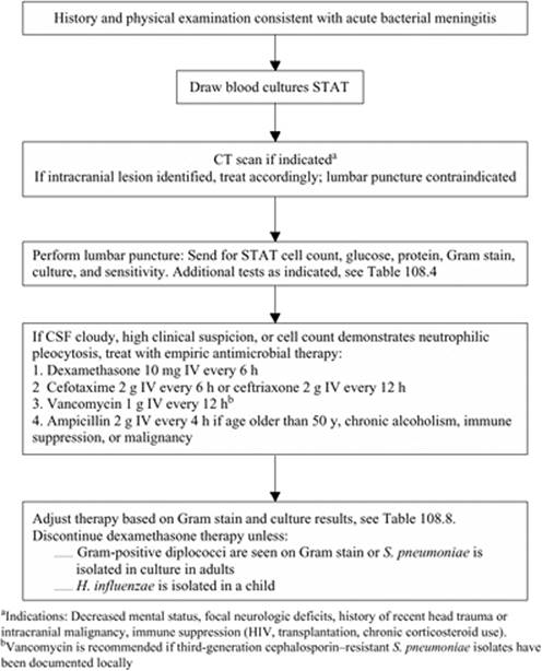
As noted above, lumbar puncture should be performed urgently in those with suspected meningitis. A protocol for the management of bacterial meningitis is presented in Figure 108.1. Imaging should be performed prior to LP in specific populations (Table 108.5) but should not result in a delay in the initiation of antimicrobial therapy. Empiric therapy should be based on age, underlying host factors, and initial CSF Gram stain results (Table 108.7).
|
|
|
Figure 108.1. Bacterial Meningitis Protocol |
The choice of antimicrobial therapy in bacterial meningitis is influenced by blood–CSF barrier penetration, effect of meningeal inflammation on penetration, and the bactericidal efficacy. In general, CSF penetration is enhanced in the setting of meningeal inflammation due to increased permeability. Additionally, high lipid solubility, low molecular weight, and low protein binding increase CSF drug levels. Bactericidal efficacy may be decreased in purulent CSF, particularly with aminoglycosides, due to the low pH. Penicillins, third-generation cephalosporins, carbapenems, fluoroquinolones, and rifampin achieve high CSF levels and are all bactericidal. Antimicrobials should be adjusted based on renal and hepatic function. Therapeutic drug monitoring may be required to ensure adequate levels and prevent toxicity (e.g., vancomycin, aminoglycosides). Antimicrobial therapy should be adjusted based on culture and susceptibility results as soon as possible (Table 108.8). In suspected meningococcal or H. influenzae meningitis, droplet isolation (single room, gowns, gloves, surgical masks, and dedicated patient care equipment) should be strictly enforced until 24 hours of effective antimicrobial therapy have been completed or an alternate diagnosis is reached. Isolation in other cases of meningitis, including pneumococcal meningitis, is not required (Table 108.9).
Streptococcus pneumoniae
Empiric therapy guidelines for pneumococcal meningitis have been recently modified due to the increasing incidence of penicillin resistance. S. pneumoniae organisms were once uniformly susceptible to penicillin; however, mutations in penicillin-binding proteins have resulted in varying levels of resistance. Empiric therapy therefore consists of a third-generation cephalosporin until susceptibility results become available. Once the minimum inhibitory concentrations (MIC) are available, therapy should be adjusted accordingly. For isolates with penicillin MIC less than 0.1 µg/mL, penicillin G (4 million units IV every 4 hours) or ampicillin (2 g IV every 4 hours) should be used. For isolates with a MIC greater than or equal to 0.1 µg/mL, treatment with a third-generation cephalosporin should be continued; either cefotaxime (2 g IV every 6 hours) or ceftriaxone (2 g IV every 12 hours). For isolates with a ceftriaxone MIC greater than or equal to 1 µg/mL, vancomycin and a third-generation cephalosporin are the recommended therapy; some clinicians administer very high doses of third-generation cephalosporins in these cases. Vancomycin should be dosed 1 g IV every 12 hours, or 500 to 750 mg IV every 6 hours, to a maximum of 2 to 3 g/day, and adjusted based on therapeutic drug monitoring to maintain a trough serum concentration of between 15 and 20 µg/mL. Meropenem is a reasonable alternative to the above agents and does not carry the theoretical risk of decreasing seizure threshold as is seen with imipenem. The efficacy of newer antimicrobials, such as linezolid and daptomycin, have not been established. Dexamethasone should be administered prior to or with the first dose of antimicrobial (see Adjunctive Therapy). Treatment duration is 10–14 days.
|
Table 108.8 Specific Therapy of Bacterial Meningitis |
||||||||||||||||||||||
|
||||||||||||||||||||||
Neisseria meningitidis
The initial treatment of meningococcal meningitis is with a third-generation cephalosporin—for example, cefotaxime (2 g IV every 6 hours) or ceftriaxone (2 g IV every 12 hours); however, therapy should be stepped down to penicillin if susceptibility is confirmed. The duration of treatment is 7 days. Chloramphenicol (25 mg/kg, to a maximum of 1 g IV every 6 hours) is a reasonable alternative in the beta-lactam–allergic patient. Meropenem (2 g IV every 8 hours) is another alternative, although there is a high degree of cross-reaction in penicillin-allergic patients. Dexamethasone is not indicated in confirmed meningococcal meningitis.
|
Table 108.9 Mortality Rates in Bacterial Meningitis by Pathogen |
||||||||||||||
|
Haemophilus influenzae
Therapy for H. influenzae meningitis is initially a third-generation cephalosporin. Therapy can be changed to ampicillin, 2 g IV every 4 hours if susceptibility is confirmed. A total of 7 days of therapy is recommended. Dexamethasone should be administered as adjunctive therapy in children.
Listeria monocytogenes
L. monocytogenes meningitis should be treated with ampicillin, 2 g IV every 4 hours. Gentamicin may be added for antimicrobial synergy, but aminoglycosides have poor penetration into CSF. When used, gentamicin should be administered as a 2 mg/kg loading dose, followed by 1.7 mg/kg every 8 hours. Trimethoprim/sulfamethoxazole (TMP/SMX), 20 mg/kg/day of the trimethoprim component, divided into 6 to 12 hourly doses, can be used in penicillin-allergic patients. Alternate therapies include meropenem and, potentially, linezolid and rifampin. Third-generation cephalosporins have no activity against L. monocytogenes and should not be used. Treatment duration is 14 to 21 days.
Aerobic Gram-negative bacilli
Aerobic Gram-negative bacilli should be treated empirically with a third-generation cephalosporin or meropenem. Susceptibility results should be obtained as soon as possible to guide therapy in consultation with an infectious diseases specialist. For Pseudomonas aeruginosa infections, ceftazidime or cefepime, 2 g IV every 8 hours, or meropenem, 2 g IV every 8 hours, with tobramycin 2 mg/kg IV every 8 hours, should be used. Cefotaxime and ceftriaxone should not be used as they do not have antipseudomonal activity. Ciprofloxacin or aztreonam are acceptable alternatives if the isolate is susceptible. The duration of therapy is prolonged, generally 21 days.
Staphylococcus
Staphylococcal meningitis therapy depends on methicillin susceptibility. Methicillin-susceptible strains should be treated with nafcillin or oxacillin, 2 g IV every 4 hours, whereas methicillin-resistant strains should be treated with vancomycin, 1 g IV every 12 hours or 500 to 750 mg IV every 6 hours, to a maximum of 2 to 3 g/day, with therapeutic drug monitoring to ensure adequate serum levels—15 to 20 µg/mL—are achieved. Vancomycin is recommended in patients with penicillin allergy. Infected prosthetic material should be removed if possible and antimicrobial therapy continued for 10 to 14 days after removal. If removal is not possible, rifampin may be added; however, cure rates are poor with hardware retention. Linezolid and daptomycin may become alternate therapies, but data on efficacy are currently lacking.
Adjunctive therapies in bacterial meningitis include corticosteroids, procedures to reduce intracranial pressure, and surgery. Corticosteroid therapy aims to decrease the inflammatory response while allowing antimicrobial therapy to eradicate infection. Although corticosteroid administration may decrease CSF penetration and bactericidal activity of antimicrobials, recent randomized controlled trials suggest benefit with its use. In children, the administration of dexamethasone has demonstrated a reduction in the incidence of hearing impairment and severe neurologic complications in H. influenzae meningitis (43). Adjunctive corticosteroid therapy has also been evaluated in adults, showing a mortality benefit in patients with pneumococcal meningitis (5). Based on these results, treatment recommendations suggest dexamethasone, 0.15 mg/kg, be given 10 to 20 minutes before, or at least concomitant with, the first dose of antimicrobial therapy and continued every 6 hours. Dexamethasone should therefore be administered to all patients with suspected meningitis until Gram stain or culture results are available. Dexamethasone should be continued for 2 to 4 days only if the Gram stain or cultures demonstrate H. influenzae in children or S. pneumoniae in adults. The potential disadvantage of decreased CSF penetration by non–beta-lactam antimicrobials with concomitant dexamethasone administration has yet to be thoroughly studied. Treatment with adjunctive dexamethasone has not been associated with an increased risk for long-term cognitive impairment (44).
Placement of an intracranial pressure monitoring device may be beneficial for patients with bacterial meningitis and elevated intracranial pressure. Admission to an ICU with expertise in this type of monitoring is most appropriate. Surgical intervention may be required in some patients, for example, those with basal skull fractures with persistent CSF leaks or dural defects.
Complications of bacterial meningitis can be divided into neurologic and nonneurologic complications. Neurologic complications include seizures, cerebral edema, cerebral infarction, cranial nerve palsies, venous sinus thrombosis, brain abscess, subdural empyema, and coma. Late complications include hearing impairment, obstructive hydrocephalus, learning disabilities, sensory and motor deficits, mental retardation, cortical blindness, and seizures. Nonneurologic complications include septic shock, coagulopathy, and the syndrome of inappropriate antidiuretic hormone secretion (SIADH).
Viral Meningitis
In general, the treatment for viral meningitis is supportive given its benign and self-limited course. Pleconaril has been evaluated for enteroviral meningitis with modest benefit but remains experimental (45,46). Intravenous immunoglobin has been used in agammaglobulinemic patients with chronic enteroviral meningitis. No specific therapy exists for arboviruses, mumps, or LCM. HIV-associated meningitis should be treated with combination antiretroviral therapy.
It is not clear whether antiviral treatment alters the course of HSV meningitis; nevertheless, primary episodes of genital herpes should be treated as per guidelines. Some physicians extend therapy to 14 days with concomitant meningitis. Intravenous acyclovir, dosed at 5 mg/kg every 8 hours, has been used in severe disease. Ganciclovir is the treatment of choice for CMV meningitis in immunocompromised hosts.
Other Less Common Causes
Syphilitic meningitis does not respond to benzathine penicillin, which is used to treat most forms of syphilis; it requires a 2-week course of high-dose IV penicillin G (4 million units every 4 hours). RPR titers should be monitored after therapy, and repeat CSF examination should be performed if titers do not decline fourfold 6 months after therapy. All HIV patients with syphilitic meningitis should have a lumbar puncture repeated 6 months following therapy. Patients with penicillin allergy should undergo desensitization, as there are no proven effective alternative therapies for syphilitic meningitis.
The treatment of Lyme meningitis is achieved with ceftriaxone, 2 g daily, or cefotaxime, 2 g IV every 8 hours for 14 to 28 days. Alternate therapy is penicillin (4 million units every 4 hours) for 14 to 28 days.
The treatment of tuberculous meningitis depends largely on the resistance pattern in the community and results of susceptibility testing; consultation with an infectious diseases specialist is recommended. In general, standard combination therapy includes isoniazid (INH), rifampin (RIF), ethambutol (ETB), and pyrazinamide (PZA). ETB may be discontinued once INH and RIF susceptibilities are confirmed. Treatment should be continued for a minimum of 12 months and up to 24 months. Adjunctive therapy with dexamethasone for the first month has been shown to decrease complications and is recommended. Pyridoxine, 25 to 50 mg daily, should also be administered to prevent INH-related neuropathy.
Therapy for fungal meningitis is complicated by the lack of standardized susceptibility testing and interpretation for many fungi. The area of antifungal therapy, however, is an evolving area with an increasing number of antifungal agents from which to choose.
Cryptococcal meningitis should be treated with a 14-day induction phase of amphotericin B, 0.7 to 1 mg/kg/day IV, with or without flucytosine, 100 mg/kg/day PO dosed every 6 hours. Consolidation therapy with fluconazole, 400 mg daily, should be continued for 8 weeks following induction. Maintenance (or suppressive) therapy with fluconazole, 200 mg per day, should be continued in patients with HIV/AIDS until immune reconstitution is achieved. Cryptococcal meningitis may require daily therapeutic lumbar punctures, an external ventricular drain, or a ventriculoperitoneal shunt to relieve increased intracranial pressure. Therapy is identical in non-HIV/AIDS patients, with the exception that consolidation therapy is continued for 10 weeks; further prolongation may be required in transplant patients. Echinocandins, such as caspofungin and micafungin, are not active in cryptococcosis.
The treatment for coccidioidal meningitis is oral fluconazole, 400 mg daily. Some clinicians initiate therapy with a higher dose of 800 mg per day or may add intrathecal amphotericin B. Treatment must be continued lifelong, as relapses are frequently lethal. Therapy for H. capsulatum meningitis consists of amphotericin B, 0.7 to 1 mg/kg/day to complete a total dose of 35 mg/kg. Fluconazole, 800 mg per day, for an additional 9 to 12 months, may be used to prevent relapse. If relapse does occur, long-term therapy with fluconazole or intraventricular amphotericin B is recommended. Itraconazole should be avoided due to poor CSF penetration. Although very rare, S. schenckii meningitis is treated with amphotericin B. Itraconazole, despite its poor CSF penetration, may be tried after initial therapy for lifelong suppression.
For candidal meningitis, the preferred initial therapy is amphotericin B, 0.7 mg/kg/d, with flucytosine, 25 mg/kg dosed every 6 hrs and adjusted to maintain serum levels of 40 to 60 µg/mL. Fluconazole therapy, in susceptible species, may be used for follow-up or suppressive therapy. The duration of therapy should continue for at least 4 weeks after resolution of symptoms. All prosthetic material must be removed to achieve cure.
Primary amoebic meningoencephalitis caused by N. fowleri is usually fatal. A few cases have been successfully treated with early diagnosis and treatment with high-dose intravenous and intrathecal amphotericin B or miconazole and rifampin. Eosinophilic meningitis caused by A. cantonensis and G. spinigerum are treated supportively. Corticosteroids are recommended to decrease the inflammatory response to intracranial larvae. Antihelminthic therapy is relatively contraindicated, as clinical deterioration and death may occur following severe inflammatory reactions to dying larvae.
Prevention
Chemoprophylaxis (medications) and immunoprophylaxis (vaccines) are available to prevent infection in contacts of cases or in times of epidemic spread. Temporary nasopharyngeal carriage with H. influenzae, N. meningitidis, and S. pneumoniae may occur following exposure to an index case and is a risk factor for the development of invasive disease. Chemoprophylaxis is recommended to eliminate nasopharyngeal carriage in some individuals at risk.
Prophylaxis is indicated in household contacts—those residing with the index case or with greater than 4 hours of close contact—and day care contacts—same day care as index case for 5 to 7 days before onset of disease—of cases of H. influenzae type b. If there is an unvaccinated contact less than or equal to 4 years of age in the household, chemoprophylaxis is recommended for all household contacts except pregnant women. Rifampin, 20 mg/kg, with a usual adult dose of 600 mg daily, for four doses, is the recommended therapy.
Prophylaxis for N. meningitidis is recommended for close contacts of cases. This includes intimate contacts (e.g., kissing) and close contacts with greater than or equal to 4 hours contact 1 week prior to the onset of illness. Most close contacts include house mates, day care center contacts, cellmates, and/or military recruits. Medical personnel exposed to oropharyngeal secretions during intubation, nasotracheal suctioning, or mouth-to-mouth resuscitation should also receive chemoprophylaxis. Rifampin, 600 mg orally every 12 hours for a total of four doses, or single doses of ciprofloxacin (500 mg orally) or ceftriaxone (250 mg intramuscularly) are all efficacious. It is recommended that ciprofloxacin be avoided in children younger than 16 years of age and in pregnant women, based on joint cartilage injury demonstrated in animal studies. Chemoprophylaxis is not indicated in S. pneumoniae infection.
Vaccination is available for the prevention of H. influenzae, N. meningitidis, and S. pneumoniae (47,48). Vaccination against H. influenzae type b is part of routine childhood immunization. Unvaccinated children less than or equal to 2 years of age exposed to an index case should receive chemoprophylaxis and vaccination.
S. pneumoniae vaccination is available in two preparations: the 23-valent polysaccharide vaccine and the 7-valent conjugate vaccine. The conjugate vaccine is recommended routinely in all children less than or equal to 23 months of age and in those at high risk of invasive disease—sickle cell disease and other hemoglobinopathies, functional or anatomic asplenia, HIV infection, immunocompromising conditions, and chronic medical conditions—who are greater than 23 months of age. The polysaccharide vaccine is recommended for all individuals greater than 65 years old and in those greater than 5 years old with the above high-risk conditions, but is of limited immunogenicity and efficacy. Studies of conjugate pneumococcal vaccine in adults are ongoing. S. pneumoniae vaccination is not an indicated as postexposure prophylaxis.
N. meningitidis vaccine is also available in two forms: conjugate and polysaccharide vaccines. Available conjugate vaccines include a quadrivalent (MCV4) vaccine, as well as the monovalent serogroup C (Men-C) vaccine. Available polysaccharide vaccines include a quadrivalent vaccine containing A, C, Y, and W-135 and a bivalent vaccine with serogroups A and C. N. meningitidis vaccination is indicated in high-risk populations, including those with specific immune deficiencies (Table 108.2), those traveling to endemic and epidemic regions, laboratory workers routinely exposed to N. meningitidis, first-year college students living in dormitories, and military recruits. Vaccination during outbreaks of meningococcal disease due to a serogroup contained in a vaccine should be performed in consultation with public health authorities.
Encephalitis
Key Points
· In distinguishing encephalitis from meningitis, the most useful finding is altered mental status.
· Encephalitis is most commonly viral or postinfectious in etiology.
· Herpes simplex encephalitis is the most common cause of sporadic encephalitis in Western countries, accounting for 10% to 20% of cases. Temporal lobe involvement on MRI and electroencephalogram (EEG) are characteristic. PCR is 75% to 98% sensitive, with false negatives occurring predominantly during the first 72 hours of illness. Mortality approaches 70% without therapy but can be significantly reduced with early antiviral therapy.
Encephalitis is defined as inflammation of the brain parenchyma. Although encephalitis and meningitis may present with similar clinical findings, the two syndromes are pathophysiologically distinct. The major distinguishing feature is the presence or absence of normal brain function. Patients with meningitis may be drowsy or lethargic but should have normal cerebral function, whereas those with encephalitis generally have altered mental status. Occasionally patients may present with a combination of findings in an overlap syndrome of meningoencephalitis. It is important to distinguish between the two syndromes, as the etiologic agents and treatments may differ.
Encephalitis is most commonly viral or postinfectious (Table 108.10). Viral encephalitis is caused by direct viral invasion of the CNS whereas postinfectious encephalitis is an immune-mediated process. Unfortunately it may be difficult to differentiate between the two; however, encephalitis with resolving infectious symptoms suggests a postviral cause. The most common viruses causing postinfectious encephalitis include mumps, measles, varicella-zoster virus, rubella, and influenza.
|
Table 108.10 Most Common Viral Causes of Encephalitis, their Vectors or Animal Hosts, and Geographic Distributions |
||||||||||||||||||||||||||||||||||||||||||||||||||||||||||||
|
||||||||||||||||||||||||||||||||||||||||||||||||||||||||||||
Access to the CNS is highly virus-specific and occurs via hematogenous or neuronal routes. In hematogenous invasion, viral infection is acquired at an initial site of entry, with primary site replication, transient viremia, and CNS seeding. Retrograde transport within motor and sensory axons to the CNS occurs in the neuronal route of entry. After CNS entry, viruses enter neural cells, causing inflammation and cell dysfunction. Clinical manifestations are the result of specific cell-type invasion. Oligodendroglial cell invasion causes demyelination, whereas cortical invasion results in altered mental status, and neuronal invasion may result in focal or generalized seizures. Thus, focal pathology is the result of specific neural tropism.
Arboviruses are acquired via vector exposure, mainly mosquitoes and ticks. These include eastern equine encephalitis (EEE), western equine encephalitis (WEE), St. Louis encephalitis, Venezuelan equine encephalitis (VEE), California encephalitis (caused by La Crosse virus), Japanese encephalitis, yellow fever, and West Nile virus (WNV). Arbovirus-related encephalitides are most prevalent during the summer and early fall months when mosquitoes and ticks are most active.
EEE has a high mortality rate and occurs in the New England area, whereas WEE is a much milder illness, occurring west of the Mississippi River. VEE occurs from Florida to South America, whereas St. Louis encephalitis virus is found throughout much of the United States. The California encephalitis viruses mainly affect children in the Midwest and Eastern states. West Nile virus, identified in North America in 1999, causes West Nile neurologic syndrome (WNNS) in less than 1% of exposed individuals. WNNS most commonly manifests as encephalitis and occurs in those with diabetes mellitus, alcoholism, and of older age (49). Muscle weakness and flaccid paralysis may present concurrently in patients with encephalitis. Japanese virus encephalitis, occurring principally in Southeast Asia, China, India, and Japan, is the most common viral encephalitis outside of the United States.
Colorado tick fever is prevalent in the western United States, and most affected individuals have a history of camping and hiking in wooded endemic areas. Malaria, in those with an appropriate travel history, should also be considered in the differential diagnosis of encephalitis.
Rabies, a zoonotic disease that requires contact with infected animals, should be considered in all cases of encephalitis. Once CNS infection is established, however, the mortality is essentially 100%. Rabies can be acquired from many sources including dogs, cats, raccoons, bats, and foxes. The history of an animal bite, although useful if present, is absent in most cases of rabies.
Herpes viruses cause disease by primary infection or reactivation. Herpes simplex encephalitis (HSE) is the most common cause of sporadic encephalitis in Western countries, accounting for 10% to 20% of cases (50). HSE is caused by type 1 virus in greater than 90% of cases, occurs year-round, and affects all age groups. Two thirds of cases are due to reactivation of the virus in the trigeminal ganglion, with retrograde transport along the olfactory tract to the orbitofrontal and mediotemporal lobes. Untreated HSE has a mortality rate of 50% to 75%, and all survivors suffer neurologic sequelae. Outcomes correlate strongly with the severity of disease at presentation, as well as the time to initiation of antiviral therapy. Varicella-zoster encephalitis generally affects immune-compromised patients and may occur with or without concomitant cutaneous lesions.
Nonviral causes of encephalitis include bacterial, rickettsial, fungal, and parasitic infections. Bacterial causes include Mycoplasma, Listeria monocytogenes, Borrelia burgdorferi (Lyme disease), Leptospira spp., Brucella, Legionella, Nocardia, Treponema pallidum (syphilis), Salmonella typhi, and mycobacterial species, Coxiella burnetii (Q-fever), and Ehrlichiae. The most common rickettsial species include R. rickettsii (Rocky Mountain spotted fever) and R. typhi (endemic typhus). Fungal causes include Cryptococcus spp., Aspergillus spp., Candida, Coccidioides immitis, Histoplasma capsulatum, and Blastomyces dermatitidis. Last, Trypanosoma brucei complex (African sleeping sickness), malaria, Toxoplasma gondii, Echinococcus granulosus, and Schistosoma species can cause encephalitis but require epidemiologic exposures or specific risk factors. For example, toxoplasma encephalitis is most common in advanced HIV.
Clinical findings of encephalitis include the classic triad of fever, headache, and altered mental status. The onset of symptoms may be acute, subacute, or chronic; the acuity and severity of symptoms at presentation correlate with prognosis. Encephalitic symptoms may be preceded by a viral prodrome consisting of fever, headache, nausea, vomiting, lethargy, and myalgias.
Disorientation, amnesia, behavioral and speech changes, movement disorders, and focal or diffuse neurologic abnormalities such as hemiparesis, cranial nerve palsies, or seizures are common presenting; neck stiffness and photophobia may also be noted. VZV, EBV, CMV, measles, and mumps may present with rash, lymphadenopathy, and hepatosplenomegaly. HSE incidence is unrelated to a history of oral or genital lesions.
Laboratory findings may include peripheral leukocytosis or leukopenia. CSF examination usually reveals a pleocytosis with lymphocytic predominance; neutrophilic predominance may be present early in infection. Red blood cells, in the absence of a traumatic tap, are suggestive of HSV but may be seen in other necrotizing viral encephalitides. Protein levels are usually elevated, and glucose may be normal or slightly decreased. Because viral cultures are rarely positive, molecular methods have become the diagnostic tests of choice. Demonstration of HSV DNA in the CSF by PCR is both sensitive and specific (75%–98% and 100%, respectively) but may miss cases in the first 72 hours of illness. PCR testing is available for WNV, VZV, enteroviruses, adenoviruses, rabies, CMV, EBV, HHV-6, and HHV-7 in most reference laboratories. Serology may be diagnostic if IgM is detected or a fourfold rise in acute and convalescent IgG titers is demonstrated. Corneal or neck (posterior, at the hairline) biopsies and saliva PCR can be diagnostic for rabies. Brain biopsy may be considered in patients with encephalitis if all other tests are nondiagnostic.
Other investigations that may aid in diagnosis include EEG, CT, or MRI. EEG is particularly helpful in HSE, showing characteristic focal changes (spiked and slow wave patterns) from the temporal regions in 80% of patients. MRI is the most sensitive imaging modality at detecting early viral encephalitis and may show virus-specific changes. CT scans are more available on an urgent basis and are useful in ruling out space-occupying lesions; however, they are rarely able to visualize encephalitic changes.
It is most unfortunate that there are few specific therapies for viral encephalitis. Treatment of HSE with acyclovir, 10 mg/kg IV every 8 hours, is the main exception. Treatment should be initiated as soon as possible, as delays in therapy correlate with mortality. Therapy should be started empirically in all patients with encephalitis until confirmatory testing is available, given the dramatic effect on outcome. Acyclovir should also be considered in VZV encephalitis even though data regarding efficacy in this form of VZV disease are only anecdotal. Supportive therapy, including ICU admission with intubation and mechanical ventilation, may be required. Ganciclovir or foscarnet for ganciclovir-resistant strains is used to treat CMV encephalitis. The role of antivirals for EBV and HHV-6 encephalitides is unproven, but the International Herpes Management Forum has recommended the use of ganciclovir or foscarnet for HHV-6 encephalitis.
Outcomes are related to multiple factors including host age and immune response, organism virulence, and time to effective therapy. Poor outcomes are more common in younger (less than 1 year of age) and older (greater than 55 years of age) populations. HSE, Japanese encephalitis, and EEE have the highest mortality rates. HSE mortality approaches 70% without therapy but can be reduced to 28% with early antiviral treatment. Most patients with HSE (62%) recover with significant neurologic deficits (paresis, seizures, cognitive and memory deficits).*
Brain Abscess
Key Points
· Brain abscess results from focal infection, trauma, or surgery.
· A solitary abscess is usually the result of contiguous infection from otitis, mastoiditis, sinusitis, or dental infection.
· Multiple abscesses commonly result from hematogenous spread from chronic pulmonary, endocardial, skin, intra-abdominal, or pelvic infections.
· The microbiology of brain abscess depends on the primary site of infection, patient age, and underlying host factors. Infections are commonly polymicrobial, and empiric therapy should include targeted anaerobic activity.
· Clinical manifestations are nonspecific and depend on the size and location of the abscess. Headache is the most common presenting feature.
· MRI is more sensitive than CT scanning and is the neuroimaging test of choice.
· Blood and abscess culture results should be used to tailor antimicrobial therapy, which is generally prolonged (6 to 8 weeks) and guided by serial imaging.
· Surgical excision may be indicated in patients with traumatic brain abscesses, fungal abscesses, and multiloculated or large (greater than 2.5 cm) abscesses.
Brain abscess is an uncommon but potentially life-threatening infection. Characterized by localized intracranial suppurative collections, brain abscesses are usually the result of extension of focal infection (45%), trauma (10%), or surgery. Bacteria may also gain entry to the CNS by hematogenous seeding in 25% of cases. Mortality rates with treatment range from 4.5% to 13%, even with new imaging techniques, antimicrobials, and surgical therapies (51,52,53). Infection begins as a localized area of cerebritis, with subsequent central necrosis, suppuration, and fibrous capsule formation (Table 108.11).
Solitary abscesses are usually the result of contiguous infection including otitis, mastoiditis, frontal or ethmoid sinusitis, or dental infection. Bullet fragments or other foreign bodies may serve as a nidus of infection and develop into abscesses even years after initial injury. Postneurosurgical brain abscesses may also present in a delayed fashion.
Multiple abscesses are more commonly the result of hematogenous seeding from chronic pulmonary, endocardial, skin, intra-abdominal, or pelvic infections. For example, patients with hereditary hemorrhagic telangiectasia (Osler-Weber-Rendu syndrome) and children with congenital cyanotic heart disease are predisposed to brain abscesses. A primary site or underlying condition cannot be identified in approximately 20% to 40% of patients with brain abscess.
The location of the brain abscess may be suggestive of the source. Temporal lobe or cerebellar abscesses commonly result from otic infections, frontal lobe abscesses from sinusitis or dental infection, and abscesses in the distribution of the middle cerebral artery from hematogenous seeding.
|
Table 108.11 Risk Factors for Brain Abscess |
||
|
||
|
|
||
|
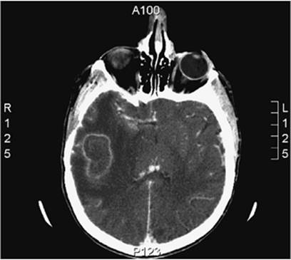
Figure 108.2. Axial contrast CT scan of a right temporal lobe abscess. This 36-year-old patient with cyanotic heart disease underwent a previous right craniotomy for subdural hematoma evacuation. Due to the presence of a pacemaker an MRI could not be performed. The image shows a 3.8 cm × 2.4 cm abscess in the posterior right temporal lobe underlying the previously noted right craniotomy. Decreased central attenuation of the lesion and surrounding vasogenic edema with uncal and subfalcine herniation are noted. There is no hydrocephalus. Abscess cultures grew Streptococcus anginosus group. |
The microbiology of brain abscesses is diverse and depends on the primary site of infection, age of the patient, and underlying host factors. Common aerobic species include streptococci (viridans, anginosus group, and microaerophilic species), which are isolated in up to 70% of cases (54). Aerobic Gram-negative bacilli—commonly Klebsiella pneumoniae, Pseudomonas spp., Escherichia coli, and Proteus spp.—and S. aureus are common pathogens with contiguous infection (55,56). Less common pathogens, such as Rhodococcus, Listeria, Nocardia, mycobacteria, and fungi—including Candida, Cryptococcus, Aspergillus, agents of zygomycosis, Pseudallescheria boydii, and the dimorphic fungi such as Histoplasma, Coccidioides, and Blastomyces—cause disease in immunocompromised hosts. Postsurgical and posttraumatic abscesses are usually due to S. aureus and aerobic Gram-negative bacilli. HIV-infected patients with advanced disease commonly present with Toxoplasma gondii infection.
Anaerobes are present in 40% to 100% of brain abscesses (55), although anaerobic cultures may not be routinely performed in all laboratories and, even if performed, may be falsely negative. Anaerobic species identified may originate from the oropharynx with contiguous head and neck infections, or from the abdomen or pelvis when infection is due to hematogenous seeding. Commonly isolated anaerobes include Peptococcus, Peptostreptococcus, Bacteroides spp, Prevotella spp., Propionibacterium, Fusobacterium, Eubacterium, Veillonella, and Actinomyces.
Helminths may occasionally cause localized brain infection in immigrant populations. Neurocysticercosis, intracranial infection with the larval cyst of T. solium or pork tapeworm, is most common and results from the ingestion of T. solium ova. Entamoeba histolytica, Schistosoma japonicum and mekongi species, Paragonimus, and Toxocara have also been described as causes of brain abscess.
|
Table 108.12 Common Presenting Features in Brain Abscess |
|
|
The clinical manifestations of brain abscess are relatively nonspecific, resulting in delays in presentation and diagnosis (Table 108.12). The onset may be acute or chronic, and most of the presenting features are related to the size and location of the abscess. Systemic toxicity is uncommon. Headache is the most common presenting symptom and is usually localized to the side of the abscess. Sudden worsening of headache may be due to rupture of the abscess into the ventricular space. Fever is present in only half of patients and thus is not a reliable sign; seizure is, however, a common presenting feature. Focal neurologic findings are relatively uncommon. Neck stiffness occurs in 15% of patients and is most commonly seen with occipital abscesses. Altered mental status and vomiting are late signs, indicating the development of elevated intracranial pressure.
|
|
|
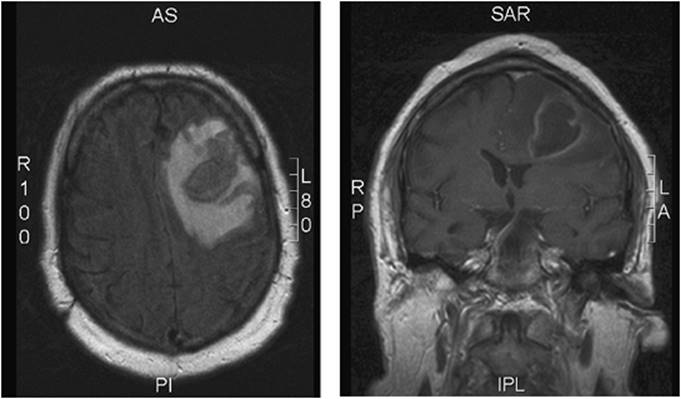
Figure 108.3. Axial and coronal MRI images of a frontal brain abscess. This 59-year-old diabetic male presented with a 10-day history of confusion, headache, right upper extremity weakness, as well as a generalized tonic-clonic seizure. Axial T2 FLAIR and coronal T1 post-gadolinium MRI images are shown above demonstrating a 3.4 cm × 3.4 cm × 4.3 cm intra-axial frontal lobe abscess with surrounding edema and mass effect. An urgent craniotomy was performed when the patient's level of consciousness decreased abruptly. Approximately 8 mL of pus were drained; cultures grew Streptococcus anginosus. Blood cultures were negative and the primary source of abscess was never identified. |
Specific presenting features correlate with abscess location. Patients with frontal lobe abscesses often present with changes in personality or mental status, hemiparesis, motor speech difficulties, and seizures. Temporal lobe abscesses may cause visual field defects or dysphasia if located in the dominant hemisphere. Patients with cerebellar abscesses may present with ataxia, nystagmus, and dysmetria. Brainstem abscesses usually extend longitudinally, with minimal compressive effect, and therefore present with few classic features. Papilledema occurs late with increased intracranial pressure.
Imaging of the brain parenchyma is the diagnostic test of choice. Lumbar puncture is contraindicated in patients with focal findings or papilledema and should be avoided in patients with suspected brain abscess. Computed tomography (CT) scanning or magnetic resonance imaging (MRI) should be performed, with the choice of test depending on the stability of the patient and availability of the imaging technique. CT scanning with contrast is not as sensitive as MRI but is generally more easily obtained on an urgent basis. MRI with gadolinium enhancement is more sensitive than CT in detecting early cerebritis and can more accurately estimate the extent of central necrosis, ring enhancement, and cerebral edema. MRI is also better able to visualize the brainstem, cerebellum, and spinal cord and can detect lesions 1.5 cm or smaller, which the CT scan may miss.
Blood cultures should be drawn in all patients with suspected or confirmed brain abscess. Abscess specimens should be obtained by stereotactic CT-guided aspiration or surgery to confirm the diagnosis and guide antimicrobial therapy. Bacterial, mycobacterial, and fungal cultures should be requested. Serology may be helpful for specific causes, such as Toxoplasma gondii and neurocysticercosis. In toxoplasma brain abscesses, IgG should be positive, as most infections are due to reactivation and not primary infection. A positive IgG antibody, however, does not prove T. gondii is the cause of a brain abscess. Brain biopsy may establish the diagnosis but is not routinely recommended given the risks involved and availability of less invasive diagnostic methods. Empiric therapy without aspiration for microbiologic samples is not generally advised except in specific situations where there is a high likelihood of a specific pathogen. For example, empiric treatment for toxoplasma infection may be warranted in a patient with advanced HIV (CD4 count less than 100 cells/µL) not receiving prophylaxis, with multiple lesions and positive IgG T. gondii serology. If clinical and radiologic responses are not evident within 7 and 14 days respectively, a microbiologic specimen should be obtained.
|
|
|
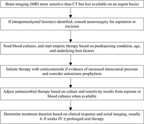
Figure 108.4. Management of Brain Abscess |
The therapy for brain abscess (Fig. 108.4) requires combination medical and surgical therapy for cure, as antimicrobial therapy alone is rarely effective. Empiric therapy should be initiated after imaging confirms the presence of an intraparenchymal lesion, pending aspiration for definitive diagnosis. Empiric therapy should be directed by the most likely source and respective pathogens.
For patients with presumed otic, mastoid, sinus, or dental sources, or temporal or cerebellar abscesses, treatment with a third-generation cephalosporin (cefotaxime, 2 g IV every 4 hours, or ceftriaxone, 2 g IV every 12 hours) and metronidazole (15 mg/kg IV load, followed by 7.5 mg/kg IV every 8 hours) is appropriate.
For patients with suspected hematogenous spread, an antimicrobial with activity against S. aureus should be used. Nafcillin or oxacillin, 2 g IV every 4 hours, is appropriate in settings with a low prevalence of methicillin resistance. Vancomycin, 15 mg/kg IV every 12 hours—adjusted for renal function and monitored with therapeutic drug levels—should be used where methicillin resistance is common or in penicillin-allergic patients. Vancomycin penetrates the CNS poorly and should be used only when indicated; a trough level of 15 to 20 µg/mL should be achieved. Metronidazole and/or a third-generation cephalosporin may be added, depending on the clinical setting.
For postneurosurgical or posttrauma patients with brain abscess, nafcillin, oxacillin, or vancomycin plus meropenem, a third-generation cephalosporin, preferably one with antipseudomonal activity such as ceftazidime, should be used.
Antimicrobial therapy should be adjusted once pathogen identification and susceptibility results are available and continued intravenously for 6 to 8 weeks, guided by clinical response and serial imaging. Prolonged oral antimicrobial therapy (2–6 months) is often administered if an appropriate regimen is available, although the efficacy of this approach has not been established. Therapy should be continued until there is complete resolution of symptoms and CT/MRI findings.
Antifungal therapy must be guided by fungal cultures and used in combination with surgical therapy. Candidal brain abscesses should be treated with amphotericin B and flucytosine. The efficacy of fluconazole has not been sufficiently evaluated in this clinical setting to recommend its use. Aspergillus brain abscesses have been historically treated with amphotericin B. However, due to recent data, voriconazole has become the treatment of choice (57), and combination antifungal therapy with voriconazole, plus either an echinocandin or an amphotericin B formulation, is increasingly used. Cerebral zygomycosis is almost invariably fatal, although amphotericin B is the treatment of choice. Posaconazole may be an alternative for zygomycoses, but voriconazole is inactive in these cases. P. boydii demonstrates in vitro resistance to amphotericin B, and, due to the lack of alternative agents, voriconazole is recommended as the antifungal of choice in these cases.
Neurosurgical consultation should be sought at the time of diagnosis. Aspiration through a burr hole or complete excision following craniotomy are both appropriate treatment options, although aspiration is generally preferred. Therapeutic aspiration may also be performed with CT or MRI guidance. Surgical excision is indicated in patients with traumatic brain abscesses, fungal abscesses, and large (greater than 2.5 cm) or multiloculated abscesses. If there is no clinical improvement within 1 week of initiation of treatment and aspiration, mental status declines, or intracranial pressure or abscess size increases despite therapy, surgical excision is also indicated. Antibiotic therapy may be shortened to 2 to 4 weeks following surgical excision.
Therapy with dexamethasone should be initiated in patients with significant edema and mass effect. Prophylactic antiseizure medications are also frequently administered. Poor prognostic factors in brain abscess include rapid progression, mental status or neurologic impairment on presentation, and rupture into a ventricle (51). Neurologic sequelae, most commonly seizures, occur in 30% to 60% of patients (51).
Cranial Subdural Empyema
Key Points
· Cranial subdural empyema and brain abscess share epidemiologic risk factors and microbiology.
· Presenting symptoms include high fever, unilateral headache, and a recent history of contiguous otic, mastoid, sinus, or meningeal infection.
· MRI is the diagnostic test of choice.
· Therapy should include prolonged antimicrobials and surgical drainage for cure.
Cranial subdural empyema is an intracranial collection of pus in the subdural space, the area between the dura and arachnoid. It is a potentially life-threatening condition, accounts for 15% to 20% of all intracranial infections (58,59,60,61), and was universally fatal prior to antimicrobial therapy. Recent data indicate that mortality with combined medical and surgical therapy now approximates 12% (61,62) with usually complete recovery in survivors.
Spread of infection to the subdural space occurs via the emissary veins or by direct extension of cranial osteomyelitis with accompanying epidural abscess. The subdural space lacks septations, so infection may spread rapidly and progressively. Most cranial subdural empyemas involve the frontal lobe, but the area of involvement is generally related to contiguous infection. Cerebral edema and hydrocephalus may develop when blood or cerebrospinal flow is disrupted by increased intracranial pressure. Cerebral infarction may also result from septic venous thrombosis.
Common predisposing infections include otic and sinus infections in up to 50% to 80% of cases (63). In patients with chronic otitis media, the middle ear and mastoids are commonly the predisposing sites of infection. Other predisposing conditions include traumatic brain injury with skull fracture, neurosurgical procedures, infection of a pre-existing hematoma, chronic pulmonary infection, or preceding meningitis.
Cranial subdural empyema is invariably polymicrobial, including streptococci, staphylococci, aerobic Gram-negative bacilli, and anaerobes. S. aureus, Enterobacteriaceae, and Pseudomonas are more common following neurosurgical procedures or neurotrauma.
The clinical presentation of cranial subdural empyema can be rapidly progressive so early diagnosis and treatment are crucial. Presenting symptoms generally include high fever and unilateral headache. A recent history of sinusitis, otitis media, mastoiditis, meningitis, cranial surgery or trauma, sinus surgery, or pulmonary infection within 2 weeks is common. Altered mental status, present in approximately 50% of patients on admission, is initially characterized by confusion and drowsiness and progresses to coma in most untreated cases. Focal neurologic signs most commonly include hemiparesis or hemiplegia, and seizures develop in up to 50% of patients (61). Other focal findings include cranial nerve palsies, homonymous hemianopsia, dysarthria or dysphasia, and ataxia. A fixed, dilated pupil portends imminent cerebral herniation and requires emergent surgical intervention.
The diagnosis of cranial subdural empyema requires a high index of suspicion and should be considered in patients presenting with meningeal signs and focal neurologic deficits, with or without systemic toxicity. A lumbar puncture is contraindicated in these cases because of the risk of cerebral herniation with increased intracranial pressure. The diagnostic imaging tests of choice are contrast CT or MRI, demonstrating a typical crescentic collection running parallel to the cranial vault. Midline shift implies significant mass effect. Gadolinium-enhanced MRI is the most sensitive, visualizing subdural empyemas too small to be detected by CT. MRI can also detect falcine, basal, and posterior fossa empyemas as well as differentiate between subdural empyemas and cystic hygromas or chronic hematomas, which CT is unable to do (61). Imaging of the sinuses, middle ear, and/or mastoids should be performed in the appropriate clinical settings to identify potential sources.
Treatment of cranial subdural empyema requires emergent combined medical and surgical therapy. Surgical drainage is mandatory, as antimicrobials alone cannot effectively cure empyemas. Cultures are, of course, required to guide antimicrobial therapy. Antiseizure treatment and/or prophylaxis may be warranted, and standard therapy for increased intracranial pressure should be instituted.
Empiric antimicrobial therapy should be initiated as soon as aspiration of the empyema is performed, or immediately on admission in unstable patients. Empiric therapy should be guided by the most likely source of primary infection. Recommended therapy includes a third-generation cephalosporin (cefotaxime, 2 g IV every 4 hours, or ceftriaxone, 2 g IV every 12 hours) or meropenem, 2g IV every 8 hours with metronidazole (15 mg/kg IV load, followed by 7.5 mg/kg IV every 12 hours). If S. aureus is suspected, nafcillin or oxacillin (2 g IV every 4 hours) should be used. Vancomycin, 1 g IV every 12 hours or 500 to 750 mg IV every 6 hours, to a maximum of 2 to 3 g/day, should be used in patients with penicillin allergy or in regions with high prevalence of methicillin resistance. Vancomycin dosing requires adjustment in patients with renal dysfunction, and serum levels should be monitored; a trough level of 15 to 20 µg/mL is desired. If Pseudomonas aeruginosa infection is suspected, ceftazidime, cefepime, or meropenem should be used in place of other third-generation cephalosporins. Intravenous antimicrobial therapy should be administered for 3 to 6 weeks, depending on clinical response and serial imaging. Prolonged therapy (6–8 weeks) may be warranted if contiguous osteomyelitis or mastoiditis is present.
Surgical therapy of cranial subdural empyema includes either burr hole drainage or craniotomy. Debridement of necrotic bone and surgical correction of sinus and otic infections are important adjuvant surgical therapies.
Epidural Abscess
Key Points
· Epidural abscesses may be cranial (between the dura and skull) or spinal (overlying the vertebral column).
· Spinal epidural abscesses are nine times more common than cranial epidural abscesses, and result most commonly from hematogenous seeding of the intervertebral disk or vertebral body. They can also occur as a complication of spinal surgery or spinal/epidural anesthesia.
· Risk factors for spinal epidural abscess include injection drug use, diabetes mellitus, bacteremia, infective endocarditis, chronic indwelling catheters, decubitus ulcers, back surgery or procedures, and trauma.
· S. aureus is the most common pathogen.
· Consider tuberculosis in those at epidemiologic risk.
· Common presenting features of spinal epidural abscess include fever, back pain, and neurologic deficits.
· MRI is the diagnostic test of choice.
· Management includes prolonged antimicrobial therapy and early surgical decompression. Surgical intervention is preferred when symptoms have been present for less than 24 hours.
An epidural abscess is a localized collection of pus between the dura and overlying skull (cranial epidural abscess) or vertebral column (spinal epidural abscess). Because severe symptoms may result due to compression of the brain or spinal cord, prompt diagnosis and treatment are crucial.
Cranial epidural abscess is commonly accompanied by subdural empyema, as emissary veins may translocate infection across the cranial dura. The microbiology is therefore identical to that of cranial subdural empyema (see previous section).
Spinal epidural abscess is nine times more common than cranial epidural abscess. The epidural space is a potential space extending from the foramen magnum down the length of the spinal canal. The space is larger in the lumbar area and is predominantly posterior, and thus most spinal epidural abscesses occur in this area. Spinal epidural abscesses most commonly originate when the intervertebral disk (diskitis) or vertebral body (osteomyelitis) become infected via hematogenous seeding. As the abscess extends, it may track longitudinally in the epidural space causing damage via direct compression of the spinal cord or local vascular damage (thrombosis, thrombophlebitis, vasculitis). Most spinal epidural abscesses extend approximately three to five vertebral spaces but can extend the entire length of the spinal canal in some cases.
Risk factors for the development of spinal epidural abscess include injection drug use, diabetes mellitus, bacteremia, infective endocarditis, chronic indwelling venous catheterization, epidural catheterization, decubitus ulcers, chronic skin conditions, paraspinal abscess, back surgery, lumbar puncture, CT-guided needle biopsies, and blunt or penetrating spinal trauma. Secondary hematogenous spread occurs in 25% to 50% of cases.
S. aureus is the most common pathogen isolated from spinal epidural abscesses, accounting for approximately 65% of cases (64). Other implicated micro-organisms include streptococci; aerobic Gram-negative bacilli, particularly Escherichia coli and Pseudomonas aeruginosa; coagulase-negative staphylococci, usually with previous spinal instrumentation; and anaerobes. Less common pathogens include Actinomyces, Nocardia, and fungi, predominantly Candida. Infections are polymicrobial in 5% to 10% of cases. Mycobacterium tuberculosis makes up approximately 25% of spinal epidural abscess and should be suspected in patients with a previous history of tuberculosis, residence in a TB-endemic region, or other TB risk factors.
Clinical manifestations in patients with cranial epidural abscess are usually insidious. Headache is the most common presenting feature. Once infection spreads to involve the meninges, subdural space, and brain perenchyma, focal neurologic signs and symptoms may develop. If the abscess is located near the petrous bone, osteomyelitis of the petrous ridge may result in Gradenigo syndrome–cranial nerve V and VI palsies with unilateral pain or otalgia.
Spinal epidural abscess presents classically with fever, back pain, and neurologic deficits. However, all three symptoms are present in only 13% of patients (65). Back pain is usually the first symptom, with paresthesias, motor weakness, and sensory changes occurring in the affected nerve roots. Bladder and bowel dysfunction, as well as paralysis, are late signs and should prompt urgent surgical consultation.
The diagnosis of epidural abscess begins with the identification of risk factors and clinical suspicion. Routine blood work may demonstrate a peripheral leukocytosis or elevated erythrocyte sedimentation rate (ESR) or C-reactive protein (CRP). MRI with gadolinium enhancement is the imaging modality of choice for both cranial and spinal epidural abscesses. CT scanning cannot visualize the spinal cord adequately and is less sensitive at identifying contiguous diskitis or osteomyelitis. Blood cultures should be collected in all patients, as they are positive in 62% of patients (64). Lumbar puncture is relatively contraindicated in the setting of epidural abscess; however, studies have shown that CSF analysis is routinely Gram stain–negative, but cultures are positive in 19% of cases. The highest-yield (90%) culture comes from the abscess itself. Ultrasound- or CT-guided drainage should be performed as soon as possible. Bacterial, mycobacterial, and fungal cultures should be requested. Additional studies to diagnose active tuberculosis should be performed—for example, sputum AFB smears and cultures, urine AFB culture, and tuberculin skin testing or QuantiFERON-TB Gold testing—in patients with suspected spinal TB.
The management of epidural abscess requires a combination of medical and surgical therapy. Empiric antimicrobial therapy for cranial epidural abscess should include a third-generation cephalosporin or meropenem plus metronidazole. Nafcillin, oxacillin, or vancomycin may be added if S. aureus is strongly suspected. Surgical drainage is crucial for cure.
The management of spinal epidural abscess similarly requires empiric antimicrobial therapy and surgical decompression, drainage, and debridement. Because of the predominance of S. aureus infection, empiric therapy is fairly targeted; vancomycin if methicillin-resistant S. aureus is likely or if the patient is penicillin-allergic; and nafcillin or oxacillin if the local prevalence of methicillin resistance is low. Early surgical intervention, specifically within the first 24 hours of presentation, results in improved outcomes (66,67). Medical therapy alone may be successful when blood or abscess aspirate cultures are available to guide therapy and there are no neurologic deficits on presentation (68,69). Serial imaging is required in these cases to confirm improvement in abscess size. Surgery should be pursued if neurologic deterioration occurs at any time, or if resolution of the abscess is not evident with medical therapy alone.
Therapy with antimicrobials is prolonged, usually 4 to 8 weeks, and should be guided by serial imaging to ensure complete resolution of the abscess. Repeat imaging should occur at approximately 4-week intervals or at any time if neurologic deterioration occurs. Although the prognosis is fair, 37% of patients experience residual neurologic deficits. The degree of residual deficit is affected by the duration of neurologic deficit prior to surgery and diagnostic delays of greater than 24 hours (65).
Suppurative Intracranial Thrombophlebitis
Key Points
· Suppurative intracranial thrombophlebitis is a complication of otic, sinus, mastoid, oropharyngeal, facial, or neurologic (bacterial meningitis, epidural abscess, or subdural abscess) infections.
· Staphylococci, streptococci, aerobic Gram-negative bacilli, and anaerobic bacteria are the most common pathogens.
· Symptoms depend on the location of septic intracranial thrombosis.
· MRI is the diagnostic test of choice.
· Management includes antimicrobials, surgical therapy, and anticoagulation.
Suppurative intracranial thrombophlebitis is septic venous thrombosis of the cortical veins. It may occur as a complication of sinus, middle ear, mastoid, oropharyngeal, or facial infections. Bacterial meningitis, epidural abscess, or subdural abscess may also result in intracranial suppurative thrombophlebitis. The absence of valves in the cerebral veins and venous sinuses aids the spread of infection from proximal sites.
Anatomically, the location of intracranial infection depends on the original source of infection. In bacterial meningitis, infection is spread via drainage of the meningeal veins into the superior sagittal sinus. The superior sagittal sinus may also be involved following facial, scalp, subdural, and epidural space infections. Otitis media and mastoiditis are the usual causes of lateral sinus and petrosal sinus thromboses. Paranasal sinus, facial, or oropharyngeal infections may result in cavernous sinus thrombosis. Risk factors for cerebral venous stasis include hypercoagulable states—specifically antiphospholipid antibody syndrome—volume depletion, polycythemia, pregnancy, the use of oral contraceptives, malignancy, sickle cell disease, and traumatic brain injury (63).
The bacterial pathogens involved in intracranial suppurative thrombophlebitis depend on the originating source of infection. S. aureus is commonly involved following facial infection; otherwise, sinusitis and otitis media pathogens cause most infections; these include staphylococci, streptococci, aerobic Gram-negative bacilli, and anaerobes such as Fusobacterium and Bacteroides. Aspergillus and the agents of zygomycosis rarely cause suppurative intracranial thrombophlebitis and are most often seen in patients with diabetes mellitus or immune deficiencies.
The clinical manifestations of suppurative intracranial thrombophlebitis depend on the anatomic site(s) involved. Septic thrombosis of the superior sagittal sinus presents with fever, headache, confusion, nausea, vomiting, and seizures. Mental status depression and progression to coma may occur rapidly. Upper motor neuron lower extremity weakness or hemiparesis may be present. When septic thrombosis is a complication of bacterial meningitis, nuchal rigidity may also be present.
Cranial nerve palsies may result from compression due to increased pressure in the cavernous sinus. Cranial nerves III, IV, V-1, V-2, and VI, as well as the internal carotid artery, travel through the cavernous sinus. Classic symptoms of septic cavernous sinus thrombosis include fever, headache, diplopia, and retro-orbital pain. Depending on the specific nerves involved, ptosis, proptosis, chemosis, hyperesthesia, and decreased corneal reflexes may be present. Venous engorgement of the retinal veins and papilledema are commonly present.
Septic transverse sinus thrombosis presents with headache and otitis. Intracranial suppurative thrombophlebitis may also be a complication of Gradenigo syndrome with spread of infection around the carotid sheath and surrounding venous plexus; patients with sigmoid sinus and internal jugular vein thrombosis may present with neck pain.
The diagnosis of suppurative intracranial thrombophlebitis is made by MRI, demonstrating absence of flow within the affected veins and venous sinuses. MR venography or angiography can be used to confirm the diagnosis, and sinus imaging should be concomitantly performed. Compared to CT scanning, MRI offers the additional benefits of detecting cerebritis, intracranial abscess, cerebral infarction, hemorrhage, or edema. Despite its lower sensitivity, CT scanning is commonly performed before MRI, as it is more easily obtained on an urgent basis.
The treatment of suppurative intracranial thrombophlebitis includes antimicrobials, surgical therapy, and anticoagulation. The choice of antimicrobial therapy depends on risk factors, the most probable source of infection, and culture results, if available. In antecedent sinusitis, empiric therapy with cefotaxime or ceftriaxone, and metronidazole is a reasonable choice. In cavernous sinus thrombosis, an agent active against S. aureus should be included. Antimicrobial therapy should be continued for 6 weeks or until radiographic resolution of thrombosis.
If antimicrobial therapy is ineffective, surgical therapy may be required for drainage of infected sinuses, ligation of the internal jugular vein, or for source control (e.g., oropharyngeal or dental infections). Anticoagulation with heparin is beneficial in cavernous sinus thrombosis, particularly if used early, and should be strongly considered (70). Intracerebral hemorrhage, if small, is not an absolute contraindication to heparin therapy; however, this form of therapy must be individualized. The efficacy of thrombolysis in septic intracranial thrombosis has not been adequately evaluated to suggest its use.
References
1. Scheld WM, Koedel U, Nathan B, et al. Pathophysiology of bacterial meningitis: mechanism(s) of neuronal injury. J Infect Dis. 2002;186(Suppl 2):S225–233.
2. Durand ML, Calderwood SB, Weber DJ, et al. Acute bacterial meningitis in adults. A review of 493 episodes. N Engl J Med. 1993;328(1):21–28.
3. Aronin SI, Peduzzi P, Quagliarello VJ. Community-acquired bacterial meningitis: risk stratification for adverse clinical outcome and effect of antibiotic timing. Ann Intern Med. 1998;129(11):862–869.
4. Tunkel AR, Scheld WM. Acute meningitis. In: Mandell GL, Bennett JE, Dolin R, eds. Mandell, Douglas, and Bennett's Principles and Practice of Infectious Diseases. 6th ed. Philadelphia, PA: Elsevier; 2005.
5. de Gans J, van de Beek D. Dexamethasone in adults with bacterial meningitis. N Engl J Med. 2002;347(20):1549–1556.
6. Attia J, Hatala R, Cook DJ, et al. The rational clinical examination. Does this adult patient have acute meningitis? JAMA. 1999;282(2):175–181.
7. Thomas KE, Hasburn R, Jekel J, et al. The diagnostic accuracy of Kernig's sign, Brudzinski's sign, and nuchal rigidity in adults with suspected meningitis. Clin Infect Dis. 2002;35(1):46–52.
8. van de Beek D, de Gans J, Spanjaard L, et al. Clinical features and prognostic factors in adults with bacterial meningitis. N Engl J Med. 2004; 351(18):1849–1859.
9. Schuchat A, Robinson K, Wenger JD, et al. Bacterial meningitis in the United States in 1995. Active Surveillance Team. N Engl J Med. 1997;337(14):970–976.
10. Weisfelt M, van de Beek D, Spanjaard L, et al. Clinical features, complications, and outcome in adults with pneumococcal meningitis: a prospective case series. Lancet Neurol. 2006;5(2):123–129.
11. Schlech WF 3rd, Ward JI, Band JD, et al. Bacterial meningitis in the United States, 1978 through 1981. The National Bacterial Meningitis Surveillance Study. JAMA. 1985;253(12):1749–1754.
12. Wenger JD, Hightower AW, Facklam RR, et al. Bacterial meningitis in the United States, 1986: report of a multistate surveillance study. The Bacterial Meningitis Study Group. J Infect Dis. 1990;162(6):1316–1323.
13. Sigurdardottir B, Björnsson OM, Jónsdóttir KE, et al. Acute bacterial meningitis in adults. A 20-year overview. Arch Intern Med. 1997;157(4):425–430.
14. Rosenstein NE, Perkins BA, Stephens DS, et al. The changing epidemiology of meningococcal disease in the United States, 1992–1996. J Infect Dis. 1999;180(6):1894–1901.
15. Wilder-Smith A, Goh KT, Barkham T, et al. Hajj-associated outbreak strain of Neisseria meningitidis serogroup W135: estimates of the attack rate in a defined population and the risk of invasive disease developing in carriers. Clin Infect Dis. 2003;36(6):679–683.
16. Fijen CA, Kuijper EJ, Tjia HG, et al. Complement deficiency predisposes for meningitis due to nongroupable meningococci and Neisseria-related bacteria. Clin Infect Dis. 1994;18(5):780–784.
17. Sjoholm AG, Kuijper EJ, Tijssen CC, et al. Dysfunctional properdin in a Dutch family with meningococcal disease. N Engl J Med. 1988;319(1):33–37.
18. Lorber B. Listeriosis. Clin Infect Dis. 1997;24(1):1–9.
19. Linnan MJ, Mascola L, Lou XD, et al. Epidemic listeriosis associated with Mexican-style cheese. N Engl J Med. 1988;319(13):823–828.
20. Bula CJ, Bille J, Glauser MP. An epidemic of food-borne listeriosis in western Switzerland: description of 57 cases involving adults. Clin Infect Dis. 1995;20(1):66–72.
21. Connolly KJ, Hammer SM. The acute aseptic meningitis syndrome. Infect Dis Clin North Am. 1990;4(4):599–622.
22. Corey L, Adams HG, Brown ZA, et al. Genital herpes simplex virus infections: clinical manifestations, course, and complications. Ann Intern Med. 1983;98(6):958–972.
23. Kent SJ, Crowe SM, Yung A, et al. Tuberculous meningitis: a 30-year review. Clin Infect Dis. 1993;17(6):987–994.
24. Klein NC, Damsker B, Hirschman SZ. Mycobacterial meningitis. Retrospective analysis from 1970 to 1983. Am J Med. 1985;79(1):29–34.
25. Mazurek GH, Jereb J, Lobue P, et al. Guidelines for using the QuantiFERON-TB Gold test for detecting Mycobacterium tuberculosis infection, United States. MMWR Recomm Rep. 2005;54(RR-15):49–55.
26. Montero A, Romero J, Vargas JA, et al. Candida infection of cerebrospinal fluid shunt devices: report of two cases and review of the literature. Acta Neurochir (Wien) 2000;142(1):67–74.
27. Nguyen MH, Yu VL. Meningitis caused by Candida species: an emerging problem in neurosurgical patients. Clin Infect Dis. 1995;21(2):323–327.
28. Bassetti M, Righi E, Costa A, et al. Epidemiological trends in nosocomial candidemia in intensive care. BMC Infect Dis. 2006;6:21.
29. Sobel JD, Vazquez J. Candidiasis in the intensive care unit. Semin Respir Crit Care Med. 2003;24(1):99–112.
30. Tortorano AM, Caspani L, Rigoni AL, et al. Candidosis in the intensive care unit: a 20-year survey. J Hosp Infect. 2004;57(1):8–13.
31. Korein J, Cravioto H, Leicach M. Reevaluation of lumbar puncture; a study of 129 patients with papilledema or intracranial hypertension. Neurology. 1959;9(4):290–297.
32. Horwitz SJ, Boxerbaum B, O'Bell J. Cerebral herniation in bacterial meningitis in childhood. Ann Neurol. 1980;7(6):524–528.
33. Hasbun R, Abrahams J, Jekel J, et al. Computed tomography of the head before lumbar puncture in adults with suspected meningitis. N Engl J Med. 2001;345(24):1727–1733.
34. Tunkel AR, Hartman BJ, Kaplan SL, et al. Practice guidelines for the management of bacterial meningitis. Clin Infect Dis. 2004;39(9):1267–1284.
35. Metersky ML, Williams A, Rafanan AL. Retrospective analysis: are fever and altered mental status indications for lumbar puncture in a hospitalized patient who has not undergone neurosurgery? Clin Infect Dis. 1997;25(2):285–288.
36. Leib SL, Boscacci R, Gratzl O, et al. Predictive value of cerebrospinal fluid (CSF) lactate level versus CSF/blood glucose ratio for the diagnosis of bacterial meningitis following neurosurgery. Clin Infect Dis. 1999;29(1):69–74.
37. Koskiniemi M, Piiparinen H, Mannonen L, et al. Herpes encephalitis is a disease of middle aged and elderly people: polymerase chain reaction for detection of herpes simplex virus in the CSF of 516 patients with encephalitis. The Study Group. J Neurol Neurosurg Psychiatry. 1996;60(2):174–178.
38. Tyler KL. Update on herpes simplex encephalitis. Rev Neurol Dis. 2004;1(4):169–178.
39. Stockstill MT, Kauffman CA. Comparison of cryptococcal and tuberculous meningitis. Arch Neurol. 1983;40(2):81–85.
40. Verdon R, Chevret S, Laissy JP, et al. Tuberculous meningitis in adults: review of 48 cases. Clin Infect Dis. 1996;22(6):982–988.
41. Miner JR, Heegaard W, Mapes A, et al. Presentation, time to antibiotics, and mortality of patients with bacterial meningitis at an urban county medical center. J Emerg Med. 2001;21(4):387–392.
42. Lu CH, Huang CR, Chang WN, et al. Community-acquired bacterial meningitis in adults: the epidemiology, timing of appropriate antimicrobial therapy, and prognostic factors. Clin Neurol. Neurosurg. 2002;104(4):352–358.
43. McIntyre PB, Berkey CS, King SM, et al. Dexamethasone as adjunctive therapy in bacterial meningitis. A meta-analysis of randomized clinical trials since 1988. JAMA. 1997;278(11):925–931.
44. Weisfelt M, Hoogman M, van de Beek D, et al. Dexamethasone and long-term outcome in adults with bacterial meningitis. Ann Neurol. 2006;60(4):456–468.
45. Desmond RA, Accortt NA, Talley L, et al. Enteroviral meningitis: natural history and outcome of pleconaril therapy. Antimicrob Agents Chemother. 2006;50(7):2409–2414.
46. Steiner I, Budka H, Chaudhuri A, et al. Viral encephalitis: a review of diagnostic methods and guidelines for management. Eur J Neurol. 2005;12(5):331–343.
47. Centers for Disease Control and Prevention. Recommended childhood and adolescent immunization schedule-United States, 2006. MMWR Morb Mortal Wkly Rep. 2005;54 (51,52):Q1–Q4.
48. Centers for Disease Control and Prevention. Recommended adult immunization schedule(United States, October 2005–September 2006. MMWR Morb Mortal Wkly Rep. 2005;54:Q1–Q4.
49. Bode AV, Sejvar JJ, Pape WJ, et al. West Nile virus disease: a descriptive study of 228 patients hospitalized in a 4-county region of Colorado in 2003. Clin Infect Dis. 2006;42(9):1234–1240.
50. Levitz RE. Herpes simplex encephalitis: a review. Heart Lung. 1998;27(3): 209–212.
51. Seydoux C, Francioli P. Bacterial brain abscesses: factors influencing mortality and sequelae. Clin Infect Dis. 1992;15(3):394–401.
52. Tattevin P, Bruneel F, Clair B, et al. Bacterial brain abscesses: a retrospective study of 94 patients admitted to an intensive care unit (1980 to 1999). Am J Med. 2003;115(2):143–146.
53. Yang SY, Zhao CS. Review of 140 patients with brain abscess. Surg Neurol. 1993;39(4):290–296.
54. Wispelwey B, Scheld WM. Brain abscess. In: Scheld WM, Whitley RJ, Durack DT, eds. Infections of the Central Nervous System. 2nd ed. Philadelphia, PA: Lippincott-Raven Publishers, 1997:463–493.
55. de Louvois J, Gortavai P, Hurley R. Bacteriology of abscesses of the central nervous system: a multicentre prospective study. Br Med J. 1977;2(6093):981–984.
56. Mandell GL, Bennett JE, Dolin R, eds. Mandell, Douglas, and Bennett's Principles and Practice of Infectious Diseases. 6th ed. Philadelphia, PA: Elsevier Churchill Livingstone; 2005.
57. Johnson LB, Kauffman CA. Voriconazole: a new triazole antifungal agent. Clin Infect Dis. 2003;36(5):630–637.
58. Silverberg AL, DiNubile MJ. Subdural empyema and cranial epidural abscess. Med Clin North Am. 1985;69(2):361–374.
59. Coonrod JD, Dans PE. Subdural empyema. Am J Med. 1972;53(1):85–91.
60. Kaufman DM, Miller MH, Steigbigel NH. Subdural empyema: analysis of 17 recent cases and review of the literature. Medicine (Baltimore). 1975;54(6):485–498.
61. Tunkel AR. Subdural empyema, epidural abscess, and suppurative intracranial thrombophlebitis. In: Mandell GL, Bennett JE, Dolin R, eds. Mandell, Douglas, and Bennett's Principles and Practice of Infectious Diseases. 6th ed. Elsevier; 2005.
62. Nathoo N, Nadvi SS, van Dellen JR, et al. Intracranial subdural empyemas in the era of computed tomography: a review of 699 cases. Neurosurgery. 1999;44(3):529–535; discussion 535–536
63. Kaufman DM, Litman N, Miller MH. Sinusitis: induced subdural empyema. Neurology. 1983;33(2):123–132.
64. Darouiche RO, Hamill RJ, Greenberg SB, et al. Bacterial spinal epidural abscess. Review of 43 cases and literature survey. Medicine (Baltimore). 1992;71(6):369–385.
65. Davis DP, Wold RM, Patel RJ, et al. The clinical presentation and impact of diagnostic delays on emergency department patients with spinal epidural abscess. J Emerg Med. 2004;26(3):285–291.
66. Rigamonti D, Liem L, Wolf Al, et al. Epidural abscess in the cervical spine. Mt Sinai J Med. 1994;61(4):357–362.
67. Liem LK, Rigamonti D, Wolf AL, et al. Thoracic epidural abscess. J Spinal Disord. 1994;7(5):449–454.
68. Wheeler D, Keiser P, Rigamonti D, et al. Medical management of spinal epidural abscesses: case report and review. Clin Infect Dis. 1992;15(1):22–27.
69. Siddiq F, Chowfin A, Tight R, et al. Medical vs surgical management of spinal epidural abscess. Arch Intern Med. 2004;164(22):2409–2412.
70. Levine SR, Twyman RE, Gilman S. The role of anticoagulation in cavernous sinus thrombosis. Neurology. 1988;38(4):517–522.
Editors: Gabrielli, Andrea; Layon, A. Joseph; Yu, Mihae