Carsten M. Schmalfuss
The Normal Pericardium
The normal pericardium forms a sac around the heart and proximal large arteries and veins. It has a thin visceral mesothelial layer that closely adheres to the epicardial surface of the heart. The parietal layer consists of the same thin mesothelium and a thicker (up to 2 mm), fairly nonelastic fibrous layer on the outside. Physiologically, the space between both mesothelial layers contains 20 to 30 mL of fluid.
The fibrous part of the pericardium is attached to the surrounding mediastinal structures and holds the heart in its position within the chest. It limits cardiac dilatation, contributes to the interventricular interaction, and primarily affects diastolic function. The smooth mesothelial surfaces and the pericardial fluid between them reduce friction and act as a barrier to inflammation from surrounding structures. The pericardium is well innervated, and pathologic processes can cause severe episodic or continuous pain.
The intrapericardial pressure is usually negative; it is approximately equal to and varies with the pleural pressure at the same hydrostatic level. Pericardial pressure affects myocardial transmural pressure by the following relationship:
Transmural pressure = cavitary pressure - adjacent intrapericardial pressure
Because the intrapericardial pressure is normally negative, this usually adds to the normal transmural pressure gradient (1).
The relationship between intrapericardial and pleural pressures causes a simultaneous fall of pressures in both spaces during inspiration and leads to an increased venous return into the right chambers (increased preload), with a subsequent increase in cardiac output. Inspiration influences filling and cardiac output of the left heart only indirectly and very little. The parietal pericardium is very resistant to acute stretching but adapts and expands to great dimension when subjected to a chronic stretching process. The pericardial pressure–volume curve is generally flat as pericardial volume increases, and when further distention is impossible, a sharp rise in the intrapericardial pressure occurs. This exponential curve accounts for the rapid clinical response when even small amounts of fluid are removed in cardiac tamponade (2).
This chapter deals with the most common clinical pericardial problems encountered in critical care medicine. They include acute pericarditis, pericardial effusion, cardiac tamponade, and pericardial constriction.
Acute Pericarditis
Etiology
Inflammation of the pericardial sac results in acute pericarditis and is either an isolated problem or part of a systemic process. Exudation of inflammatory fluid into the pericardial space can result in pericardial effusion. Depending on the frequency and time course, pericarditis can be acute, recurrent, or chronic. Common causes of acute pericarditis are shown in Table 125.1. Most often, clinically recognizable pericarditis in the adult is idiopathic. In these cases, various viruses are often the suspected causes; an etiologic agent is infrequently demonstrated. The most commonly demonstrated virus is the Coxsackie B group, which causes myopericarditis in children and pleuropericarditis in adults—also called Bornholm disease (3). ECHO, influenza, Epstein-Barr, varicella, hepatitis, mumps, and human immunodeficiency viruses can also cause pericarditis.
Up to one third of patients with end-stage renal disease will develop uremic pericarditis. Most of them have not started dialysis when they present with pericarditis, and the symptoms usually disappear after beginning or increasing the frequency of dialysis (4). There is no direct association with serum blood urea nitrogen level or serum creatinine and the acute illness. However, it is suspected that increased toxin levels from declining renal function cause the inflammatory process. It is important to remember that the uremic patient is susceptible to infections and that the pericarditis may be infectious. Last but not least, the underlying disease process leading to the renal insufficiency (i.e., lupus erythematosus) may also be the cause for the pericardial inflammation.
Acute pericarditis after myocardial injury is thought to be due to direct irritation of the visceral mesothelium. In the past, pericarditis occurred in up to 20% of patients after transmural myocardial infarction; recently, the frequency has decreased due to the increased use of reperfusion therapy—that is, thrombolytic therapy or angioplasty (5). Typically, symptoms occur 1 to 3 days after the myocardial damage and can mimic recurrent angina pectoris. If the patient is receiving anticoagulants, the inflammation can lead to hemorrhagic intrapericardial effusion and possibly cardiac tamponade. Acute pericardial inflammation is also found after open heart surgery, implantation of cardiac pacemakers, percutaneous coronary interventions, or external cardiac trauma, and the presentation is similar to that after transmural infarction.
|
Table 125.1 Common Causes of Pericarditis |
|||||||||||||||||||||||||||||||||||
|
The postpericardiotomy syndrome was originally described as postmyocardial infarction pericarditis. Later, Engle and Ito (6) noted the same clinical syndrome in children and adults who experienced an opening of the pericardium. The syndrome occurs in 10% to 30% of patients who have undergone pericardiotomy and is thought to be an immune complex reaction to the patient's own pericardium (7). In contrast to pericarditis caused by myocardial injury, these patients usually have symptoms of chest pain and fever beginning several weeks to months after cardiac surgery or other myocardial injury.
Neoplastic pericarditis is most often caused by cancer of the lung, breast, or esophagus as well as lymphoma and melanoma (8). The tumor directly invades the pericardial space or metastasizes through lymphatics or blood vessels; primary pericardial tumors like mesothelioma are rare. The likelihood of finding previously undiagnosed cancer in a patient presenting with pericarditis is about 6% to 7% (9). The cause of pericarditis in patients with known malignancy is neoplastic in only 50% to 60%; idiopathic pericarditis and radiation-induced pericarditis are the most common benign causes (10,11). The prognosis of neoplastic pericarditis and effusion is poor.
Pericarditis is also seen in patients with systemic lupus erythematosus, rheumatoid arthritis, and scleroderma. Inflammation of the pericardium is often the first manifestation of lupus in female patients; this diagnosis should be ruled out during the workup for a first episode of idiopathic pericarditis. Radiation pericarditis often follows a mediastinal dose of 4,000 rad or more and can lead to pericardial effusion and acute cardiac tamponade. The long-term effects of radiation also can lead to constrictive pericarditis.
Pericarditis caused by infectious organisms other than viruses is less frequent now than it was in the preantibiotic era. Pneumonia is still the most common cause; others include sepsis from peritonitis and urinary tract infection, or direct spread of the infectious process from mediastinitis or necrotizing fasciitis of the head or neck. Immunocompromised and elderly patients are more prone to infectious pericarditis than the general population. In adults, Staphylococcus aureus is still the most common organism, and there is an apparent decline in infections with Streptococcus spp., Pneumococcus spp., and Haemophilus influenzae.
Tuberculous pericarditis was once a common cause of acute and constrictive pericarditis, but with the overall decline of tuberculosis, it has become a rare entity in the United States (12). More recently, states with a high percentage of immigrants have again reported rising numbers of tuberculous pericarditis (13). One to two percent of patients with pulmonary tuberculosis will develop tuberculous pericarditis (14). Mycobacterial infection must be ruled out in any case of suspected purulent pericarditis.
The most common fungal organism to cause pericarditis is Histoplasma capsulatum. Histoplasmosis in the United States is most common in the Mississippi and Ohio River Valleys (15). Diagnosis is usually delayed and made by positive fungal culture of the pericardial fluid and/or a significant rise of serologic antibody titers (greater than 1:32) against Histoplasma capsulatum.
Clinical Presentation
The typical patient presenting with pericarditis is young and was previously healthy. Symptoms of acute pericarditis include sharp and, usually, persistent chest pain that is generally increased with respiration and motion. It is worse in the supine position and usually improves sitting up and/or with shallow breathing. The pain can radiate to the neck, and dyspnea may also be present. Other common findings preceding or accompanying pericarditis are fever, myalgia, malaise, and tachycardia. The characteristic and pathognomonic three-component friction rub is best described as coarse, leathery, superficial—like “pulling Velcro”. The rub is only intermittently heard during the episode of pericarditis, and therefore, it is important to auscultate frequently. The patient is ideally examined in a quiet setting in an upright position leaning forward; the rub is best heard at the left sternal border or the cardiac apex.
An electrocardiogram (ECG) should be obtained in every patient presenting with chest pain. Four ECG stages, evolving over hours to days and weeks, have been described:
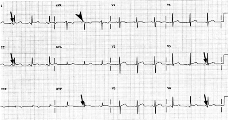
· Stage I includes classic and diffuse ST elevations with a concave ST segment and significant PR-segment depression (Fig. 125.1).
· Stage II is normalization of the ECG.
· Stage III is the development of diffuse T-wave inversion that may persist or normalize.
· Stage IV is final normalization of the ECG (16).
|
|
|
Figure 125.1. Electrocardiogram of a patient with acute pericarditis. Note the PR-segment depressions (arrows) and convex ST-segment elevations in the inferolateral leads and PR-segment elevation (arrowhead) and ST-segment depression in lead aVR consistent with stage I of electrocardiogram findings in acute pericarditis. |
The ECG may show all, several, or none of the stages during an episode of acute pericarditis. Atrial arrhythmias are rare but do occur in acute pericarditis (17) and can be the first manifestation of acute pericarditis. However, sustained atrial or ventricular arrhythmias are suggestive of concomitant myocarditis.
The key to an ECG diagnosis of pericarditis is the diffuse nature of the ECG changes, the absence of localization to a particular ECG anatomic area, PR-segment depression, and the absence of ST depression except in lead aVR.
Every patient suspected to have pericarditis should have a chest radiograph taken. It will be normal in most cases of pericarditis; a new finding of an enlarged cardiac silhouette is, however, suggestive of a pericardial effusion (greater than 200 mL) and should be further evaluated.
The laboratory may report positive acute-phase reactants (especially the erythrocyte sedimentation rate) and an elevated white blood cell count; however, these are nonspecific findings. Cardiac troponin T or I and CK-MB isoenzymes are cardiac—but not pericardium—specific and are often found minimally elevated in acute pericarditis. Viral studies may confirm a viral cause of the pericarditis; however, their yield is low, and the result does not change management. In cases of suspected infectious etiology, cultures from blood and pericardial fluid, if available, should be examined for bacterial and mycobacterial pathogens.
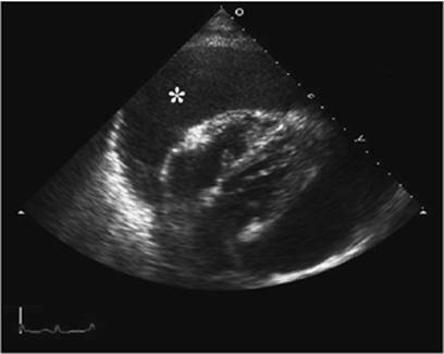
Echocardiography should be performed in every patient with the suspected diagnosis of pericarditis to evaluate for and follow pericardial effusion (Fig. 125.2) and to help diagnose cardiac tamponade.
The triad of typical chest pain, pericardial friction rub, and the aforementioned ECG changes confirms the diagnosis of acute pericarditis. However, this diagnosis should be made only after life-threatening conditions with similar presentation (Table 125.2) have been ruled out. Electrocardiographic differential diagnosis also includes variant angina, hypertrophic cardiomyopathy, and the benign finding of early repolarization—all of which can mimic the ECG changes described earlier.
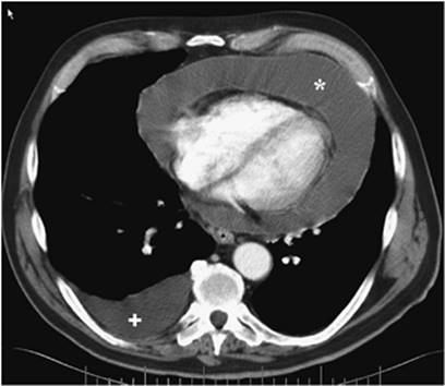
Aside from history and physical exam, ECG, chest radiographs, blood work, and echocardiogram, it may be necessary to evaluate the patient with computed tomography (CT) of the chest to rule out pulmonary embolism or aortic dissection (Fig 125.3). With the recent development of 64-slice or dual-head CT scanners, it has become possible to perform a so-called triple rule-out, at least in specialized centers. During a short (20–25 seconds), single breath-hold, contrasted, ECG-gated CT scan, all data can be acquired to evaluate the pulmonary artery tree, the thoracic aorta, and the coronary arteries as well (18).
|
Table 125.2 Life-Threatening Differential Diagnosis of Acute Pericarditis |
|
|
|
|
|
|
|
Figure 125.2. Subcostal echocardiogram showing a giant pericardial effusion (asterisk) in a patient with neoplastic pericarditis. |
Treatment
Patients with acute pericarditis have a high likelihood of uncomplicated recovery and can be treated outside the hospital. However, several factors are described as being associated with a complicated course (Table 125.3), and patients with any of these factors should be hospitalized for their initial treatment (19).
Acute idiopathic or viral pericarditis usually responds to nonsteroidal anti-inflammatory drugs (NSAIDs). The drug regimen consists of high-dose aspirin (325–975 mg three to four times daily for 4 weeks), with the addition of a proton pump inhibitor to lessen gastrointestinal effects. Alternatively, indomethacin (25–50 mg four times daily) or ibuprofen (400–600 mg four times daily) can be given. Colchicine has been shown to be effective as a second-line treatment for patients who do not respond to NSAIDs or who have recurrence of their acute pericarditis (20,21). Recently, Imazio et al. (22) found that routine use of colchicine (1–2 mg on day 1 followed by 0.5–1 mg/day for 3 months) in addition to aspirin—compared to aspirin alone—in patients with a first episode of acute pericarditis significantly reduced symptoms at 72 hours and recurrence at 18 months. Diarrhea is a known side effect of colchicine and may cause discontinuation of drug therapy in about 5% of patients.
|
|
|
|
Figure 125.3. Computed tomography (CT) of the chest with large pericardial effusion (asterisk) and small right-sided pleural effusion (plus sign). The dark rim between the pericardial effusion and the right heart represents epicardial fat. |
|
|
Table 125.3 Presenting Factors Predicting Complicated Course |
|
|
Symptoms of acute pericarditis respond rapidly to systemic steroids, but there seems to be an increase in relapse after tapering (23). Therefore, corticosteroid therapy should be reserved only for patients with recurrent pericarditis not responding to NSAIDs and colchicine. The recommended regimen is 1 to 1.5 mg/kg of prednisone for at least 1 month before slowly tapering the dose by 5 mg/week until the drug is withdrawn (24). The possible side effects of corticosteroid treatment include peptic ulcer disease, sodium retention, hypokalemia, hyperglycemia, Cushing syndrome, and suppression of the adrenal axis. Treatment with corticosteroids also requires the exclusion of infection or an appropriate antibiotic regimen before initiation of therapy.
The treatment of choice for uremic pericarditis consists of intensive, initially daily dialysis therapy. Heparin should be used sparingly during dialysis to reduce the risk of intrapericardial hemorrhage and possible tamponade. The presence of acute pericarditis in acute myocardial infarction also requires caution with the use of intravenous anticoagulants. These drugs are not, however, absolutely contraindicated. Thrombolytic agents have been reported to lead to cardiac tamponade and should be used with caution in the patient with acute myocardial infarction and acute pericarditis.
The postpericardiotomy syndrome is usually self-limited if left untreated; however, the disease may increase the risk of early coronary artery bypass graft closure. Therefore, aggressive treatment has been recommended; NSAIDs often decrease symptoms and speed recovery (25). Refractory cases may occur but usually respond rapidly to systemic corticosteroids as outlined above. Advocates of corticosteroid therapy claim that this treatment reduces the incidence of late constrictive pericarditis.
Nonviral infectious etiology of pericarditis requires prompt evacuation of pus from the pericardium, usually by operative intervention, because of the need to establish a definitive diagnosis, eradicate the infection, and prevent constrictive pericarditis.
Recurrence of acute pericarditis is quite common and often requires long-term drug therapy as noted above. In a few selected cases refractory to medical therapy, radical pericardectomy may need to be considered (23).
|
|
|
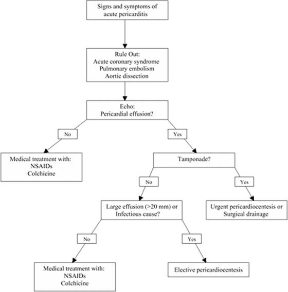
Figure 125.4. Diagnostic and treatment algorithm for patients presenting with signs and symptoms of acute pericarditis. |
In general, acute pericarditis symptoms subside within several days to weeks. The major immediate complication is cardiac tamponade, which occurs in less than 5% of patients. For diagnostic and treatment approach in patients with suspected acute uncomplicated or complicated pericarditis, see Fig. 125.4.
Pericardial Effusion and Cardiac Tamponade
Etiology
Pericardial effusion often develops with acute pericarditis. It is caused by an inflammatory exudation and an occlusion of the normal drainage through epicardial venous and lymphatic systems by the inflammatory process. The most common causes of tamponade include idiopathic pericarditis, cancer of the lung and breast, lymphoma, renal failure, and tuberculosis (26). Pericardial effusion may also occur in the absence of pericardial inflammation—for example, as a hemorrhagic effusion from internal sources such as a pacemaker, angioplasty, coronary artery bypass grafting (CABG) surgery, aortic dissection, ventricular rupture—or external cardiac trauma. Regardless of their size, pericardial effusions can either be clinically silent or cause hemodynamic compromise. The latter situation is called cardiac tamponade. The most important factor contributing to the development of cardiac tamponade is not the total amount of pericardial fluid but the rate at which it accumulates. The pericardium resists sudden stretching but can gradually expand in response to a chronic distending force. A small but rapidly developing effusion—less than 200 mL—in a trauma patient can cause tamponade, because the fibrous pericardial membrane does not have enough time to stretch and accommodate the increased volume. Conversely, a patient with a very large pericardial effusion—1,000 mL—developing over weeks or months may be completely asymptomatic, given that the parietal pericardium has had time to adjust to the increased volume.
It is also important to note that the pressure–volume curve for the stretchable pericardium is curvilinear; a large amount of fluid accumulating over a long time raises the intrapericardial pressure very little. However, at some point, the ability of the pericardium to stretch further is exceeded, and the addition of a very small volume raises the intrapericardial pressure significantly. Once the intrapericardial pressure exceeds the filling pressures of the right atrium and/or the right ventricle, central venous pressure rises, cardiac output drops, and cardiac tamponade—and consequently cardiogenic shock—occur (27,28). Physiologically, the total cardiac volume is limited, and volume in one chamber can increase only if volume in another chamber decreases. This physiologic interdependence of the ventricles is accentuated in cardiac tamponade when right ventricular filling during inspiration causes a significant decrease in left ventricular filling and a significant decrease in stroke volume. This inspiratory drop in systolic blood pressure of greater than 10 mm Hg, termed pulsus paradoxus, is very suggestive of cardiac tamponade in a patient presenting with hypotension and tachycardia, but it is not specific and can also present in other conditions such as chronic obstructive pulmonary disease (COPD), pulmonary embolism, pneumothorax, acute asthma, and hypovolemic shock (29).
Cardiac tamponade is a serious problem that, if not treated aggressively and rapidly, may be fatal. Treatment may be successful if diagnosed in a timely fashion, and thus cardiac tamponade needs be included in the initial differential diagnosis of cardiogenic shock or pulseless electrical activity.
Clinical Presentation
The patient with early pericardial tamponade is often confused, agitated, pale, and diaphoretic and complains of chest pain and dyspnea. Initially, compensatory catecholamine release, caused by a decreased cardiac output, leads to sinus tachycardia and often to peripheral vasoconstriction. Later in the course, bradycardia occurs, indicating imminent pulseless electrical activity and cardiorespiratory arrest unless the effusion is immediately decompressed.
Classic clinical signs include jugular venous distension (JVD), demonstrating a rapid x-descent but no y-descent because of right atrial and ventricular compression throughout the entire diastolic cycle. In this situation, the central venous pressure is usually greater than 15 mm Hg, but jugular venous distention may be missing in trauma patients with rapidly developing hemorrhagic tamponade or in patients with uremic pericarditis due to volume depletion from blood loss or dialysis, respectively (low-pressure tamponade).
Pulsus paradoxus is often present and can be ascertained through invasive arterial pressure tracing by palpation of an artery or with a sphygmomanometer. The amount of paradox is gauged by measuring the systolic blood pressure and observing the difference in the level at which the Korotkoff sounds are heard only during expiration and the level at which they are heard throughout the respiratory cycle. A paradoxical pulse greater than 10 mm Hg is abnormal; patients with tamponade physiology often drop their systolic blood pressure more than 20 mm Hg with inspiration. Paradoxical pulse is absent in patients with severe aortic insufficiency or atrial septal defect and is difficult to assess in acute cardiac tamponade with hypotension, as the pulse may be unobtainable or disappear completely with inspiration. Other clinical signs of cardiac tamponade are distant and muffled heart sounds and clear lungs. The patient with chronic tamponade may present with a low-output state, right upper quadrant pain caused by swelling of the hepatic capsule, or even ascites and lower extremity edema. The two main differential diagnoses for cardiac tamponade are tension pneumothorax and pulmonary edema; both conditions can present with tachycardia, hypotension, and JVD.
The ECG usually shows signs of acute pericarditis, including sinus tachycardia, PR depression, and abnormal T-wave changes. Electrical alternans of the ECG is almost pathognomonic of pericardial tamponade; it represents a change of direction and amplitude of the P, QRS, and T vectors with every other heart beat, and probably results from the “swinging” movement of the heart in a large volume of fluid. A change of the electrical QRS alternans alone is the most common finding (30). As mentioned above, the finding is highly specific for cardiac tamponade, but its absence does not rule out large effusion or tamponade, particularly if they develop rapidly.
|
|
|
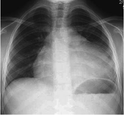
Figure 125.5. Anteroposterior chest radiograph in patient with a very large pericardial effusion. The extensive widening of the cardiac silhouette resembles a water bottle. |
The chest radiograph may show a globular heart hanging down in the mediastinum—the water bottle heart (Fig. 125.5). It is also helpful to rule out diagnoses presenting similarly to tamponade, such as tension pneumothorax and pulmonary edema.
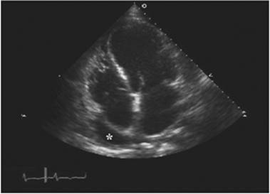
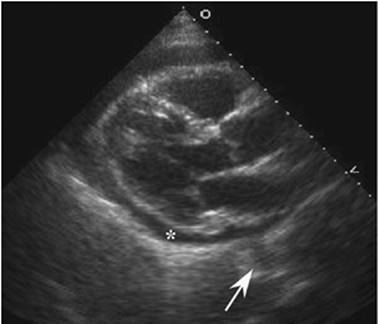
Cardiac tamponade is a clinical diagnosis based on the previously described symptoms and findings. However, the best adjunctive test to assess for pericardial effusion and cardiac tamponade is the echocardiogram (31). Pericardial effusion is seen as an echo-free space surrounding the heart. An echocardiogram can detect even very small amounts of pericardial effusion (less than 20 mL), helps to estimate amount and distribution of the effusion, and visualizes clot or tumor in the fluid if present. Small effusions (less than 1 cm) are seen only inferolaterally and around the right atrium (Fig. 125.6). Effusions causing tamponade are mostly large (greater than 2 cm) and circumferential. Effusions seen with acute tamponade are usually smaller than those seen with chronic tamponade. An echocardiogram can distinguish pericardial from pleural effusion; pericardial effusion tracks between the inferolateral wall and the descending thoracic aorta in the parasternal long axis view and separates both structures (Fig. 125.7), whereas pleural effusion is found only posterior to the aorta in this view.
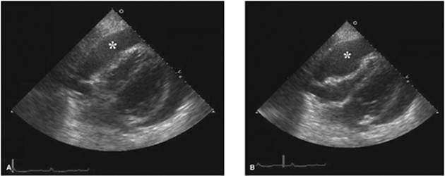
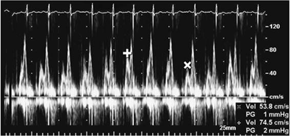
Once a pericardial effusion begins to compromise the hemodynamics, there are several characteristic echocardiographic findings. There is diastolic collapse, first of the right atrium, and then right ventricle, both of which worsen during expiration when right-sided filling is reduced (32,33). Diastolic collapse lasting more than one third of diastole is considered indicative for tamponade (Fig. 125.8). Reciprocal respiratory variation of greater than 20% to 25% of the peak transmitral and transtricuspid Doppler velocities is another very specific indicator for hemodynamically significant pericardial effusion (Fig. 125.9). Echocardiography is a very useful tool to assess pericardial effusion and determine its hemodynamic significance. However, life-saving treatments for an unstable or deteriorating patient suffering from cardiac tamponade should not be delayed by waiting for an echocardiogram to be performed.
|
|
|
Figure 125.6. Apical four-chamber view echocardiogram in patient presenting with acute pericarditis. The very small pericardial effusion is seen only around the right atrial wall (asterisk). The right atrium is not compressed by the effusion. This location may be the only one in which an early or small pericardial effusion can be seen in a patient with poor subcostal windows. |
The invasive hemodynamic profile of acute cardiac tamponade is characteristic and can be assessed by placement of a pulmonary artery catheter. The right-sided cardiac pressures are elevated, and the diastolic pressures equilibrate. The mean right atrial pressure, right ventricular diastolic pressure, pulmonary artery diastolic pressure, and the pulmonary capillary wedge pressure are elevated and are measured within 2 to 3 mm Hg of each other. The pressure contour does not show a dip and plateau sign as seen in constrictive pericarditis. The pressures in chronic congestive heart failure are elevated but do not equilibrate in diastole. These measurements made in the intensive care unit (ICU) setting can confirm the diagnosis of cardiac tamponade.
|
|
|
Figure 125.7. Parasternal long-axis echocardiographic view in a patient presenting with clinical symptoms of acute pericarditis. There is a small to moderate-sized concentric effusion (asterisk). It tracks between the inferolateral wall and the descending thoracic aorta (arrow) and thereby confirms the diagnosis of pericardial effusion. A pleural effusion may be seen in the same inferolateral location but would not separate the heart from the aorta. |
Treatment
Patients with newly diagnosed pericardial effusion, without tamponade physiology, should be monitored in the hospital for 24 to 48 hours, with at least one repeat echocardiogram prior to discharge. A repeat echocardiogram should be performed in patients with large pericardial effusions 4 to 6 weeks after the initial presentation or with change in symptoms suggestive of beginning hemodynamic significance of the pericardial effusion.
Patients diagnosed with hemodynamically significant cardiac tamponade, defined as systolic blood pressure less than 110 mm Hg or pulsus paradoxus greater than 10 mm Hg, should receive immediate aggressive fluid resuscitation with normal saline to increase right-sided filling pressures to at least temporarily stabilize hemodynamics. Additionally, inotropic support with dobutamine, dopamine, isoproterenol, or norepinephrine may be needed to further stabilize the blood pressure. More definitive treatment with percutaneous pericardiocentesis or surgically created pericardial window should follow promptly. Any patient presenting with traumatic hemopericardium should be treated surgically (34).
Pericardiocentesis should be performed in any patient with acute tamponade and hemodynamic compromise, or when an infectious or malignant cause of the pericardial effusion is suspected. The procedure is usually performed in the cardiac catheterization laboratory, but in an emergency, may be done at the bedside if clinically necessary (34).
The patient should be placed in a supine position at a 45-degree angle. The area between the xiphoid and the left costal arch should be sterilely prepped using a tinted (so clinicians can see where they have prepped) chlorhexidine–alcohol solution and draped in the usual fashion, and anesthetized with a 1% or 2% lidocaine solution. A 7.6-cm (3-inch) aspiratory needle (16–18 gauge) with a short bevel should be directly attached to a three-way stopcock and a 50-mL syringe, and the needle advanced with negative pressure at an angle of 30 to 45 degrees to the abdominal wall and oriented in a posterocephalad direction toward the left shoulder (35). Once it enters the pericardial space, fluid can be easily removed. Removal of a small amount of fluid may provide significant clinical improvement. A temporary catheter is then placed into the pericardial space via Seldinger technique and connected to a bag draining to gravity for several days. This approach provides more complete drainage and reduces the risk for reaccumulation of the effusion.
The first sample of fluid retrieved should be sent for microbiologic studies and several other diagnostic tests (Table 125.4).
Echocardiography can be used to locate the ideal spot for percutaneous puncture (36). It is helpful to determine the distance from the surface to the effusion and demonstrates liver or lung tissue that may be in the projected path of the needle. After the puncture, an echocardiogram confirms the correct position when bubbles generated with sterile, agitated saline injected through the needle are demonstrated within the pericardial effusion. Alternatively, ECG can be used to determine the position of the needle; the needle is connected to a V lead of the ECG via an alligator clamp. If the tip of the needle contacts the epicardium, the ECG will suddenly demonstrate ST-segment elevation. The needle should then be withdrawn until the changes disappear. Complications of this procedure include pneumothorax, myocardial and coronary artery laceration, dysrhythmias, and death. Many authors have pointed out the significant risks associated with this procedure, which should be approached with experience and caution.
|
|
|
Figure 125.8. Subcostal echocardiogram in a patient with cardiogenic shock. Compared to early systole (A), there is significant collapse of the right ventricular free wall until very late into the diastolic filling phase (B), consistent with pericardial tamponade. The treatment of choice is immediate decompression of the effusion (*) by percutaneous or surgical drainage. |
If the patient with acute cardiac tamponade can be stabilized by volume expansion and vasopressor support, a safer and equally effective drainage of pericardial fluid can be accomplished by surgical subxiphoid pericardial resection and drainage (34). Subxiphoid resection can be performed in a sterile environment under local anesthesia; pericardial fluid can be removed and pericardial tissue obtained for biopsy and culture (37).
|
|
|
Figure 125.9. Transmitral pulse-wave Doppler tracing with significant (greater than 20%) decrease of E-wave velocity during inspiration (x) when compared to velocity during expiration (+). This finding indicates hemodynamic significance if found in conjunction with a pericardial effusion, and immediate drainage of the effusion via percutaneous or surgical route should be considered. |
Malignant recurrent pericardial effusions often require surgical creation of a pericardial window to allow fluid to drain into the adjacent pleural space. More recently, percutaneous balloon pericardiotomy has been developed as a nonsurgical approach to create such a window and to drain large pericardial effusions (38).
Constrictive Pericarditis
Etiology
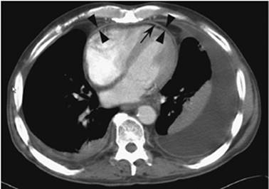
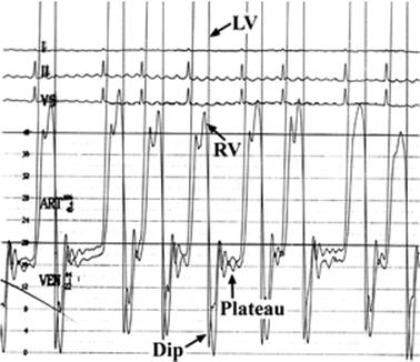
Constrictive pericarditis occurs when chronic inflammation leads to scarring and, in some cases, calcification of the pericardium. Tuberculosis is the most common cause of constrictive pericarditis worldwide; the leading causes in the United States are idiopathic pericarditis, previous mediastinal radiation, or cardiac surgery (34). The thickened (greater than 2 to 3 mm) and shrunken pericardium leads to compromise of diastolic filling and elevation and equalization of end-diastolic pressures in all four cardiac chambers (39). Of note is that up to 20% of patients with surgically proven constriction may present without any pericardial thickening (40) (Fig. 125.10). Contrary to pericardial tamponade, the initial diastolic ventricular filling is not inhibited and is very rapid (dip on central pressure tracing). However, once the ventricular volume has reached the limits allowed by the constricted pericardium, filling abruptly ceases (plateau on central pressure tracing). Together, these two findings compose the dip and plateau or square root sign of constrictive pericarditis seen during pulmonary artery catheterization (Fig. 125.11). The signs and symptoms of constrictive pericarditis generally develop over a prolonged period and are similar to those of biventricular congestive heart failure, restrictive cardiomyopathy, cor pulmonale, cirrhosis, and pericardial tamponade. Features of cardiac tamponade and constrictive pericarditis can occur simultaneously, referred to as effusive-constrictive pericarditis. This likely represents a transitional state from effusive to constrictive pericarditis, and is commonly seen in patients with thoracic neoplasm who present with malignant pericardial effusion and constrictive pericarditis after radiation to the chest.
|
Table 125.4 Diagnostic Tests on Pericardial Fluid |
||
|
||
|
|
||
|
Figure 125.10. Axial slice of chest computed tomography in a patient with clinical findings of severe right heart failure. The pericardium is well seen (arrowheads) due to the separation from the myocardium by a very small pericardial effusion (arrow). Of note is that the pericardium is neither thickened (maximally 3 mm in thickness) nor calcified; however, preoperative cardiac catheterization results and the intraoperative findings during surgical pericardial stripping confirmed the diagnosis of constrictive pericarditis. |
|
|
|
Figure 125.11. Right (RV) and left ventricular (LV) pressure tracings in a patient with clinical findings consistent with constrictive pericarditis. The early diastolic dip is followed by plateau, with elevation and equalization of right and left ventricular pressures. Note that contrary to ventricular discordance, the dip and plateau sign is classic but not specific for constrictive pericarditis and can be found in several other medical conditions (see text for details). The ECG tracing shows atrial fibrillation seen often in constrictive pericarditis. |
Clinical Presentation
Patients often complain of fatigue, increasing dyspnea on exertion, abdominal discomfort, and abdominal and lower extremity swelling.
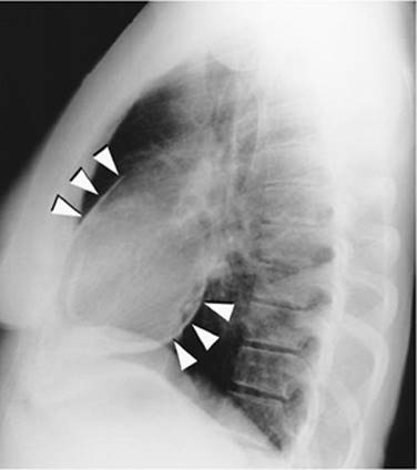
Physical findings include increased jugular venous pressure with a prominent x- and y-descent. A loud early diastolic sound, a “pericardial knock,” is heard in up to 50% of patients. It is caused by the sudden cessation of ventricular filling and is pathognomonic for pericardial constriction. The lungs remain clear initially, and later in the course, left-sided or bilateral pleural effusions develop. The liver is enlarged secondary to congestion; ascites, splenomegaly, and significant lower extremity edema are also present. Unlike in cardiac tamponade, blood pressure is maintained, and less than 20% of patients have a significant pulsus paradoxus. A lateral chest radiograph may show pericardial calcium in up to 50% (Fig. 125.12).
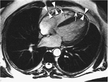
The ECG findings are nonspecific and include T-wave inversions, low voltage, and atrial fibrillation. Echocardiography is helpful to distinguish right heart failure from pericardial constriction; however echocardiographic findings are not specific for the diagnosis of pericardial constriction. They include paradoxical septal motion, rapid deceleration of the early diastolic mitral inflow velocity (E wave), significant respiratory variation of mitral inflow velocity (more than 25%), and normal mitral valve annular tissue Doppler velocity. In the absence of a pericardial effusion, a thickened pericardium is often hard to distinguish from the myocardium. On the other hand, cardiac CT and cardiac magnetic resonance imaging (MRI) are very useful imaging modalities to precisely determine the pericardial thickness. Cardiac MRI is better suited to image soft tissues and can, in contrast to CT, show normal pericardium, even in the absence of pericardial effusion (Fig. 125.13). Furthermore, tagged cine MRI is able to demonstrate adhesions between the pericardium and myocardium (41).
|
|
|
Figure 125.12. Lateral chest radiograph showing calcifications of the anterior and inferolateral pericardium in a patient with constrictive pericarditis (arrowheads). |
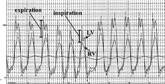
Cardiac catheterization pressure tracings are still the gold standard to diagnose constrictive pericarditis and to differentiate it from restrictive cardiomyopathy. The hemodynamic findings of elevated and equalized diastolic pressures in all four chambers and the dip and plateau sign are classic, although not very specific for constriction. In contrast, respiratory variation of ventricular pressures (right ventricular [RV] pressure rises and left ventricular [LV] pressure falls during inspiration and vice versa during expiration, causing ventricular discordance) has been shown to be highly sensitive and specific for the diagnosis of constrictive pericarditis (42) (Fig. 125.14). Hemodynamically silent constrictive pericarditis can be evaluated by performing volume loading during catheterization; the initially normal right and left heart pressures may elevate and equilibrate in diastole.
|
|
|
Figure 125.13. Four-chamber view of electrocardiogram (ECG)-gated cardiac magnetic resonance imaging (MRI) in a patient with suspected pericardial constriction. The normal-appearing pericardium (arrowheads) is very precisely visualized in areas with pericardial effusion (arrows) and with pericardial fat (asterisk) only. The outstanding soft-tissue imaging capabilities of MRI make it preferable over cardiac computed tomography (CT) to evaluate the pericardium in patients with no or very little pericardial effusion. |
Treatment
Patients with acute onset of constrictive symptoms may improve significantly with medical treatment that includes NSAIDs, colchicine, and steroids (43). Chronic constrictive pericarditis can be treated initially with diuretics, and sodium and fluid restriction, if symptoms are mild. Moderate to severe disease requires definitive treatment with complete removal of the pericardium by surgical stripping (34). This major procedure is associated with a perioperative mortality of greater than 6%; it can be life-saving and improves symptoms drastically in most patients; however, poor outcome is likely if the constriction is radiation induced.
|
|
|
Figure 125.14. Simultaneous invasive left (LV) and right ventricular (RV) pressure tracings. Note the ventricular discordance with respiration (RV pressure rises and LV pressure falls during inspiration and vice versa during expiration). This finding is highly sensitive and specific for the diagnosis of constrictive pericarditis; its presence rules out the differential diagnosis of restrictive cardiomyopathy. |
Summary
Disease processes affecting the pericardium are relatively rare. Inflammation and accumulation of fluid in the pericardial space occur first; in some patients, these conditions can progress to pericardial constriction or tamponade with hemodynamic compromise. The presentation can be acute or chronic; the signs and symptoms are often nonspecific and can mimic other acute conditions as described in the chapter. It is therefore important to include pericardial diseases in the differential diagnosis during the initial workup of a patient presenting with chest symptoms or hemodynamic compromise to avoid inappropriate (i.e., lytic therapy for presumed myocardial infarction in a patient with acute hemorrhagic pericarditis) or delayed treatment (delayed pericardiocentesis in a patient with pericardial tamponade) with possible fatal outcome.
Acknowledgments
I would like to thank Dr. Ilona Schmalfuss, Radiology and the Medical Media Service, and particularly Mr. John Richardson, both at the Malcom Randall VA Medical Center, for their assistance with the figures for this chapter.
References
1. Morgan BC, Guntheroth WG, Dillard DH, et al. Relationship of pericardial to pleural pressure during quiet respiration and cardiac tamponade. Cir Res. 1965;16:493.
2. Shabetai R. Function of the pericardium. In: Fowler NO, ed. The Pericardium in Health and Disease. Mount Kisco, NY: Futura Publishing; 1985:19.
3. Shabetai R. Acute viral and idiopathic pericarditis. In: Shabetai R, ed. The Pericardium. New York, NY: Grune & Stratton; 1981:348.
4. Gunukula SR, Spodick DH. Pericardial disease in renal patients. Semin Nephro. 2001;21:52.
5. Coreale E, Maggioni AP, Romano S, et al. Comparison of frequency, diagnostic and prognostic significance of pericardial involvement in acute myocardial infarction treated with and without thrombolytics. Am J Cardiol. 1993;71:1377.
6. Engle MA, Ito T. The post-pericardiotomy syndrome. Am J Cardiol 1961;7:73.
7. Engle MA, McCabe JC, Ebert PA, et al. The post-pericardiotomy syndrome and antiheart antibodies. Circulation. 1974;49:401.
8. Abraham KP, Reddy V, Gattuso P, et al. Neoplasms metastatic to the heart: review of 3314 consecutive autopsies. Am J Cardiovasc Pathol. 1990;3:195.
9. Permanyer-Miralda G, Sagrista-Sauleda J, Soler-Soler J, et al. Primary acute pericardial disease: a prospective series of 231 consecutive patients. Am J Cardiol. 1985;56:623.
10. Gornik HL, Gerhard-Herman M, Beckman JA, et al. Abnormal cytology predicts poor prognosis in cancer patients with pericardial effusion. J Clin Oncol. 2005;23:5211.
11. Posner MR, Cohen GI, Skarin AT, et al. Pericardial disease in patients with cancer. The differentiation of malignant from idiopathic and radiation-induced pericarditis. Am J Med. 1981;71:407.
12. McCaughan BC, Schaff HV, Piehler JM, et al. Early and late results of pericardiectomy for constrictive pericarditis. J Thorac Cardiovasc Surg. 1985;89:340.
13. Trautner BW, Darouiche RO: Tuberculous pericarditis: optimal diagnosis and management. Clin Infect Dis. 2001;33:954.
14. Larrieu AJ, Tyers GFO, Williams EH, et al. Recent experience with tuberculous pericarditis. Ann Thorac Surg. 1980;29:464.
15. Hammerman KJ, Powell KE, Tosh FE, et al. The incidence of hospitalized cases of systemic mycotic infections. Sabouraudia. 1974;12:33.
16. Spodick DH. Diagnostic electrocardiographic sequences in acute pericarditis. Circulation. 1973;48:575.
17. Spodick DH. Arrhythmias during acute pericarditis (100 cases). JAMA. 1976;235:39.
18. Johnson TR, Nikolaou K, Wintersperger BJ, et al. ECG-gated 64-MDCT angiography in the differential diagnosis of acute chest pain: AJR Am J Roentgenol. 2007;188:76.
19. Imazio M, Demichellis B, Parrini I, et al. Day-hospital treatment of acute pericarditis: a management program for outpatient therapy. J Am Coll Cardiol. 2004;43:1042.
20. Guindo J, de la Serna AR, Ramio J, et al. Recurrent pericarditis: relief with colchicine. Circulation. 1990;82:1117.
21. Adler Y, Finkelstein Y, Guindo J, et al. Colchicine treatment for recurrent pericarditis: a decade of experience. Circulation. 1998;97:2183.
22. Imazio M, Bobbio M, Cecchi E, et al. Colchicine in addition to conventional therapy for pericarditis: results of the COlchicine for acute PEricarditis (COPE) Trial. Circulation. 2005;112:2012.
23. Shabetai R. Recurrent pericarditis: recent advances and remaining questions. Circulation. 2005;112,1921.
24. Maisch B. Recurrent pericarditis: mysterious or not so mysterious? Eur Heart J. 2005;26:631.
25. Urschel HC, Razzuk MA, Gardner M, et al. Coronary artery bypass occlusion secondary to postpericardiotomy syndrome. Ann Thorac Surg. 1976;22:528.
26. Guberman BA, Fowler NO, Engel PJ, et al. Cardiac tamponade in medical patients. Circulation. 1981;64:633.
27. Fowler NO. Physiology of cardiac tamponade and pulsus paradoxus. Mod Concept Cardiovasc Dis. 1978;48:115.
28. Shabetai R. Cardiac tamponade. In: Shabetai R, ed. The Pericardium. New York, NY: Grune & Stratton; 1981:224.
29. Fowler NO. The paradoxical pulse (pulses paradoxus). In: Fowler NO, ed. The Pericardium in Health and Disease. Mount Kisco, NY: Futura Publishing; 1985:235.
30. Spodick DH. Electric alternation of the heart. Am J Cardiol. 1962;10:155.
31. Horowitz MS, Schultz CS, Stinson EB, et al. Sensitivity and specificity of echocardiographic diagnosis of pericardial effusion. Circulation. 1974;50:239.
32. Armstrong WF, Schilt BF, Helper DJ, et al. Diastolic collapse of the right ventricle with cardiac tamponade: an echocardiographic study. Circulation. 1982;65:1491.
33. Singh S, Wann S, Schuchard GH, et al. Right ventricular and right atrial collapse in patients with cardiac tamponade: a combined echocardiographic and hemodynamic study. Circulation. 1984;70:966.
34. Maisch B, Seferovic PM, Ristic AD, et al. Task force on the diagnosis and management of pericardial diseases of the European Society of Cardiology. Guidelines on the diagnosis and management of pericardial disease: executive summary. Eur Heart J. 2004;25:587.
35. Lorell BH, Braunwald E. Pericardial disease. In: Braunwald E, ed. Heart Disease: A Textbook of Cardiovascular Medicine. Philadelphia, PA: WB Saunders; 1984: 1487.
36. Tsang TS, Enriquez-Sarano M, Freeman WK, et al. Consecutive 1127 therapeutic echocardiographically guided pericardiocenteses: clinical profile, practice patterns, and outcomes spanning 21 years. Mayo Clin Proc. 2002;77:429.
37. Santos GH, Frater RW. The subxiphoid approach in the treatment of pericardial effusion. Ann Thorac Surg. 1977;23:468.
38. Wang HJ, Hsu KL, Chiang FT, et al. Technical and prognostic outcomes of double-balloon pericardiotomy for large malignancy-related pericardial effusions. Chest. 2002;122:893.
39. Shabetai R. Constrictive pericarditis. In: Shabetai R, ed. The Pericardium. New York, NY: Grune & Stratton; 1981:154.
40. Talreja DR, Edwards DW, Danielson GK, et al. Constrictive pericarditis in 26 patients with histologically normal pericardial thickness. Circulation. 2003;108:1852.
41. Kojima S, Yamada N, Goto Y, et al. Diagnosis of constrictive pericarditis by tagged cine magnetic resonance imaging. N Engl J Med. 1999;341:373.
42. Hurrell DG, Nishimura RA, Higano ST, et al. Value of dynamic respiratory changes in left and right ventricular pressures for the diagnosis of constrictive pericarditis. Circulation. 1996;93:2007.
43. Haley JH, Tajik J, Danielson GK, et al. transient constrictive pericarditis: causes and natural history. J Am Coll Cardiol. 2004;43:271.