Robin D. Kim
Kristin L. Mekeel
Alan W. Hemming
Patients are admitted to the intensive care unit (ICU) for a variety of reasons following hepatopancreatobiliary (HPB) surgery, including maintenance or restoration of normal physiology immediately after extensive surgery and the subsequent management of complications that develop. Many of the issues that require ICU management are common to all ICU patients and will not be discussed in this chapter; however, there are recurring issues that are relatively specific to HPB surgery patients that will be discussed. The role of normal liver physiology and its alteration during HPB surgery and disease states will be discussed, as well as the management of common problems that arise after HPB surgery.
HPB surgery is composed of surgery of the liver, bile duct, and pancreas, and may include portal decompressive procedures for complications of portal hypertension. Surgical procedures on the pancreas and bile duct alone generally do not require care in a critical care setting immediately after leaving the operating room unless complications occur. Liver resections, particularly when extensive, can require admission to a critical care unit immediately following surgery due to the alterations in normal physiology that occur during the procedure itself. An understanding of normal liver physiology and the alterations it undergoes during surgery is important when managing these patients.
Liver Anatomy and Physiology
The liver is approximately 4% to 5% of total body weight and has multiple complex functions. The anatomy of the liver has been described using various and different methods (1,2,3,4,5); however, surgical anatomy is based on the segmental nature of vascular and bile duct distribution. The liver receives a dual blood supply from both the portal vein and hepatic artery that run, along with the bile duct, within the glissonian sheath or main portal pedicle. The portal pedicle divides into right and left branches and then supplies the liver in a segmental fashion. Venous drainage is via the hepatic veins, which drain directly into the inferior vena cava. Hepatic segmentation is based on the distribution of the portal pedicles and their relation to the hepatic veins (Fig. 78.1). The three hepatic veins run in the portal scissurae and divide the liver into four sectors, which are, in turn, divided by the portal pedicles running in the hepatic scissurae. The liver is divided into right and left hemilivers by the middle hepatic vein. The right hemiliver is divided by the right hepatic vein into anterior and posterior sectors. The anterior sector is divided by the plane of the portal pedicle into an inferior segment V and a superior segment VIII. The posterior sector is divided by the plane of the portal pedicle into an inferior segment VI and a superior segment VII. The left hemiliver lies to the left of the middle hepatic vein and is divided into anterior and posterior sectors by the left hepatic vein. The anterior sector is divided by the umbilical fissure into segment IV medially and segment III laterally. The segment posterior to the left hepatic vein is segment II. Segment IV can be divided by the plane of the portal pedicle into a superior segment IVa and an inferior segment IVb. Segment I is the caudate lobe, which lies between the inferior vena cava and the hepatic veins. The caudate lobe has variable portal venous, hepatic arterial and biliary anatomy, and is essentially independent of the portal pedicle divisions and hepatic venous drainage.
|
|
|
Figure 78.1. Diagrammatic representation of liver segments with standard liver resections demonstrated. IVC, inferior vena cava; PV, pulmonary vein. |
Segmental anatomy becomes important in considering surgical resection when essentially any segment or combination of segments can be resected if attention is paid to maintaining vascular and biliary continuity to the remaining segments.
Common liver resections performed that may require ICU admission after surgery are left or right hepatectomies, in which approximately 50% of liver volume is removed, or more extensive procedures such as right or left trisectionectomy, in which up to 80% of the liver is removed (Fig. 78.1). If less than 40% of the liver is resected in patients with normal underlying liver function, then relatively little derangement of liver physiology is noted.
The liver performs many functions, including uptake, storage, and eventual distribution of nutrients from the blood or gastrointestinal tract, as well as synthesis, metabolism, and elimination of a variety of endogenous and exogenous substrates and toxins (including narcotics and other drugs). Although the liver is only 4% to 5% of total body weight, it is responsible for 20% to 25% of body oxygen consumption and 20% of total energy expenditure (6). The liver receives a dual blood supply, with 75% of flow from the portal vein and 25% from the hepatic artery. Total blood flow (7) to the liver is approximately 1.5 L/minute/1.73 m2. While decreasing portal venous flow causes a subsequent increase in hepatic arterial flow, with complete portal occlusion or diversion, hepatic arterial flow does not completely compensate, and total liver blood flow is diminished (8). The opposite is not true, however (i.e., decreasing flow in the hepatic artery does not increase flow in the portal vein). There is autoregulation of hepatic arterial flow but not of the portal venous system. Portal flow is increased by food intake, bile salts, secretin, pentagastrin, vasoactive intestinal peptide (VIP), glucagon, isoproterenol, prostaglandin E1 and E2, and papaverine. Portal flow is decreased by serotonin, angiotensin, vasopressin, nitrates, and somatostatin.
Bile, composed of inorganic ions and organic solutes, is formed at the canalicular membrane of the hepatocyte, as well as in the bile ductules, and is secreted by an active process that is relatively independent of blood flow (9). The major organic components of bile are the conjugated bile acids, cholesterol, phospholipid, bile pigments, and protein. Under normal conditions, 600 to 1,000 mL of bile is produced per day (10). Bile secretory pressure is approximately 10 to 20 cm saline, with maximal secretory pressures of 30 to 35 cm in the presence of complete biliary obstruction.
Bilirubin, a degradation product of heme, is eliminated almost entirely in the bile. Bilirubin circulates bound to albumin and is removed from plasma by the liver via a carrier-mediated transport system. In the hepatocyte, bilirubin is bound to glucuronic acid before being secreted in bile. The liver maintains the ability to clear bilirubin with partial duct obstruction. Complete obstruction of one of the right or left hepatic ducts alone will cause marked liver enzyme abnormalities, but rarely causes jaundice.
The liver synthesizes many of the major human plasma proteins including albumin, γ-globulin, and many of the coagulation proteins. Liver dysfunction can have a profound effect on coagulation through the decreased production of coagulation proteins or, in the case of obstructive jaundice, there is decreased activity of factors II, V, VII, IX, and X, secondary to a lack of vitamin K–dependent posttranslational modification. Reversal of coagulation abnormalities by exogenous administration of vitamin K allows differentiation between synthetic dysfunction and lack of vitamin K absorption secondary to obstructive jaundice.
After liver resection, liver function is altered through both a reduction in functional liver mass and potential ischemia/reperfusion injury to the liver remnant. With extensive liver resection in patients with normal presurgical underlying liver function, reduction of functional liver volume below 25% has been associated with an increased risk of both liver failure and mortality (11). To reduce the risk of liver failure in this setting, preoperative portal vein embolization (PVE) has been developed. During PVE, the portal vein of the side of the liver to be resected is embolized percutaneously. Diversion of portal flow and its hepatotrophic factors to the future liver remnant (FLR) causes growth and hypertrophy of the FLR of about 30% (Fig. 78.2) over a 6-week period and has been shown to reduce the complications associated with subsequent extended liver resections (12).
|
|
|
Figure 78.2. Right portal vein embolization preoperatively allows an increase of functional liver remnant of approximately 30% from prior to embolization (A) to postembolization (B). MHV, middle hepatic vein. (Reused with permission from Hemming AW, Reed AI, Howard RJ, et al. Preoperative portal vein embolization for extended hepatectomy. Ann Surg. 2003;237[5]:686–691.) |
Surgical Procedures
Liver Resection
The liver is a tremendously vascular organ. Since intra- or postoperative complications are often related to excessive blood loss (13), a number of techniques have been developed to achieve preresection vascular control and decreased bleeding. While liver resection may be performed, in many cases without the need for interruption of blood flow to the liver, it is sometimes necessary to reduce blood flow to prevent excessive blood loss. Selective inflow control can be established by division or occlusion of the vascular structures supplying the segment(s) of liver to be removed. The right or left portal pedicle containing the respective portal vein, hepatic artery, and bile duct are controlled with a vascular clamp. This technique has the advantage of preserving blood flow to the segment of the liver being preserved, but is generally only useful in smaller resections.
Total inflow occlusion (Pringle maneuver) clamps the entire inflow of the liver at the hepatoduodenal ligament, and has been shown to reduce blood loss during the parenchymal transection phase of the resection (14). While there is some concern regarding warm ischemic injury, abundant data show that the normal liver can tolerate inflow occlusion for up to 1 hour, and there are reports suggesting that some cirrhotic livers can safely tolerate 60 minutes of inflow occlusion as well (15). We use total inflow occlusion when selective occlusion provides insufficient control. Clamp times are expected to be less than 30 minutes for formal hepatectomies, but may be higher for more complex parenchymal transections. In such cases, total occlusion is carried out in 15-minute increments with 5-minute reperfusion intervals. An alternative to the intermittent clamping technique is to use ischemic preconditioning, during which the liver inflow is occluded for 10 minutes, after which it is allowed to reperfuse for 15 minutes prior to clamping again for a sustained time period up to 1 hour. Intermittent clamping is associated with more blood loss than ischemic preconditioning; however, the protective results of ischemic preconditioning in ischemia reperfusion injury have not been uniform across age groups, and may not be as effective in livers that have been exposed to preoperative chemotherapy (16).
Total vascular isolation of the liver with both inflow occlusion and occlusion of the supra- and infrahepatic vena cava can be useful for technically demanding cases where the vena cava or proximal hepatic veins are involved with tumor (Fig. 78.3). Total isolation has been shown to be safe for up to 60 minutes in normal liver, but can be accompanied by varying degrees of hemodynamic instability (17). In cases where this is required, we carry out as much of the operation as possible prior to isolation of the liver to reduce the ischemic time and the period of hemodynamic instability.
The most troublesome bleeding sources during liver resection are usually from hepatic vein branches, which may be minimized by maintaining the central venous pressure (CVP) below 5 mm Hg during the period of hepatic transection. Cooperation of the anesthetist in minimizing volume loading, and occasionally using pharmacologic agents to reduce CVP, is essential. However, if total vascular isolation is to be used, volume loading prior to caval clamping is required to avoid an acute decrease in cardiac output at the time the clamps are applied.
|
|
|
Figure 78.3. Tumors that involve the inferior vena cava or hepatic veins may require total vascular isolation of the liver. Both the infrahepatic and suprahepatic inferior vena cava are clamped along with the portal vein and hepatic artery. (Reused with permission from Hemming AW, Reed AI, Langham MR, et al. Combined resection of the liver and inferior vena cava for hepatic malignancy. Ann Surg. 2004;239[5]:712–721.) |
Knowledge of the details of intraoperative conduct of the operation is therefore important to the physicians who are to manage the postoperative care of the liver resection patient in the ICU setting. Was inflow occlusion or vascular isolation required, and for how long? Prolonged clamp times are associated with greater liver dysfunction. Was the patient maintained with a low CVP throughout the course of the surgery? If so, then the patient may need volume expansion on arrival to the ICU. How much liver remains, and is it normal? If the percentage of remaining liver is less than 25% in normal livers or less than 40% in cirrhotic livers or livers with bile duct obstruction, then the chance of liver failure and the need for its management are higher. Was there significant blood or fluid requirements? Patients may need a period of ventilation while fluid shifts and equilibrates.
|
|
|
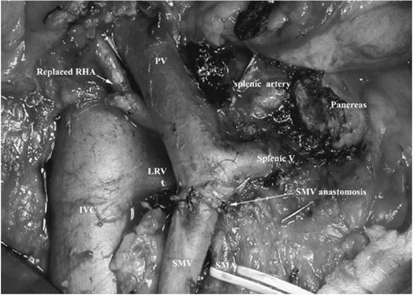
Figure 78.4. Pancreaticoduodenectomy demonstrating resection of the portal vein. IVC, inferior vena cava; LRV, left renal vein; PV, portal vein; RHA, right hepatic artery; SMA, superior mesenteric artery; SMV, superior mesenteric vein; splenic V, splenic vein. (Reused with permission from Hemming AW, Reed AI, Langham MR, et al. Combined resection of the liver and inferior vena cava for hepatic malignancy. Ann Surg. 2004;239[5]:712–721.) |
Pancreatic and Bile Duct Surgery
The majority of patients who undergo pancreatic or bile duct surgeries do not require admission to an ICU setting in the immediate postoperative period because of issues specific to the pancreaticobiliary surgery itself. In general, procedures on the pancreas or biliary tree should not be associated with major intraoperative hemodynamic changes or alterations in physiology. Tumors of the head of the pancreas or bile duct may involve the portal vein or cause extensive fibrotic reaction in the area. Technical difficulties can arise in which damage occurs to, or resection is required of, the portal vein (Fig. 78.4). If portal vein resection or repair is required, it is more likely that the patient will require ICU care. Portal vein resection, when planned, requires variable durations of portal venous outflow obstruction from the gut, which are usually short and well tolerated, but can increase the amount of fluid third-spaced into the bowel wall. Portal vein injury, however, can lead to massive transfusion requirements and hypotension that can require postoperative ICU care. The more common indications for admission to the ICU after pancreatic or biliary surgery are either an underlying medical condition or the development of a postoperative complication. Pancreatic or bile leaks, which can lead to sepsis, will be discussed later in the chapter.
Portal Decompressive Procedures
Surgical portal decompressive procedures, although a rarity since the introduction of the transjugular intrahepatic portosystemic shunt (TIPS), remain indicated in select patients with variceal bleeding and preserved liver function who have failed medical management and are not transplant candidates. The myriad technical variations of surgical portosystemic shunts are beyond the scope of this chapter, but certain commonalities exist. Whether total or partial shunts, selective or nonselective, patients will have had the high-pressure portal system surgically connected to the low-pressure caval circulation to lower the pressure in the portal venous system and stop variceal bleeding. Reduction of portal flow in patients who have borderline liver function can precipitate liver dysfunction or failure. Additionally, the fraction of portal flow that is diverted into the systemic circulation through the shunt is not cleared by the liver until it returns to the liver via the arterial circulation. This may induce encephalopathy; shunts that divert most or all of the portal flow into the systemic circulation are more likely to induce encephalopathy than those shunts that are selective or partial. One special case scenario is Budd-Chiari syndrome, in which the hepatic venous outflow is obstructed, usually due to thrombosis secondary to a hypercoagulable state. In this disorder, blood flow perfuses the hepatic sinusoids from both the hepatic artery and portal vein but cannot exit through the blocked hepatic veins. A functional side-to-side shunt is performed (portacaval, mesocaval) that allows hepatic arterial blood to flow into the sinusoids and then exit via the portal vein, and through the shunt into the systemic circulation. It is not uncommon for liver function to deteriorate initially after the shunt is performed, with subsequent gradual improvement and liver regeneration. Support of liver function may be required immediately after the shunt while liver function stabilizes. In some cases, the shunt may precipitate acute liver failure, making urgent liver transplantation the only option.
Immediate Postoperative Management
Postoperative fluid management is important in the care of patients after major hepatobiliary surgery. In particular, postoperative fluid shifts in patients who have had major liver resection can be difficult to manage. Intraoperatively, most liver resections are performed with low central venous pressure and low intravascular volume. While this practice minimizes bleeding during the hepatic parenchymal transection phase of the procedure, it may pose some difficulty postoperatively, as these patients may have signs of hypovolemia with low urine output and low blood pressure. Volume reexpansion should be gentle, as partial liver resection leads to hypoalbuminemia, and pulmonary edema and ascites can develop with aggressive resuscitation. Although the use of albumin infusions is generally frowned upon in critical care medicine, albumin and fresh frozen plasma may be useful in the resuscitation of patients after liver resection, as the physiology is similar to patients with cirrhosis. We use albumin-containing fluids for volume expansion if the serum albumin is less than 2.9 mg/dL. Fresh frozen plasma can be used for volume expansion; however, it is generally reserved for abnormalities in coagulation. Serial lactate levels are helpful in the postoperative management of patients after liver resection. Elevated lactic acid levels may be a sign of hypovolemia, but the lack of response to volume can indicate liver dysfunction.
After liver resection, glucose metabolism is altered due to both a reduction in functional liver mass and the relative dysfunction of the remaining liver secondary to ischemia reperfusion injury if vascular control has been used during the procedure. As glycogen stores are depleted, the liver uses gluconeogenesis to provide glucose. Resulting from this alteration in hepatic physiology, hypoglycemia may occur, although lethal hypoglycemia is rare. It has become standard practice in most critical care units to tightly control blood glucose levels. While the advantages of this approach, particularly the reduced risk of sepsis, remain for patients after major liver resection, aggressive blood glucose control with insulin infusions requires closer monitoring and may necessitate reduced insulin dosing to prevent hypoglycemia.
Patients who have undergone shunt surgery require a different approach than patients undergoing other types of hepatobiliary surgery. These individuals need more aggressive fluid management immediately postoperatively to maintain circulating intravascular volume and reduce the risk of shunt thrombosis. Maintenance fluid should be 0.45% saline solution with 5% dextrose to provide the liver with carbohydrate. After the immediate postoperative period, patients are also at risk for ascites formation, so excessive sodium should be minimized and additional volume expansion—if needed—should be albumin or fresh frozen plasma. Diuretics can be reinstituted after the immediate postoperative period. A general rule is to use a combination of Lasix and spironolactone, with 100 mg of spironolactone for every 40 mg of Lasix. Antibiotics are administered for 48 hours postoperatively to minimize infection from bacterial translocation.
Encephalopathy is rare in patients after liver resection, unless they are in liver failure or have pre-existing liver disease. The presence of asterixis can be an early sign of encephalopathy. Encephalopathy is treated with lactulose and dietary protein restriction, as in other patients with end-stage liver disease. Infection, dehydration, and bleeding, as well as narcotic use, must be evaluated, as they can trigger decompensation that leads to encephalopathy.
Hypophosphatemia
While the exact mechanism of the hypophosphatemia seen after hepatic resection remains unclear, care must be taken to aggressively replace the low serum phosphate, since increased utilization during liver regeneration and a renal wasting mechanism have been proposed (18).
Regardless of the etiology, the clinical consequences of hypophosphatemia are well established and include respiratory depression, diaphragmatic insufficiency, seizures, and cardiac irritability. In addition, hepatocellular regeneration is dependent on adenosine triphosphate (ATP), and after liver resection, regeneration may be impaired if phosphate is not repleted (19). In a series of 35 liver resections, 21% had significant postoperative hypophosphatemia (less than 2.5 mg/dL) after surgery. This group had a significant increase in complications (80%) compared to the normophosphatemic group (28%) (20). Phosphate should be replaced with potassium or sodium phosphate preparations, or added to parenteral nutrition solutions. Recent data in living donor right hepatic lobectomies suggest that replacement up to two times the recommended daily allowance (60 mmol) is necessary to replete severe hypophosphatemia and prevent complications associated with hypophosphatemia (21).
Liver Function: Assessment and Support
Liver function should be carefully monitored after major liver resections and shunt surgery, as liver failure is a risk in any major hepatobiliary surgery. The risk of liver failure increases with the extent of hepatectomy and in patients with preoperative liver disease or cirrhosis (22,23). Although standard liver function tests are helpful after major liver resection or shunt surgery, they may not show elevation until the patient has significant liver failure. Transaminases are frequently elevated into the 200 to 300 units/dL range post resection due to the direct effect of mechanical injury to the liver during transection, as well as to partial devascularization of areas of the liver. Measurements of liver function, including the prothrombin time and lactate, are more helpful in evaluating early postoperative liver dysfunction.
Elevated total and indirect bilirubin are also useful indicators of postoperative liver dysfunction. However, isolated elevation of total bilirubin in the presence of normal liver function can have other etiologies. Perioperative blood transfusions can lead to hemolysis and hyperbilirubinemia, with a predominance of direct hyperbilirubinemia, and can be diagnosed with a standard hemolytic workup. Bile leaks or obstruction can also lead to an elevated serum bilirubin. The diagnosis and treatment of bile leaks is covered later in this chapter. Many popular anesthetics, antibiotics, and other drugs can cause hepatotoxicity and elevation of the serum bilirubin and need to be reduced or stopped if liver failure occurs.
When postoperative liver dysfunction does develop, it is important to exclude sepsis and anatomic causes of liver failure. A postoperative ultrasound can evaluate for portal vein, hepatic arterial, or hepatic vein thrombosis or obstruction, which may be amenable to surgical intervention. If the patient does not have sepsis, drug toxicity, biliary obstruction or leak, or vascular occlusion, liver failure is likely related to a pre-existing liver disease and/or the extent of resection. Treatment is then supportive, with correction of coagulopathy, encephalopathy, and ascites as described above. Systemic antibiotics or gut decontamination may be beneficial, since the liver Kupffer cells play a role in decreasing bacterial translocation from the portal blood flow, and patients with liver failure or biliary leak or obstruction may have an increased risk of bacteremia and sepsis.
N-acetylcysteine has been shown to decrease liver injury after acetaminophen overdose (24) and lessen ischemia reperfusion injury of the liver (25). Intravenous infusions of prostaglandin have also been linked to improvement of ischemia reperfusion injury and liver damage (26). Although definitive clinical data are lacking, both N-acetylcysteine and prostaglandin (PG) E1 have been used to ameliorate postoperative liver damage in both liver resection and transplant patients. N-acetylcysteine is given as a continuous infusion of 40 mL of 10% solution mixed in 250 mL of D5W and given over 16 hours. Prostaglandin is also given as a continuous intravenous infusion, starting at 0.15 µg/kg/hour. It is titrated up to 1 µg/kg/hour based on systemic hypotension.
Coagulopathy
Coagulopathy is common after liver resection, and several studies have demonstrated an increase in prothrombin time directly proportional to the extent of liver resection (27,28). This coagulopathy has been attributed to impaired synthesis and clearance of clotting factors, inhibitors, and regulatory proteins (29,30). Patients with underlying liver disease and cirrhosis also often have thrombocytopenia and qualitative platelet defects. In addition, intraoperative hypothermia and perioperative transfusions, while not routine, are not uncommon during major hepatobiliary surgery, and can contribute to postoperative coagulopathy.
Serial hemoglobin and prothrombin levels should be measured. Because of the vascular nature of hepatobiliary surgery combined with postoperative coagulopathy from decreased liver function, as well as the frequent need for intravascular volume expansion, serial hemoglobin levels should be followed for postoperative bleeding. In general, we would obtain a hematocrit and international normalized ratio (INR) on ICU arrival and then repeat them, every 6 hours, for the next 24 hours. The surgeon should be notified of excessive bloody output from the drains, increasing abdominal distention, or hemodynamic instability. If coagulopathy does develop, it should be corrected if the INR goes above 2.0 (31), both with vitamin K and fresh frozen plasma. Any patient who is bleeding should have his or her coagulopathy completely corrected. For severe bleeding, both aprotinin and activated factor VII are safe in patients during and after liver resection (30,32). Patients who fail to stop bleeding after correction of their coagulopathy require return to the operating room for exploration. The surgical team should be made aware of any patient immediately postsurgery who requires transfusion. Once the postoperative coagulopathy has resolved or stabilized, all patients should be given subcutaneous heparin or low-molecular-weight heparin with sequential compression devices to prevent the formation of deep venous thrombosis.
Pain Management and Sedation
The large subcostal incision needed for major hepatobiliary surgery can result in significant pain after surgery. However, altered pharmacokinetics and coagulopathy, in particular after partial liver resection or shunt surgery, can make postoperative pain management a challenging proposition. Patients with liver failure or compromised liver function secondary to hepatectomy have altered metabolism of many common medications, in particular narcotics and sedatives that require hepatic clearance.
One of the more common problems that arises in the ICU after liver resection is oversedation of patients. A standard dose of narcotics given to a patient who has had 80% of the liver resected may well cause prolonged respiratory depression and signs and symptoms of encephalopathy. Narcotics and benzodiazepines should be used at the minimum dose required to achieve pain control. After liver resection, it is recommended that basal rates on patient-controlled anesthesia pumps be avoided, as metabolism of narcotics is difficult to forecast. Benzodiazepines also have altered clearance after liver resection, and should be administered at a lower dose or, if possible, avoided altogether. In patients requiring ongoing endotracheal intubation and mechanical ventilation, we have found it useful to use sedative agents such as propofol rather than narcotics since the level of sedation can be more easily titrated and reversed. Our institution has, at present, no experience with dexmedetomidine.
Epidural pain management may be the optimal analgesic technique after liver resection. Unfortunately, it is contraindicated in many patients because of postoperative coagulopathy. Recent literature has examined the use of epidural catheters in patients undergoing living donor partial hepatic resection. In a review of eight patients with epidural catheters, good pain control was achieved, with only one case of oversedation requiring naloxone. Although postoperative coagulopathy did occur, it was not to the extent that factor transfusion was needed prior to catheter removal, and there were no cases of hemorrhage (33). Epidural analgesia may be useful in select patients who do not have underlying liver disease and who are not undergoing extensive resections.
Nutrition
Although nutrition plays an important role in the care of any critically ill patient, the role of the liver in protein and carbohydrate metabolism makes proper postoperative nutrition imperative in the management of patients after major hepatobiliary surgery, in particular after partial liver resection when liver function is temporarily reduced. Patients with preoperative biliary obstruction, malignancy, and cirrhosis are at a higher risk for nutrition-related complications after major liver or bile duct surgery. Preoperative nutritional risk factors associated with postoperative complications in hepatobiliary surgery include weight loss greater than 14% lean body mass over 6 months, serum albumin less than 3 g/dL, hematocrit of less than 30%, total body potassium less than 85% of normal, less than the 25th percentile for midarm circumference, and skin test anergy (34). Preoperative bilirubin, albumin, prealbumin, prothrombin time, transferrin, as well as replacement of vitamin and trace mineral deficits may also be important preoperatively.
As with most critically ill patients, early enteral nutrition has been associated with improved outcomes. In hepatobiliary surgery, both enteral and parenteral nutrition have been associated with improved outcomes, especially in high-risk patients (34,35). However, parenteral nutrition has been clearly associated with an increased risk of infection (36). Enteral nutrition has been shown to improve gut flora, preventing gastrointestinal atrophy and loss of immunocompetence. A review of five prospective randomized trials on enteral and parenteral nutrition in patients after liver resection found a decrease in wound infection and catheter sepsis in patients receiving enteral nutrition (37). As one might expect, there were no mortality differences.
In patients who have undergone routine liver resection or shunt surgery, low-volume enteral feeds can be started almost immediately post surgery. Those patients having undergone hepaticojejunostomies or pancreatic surgery must await return of bowel function prior to starting feeds, unless the feeding tube is placed distal to the anastomosis. It is best to consult with the operating surgeon before starting enteral feeds in any patient, particularly those with enteric reconstruction. Patients with major pancreatic surgery (pancreaticoduodenectomy, subtotal or total pancreatectomy) may require pancreatic enzyme supplementation with enteral feeds or when resuming oral intake.
Patients with chronic liver disease or cirrhosis often have severe metabolic derangements that make nutritional management difficult. The depletion of the fat-soluble vitamins, in particular a loss of vitamin K, leads to coagulopathy and diminished antioxidant response. Chronic liver disease also stimulates a catabolic state with proteolysis and cachexia. Protein loss can be exacerbated by dietary restriction to help decrease encephalopathy. Branched-chain amino acids were initially thought to reduce the development of encephalopathy in catabolic patients with advanced liver failure, but this has not borne out in clinical data. Patients with cirrhosis also have abnormal glucose tolerance and insulin levels, along with elevated ammonia levels, hypophosphatemia, and hypoalbuminemia, all which influence perioperative nutrition. All Child's B or C cirrhotic patients should be fed enterally when hospitalized. The caloric needs of these patients are increased, and goal kcal is 25 to 25 kcal/kg/day, with administration of protein at 1 to 1.5 g/kg dry weight in nonencephalopathic patients and 0.5 g/kg dry weight in encephalopathic patients (37). Patients with ascites need sodium restriction of 2 g/day and a fluid restriction of 1 to 1.5 L/day, in combination with diuretics if tolerated.
Patients with preoperative obstructive jaundice often have chronic, low-grade endotoxemia and sepsis. This can lead to weight loss and anorexia, often due to malabsorption of fat and fat-soluble vitamins from obstruction of bile flow, which leads to coagulopathy and a diminished antioxidant response. Endotoxemia also results in decreased hepatic protein synthesis and catabolism (38). Most patients with biliary obstruction and resultant sepsis should undergo biliary decompression prior to major hepatobiliary surgery to allow malnutrition secondary to biliary obstruction and sepsis to resolve. Although it is controversial as to whether preoperative biliary decompression is required prior to surgery, one prospective, randomized trial looked at patients with obstructive jaundice who underwent biliary decompression and then were randomized to immediate operation or 2 weeks of alimentation (both parenteral and enteral) followed by operation. The second group had a lower risk of infection, morbidity, and mortality (39). Since these patients usually do not have hepatic dysfunction, standard enteral or parenteral nutrition is acceptable. Patients in whom bile flow has not been restored should have a low-fat diet—as fat absorption is impaired—and replacement of the fat-soluble vitamins. Medium-chain triglycerides may be helpful, because their absorption is not bile salt-dependent and may avoid diarrhea until bile flow is reestablished.
Partial liver resections also cause metabolic abnormalities secondary to the regenerating liver. Hepatic mitochondria switch to fat from glucose as their preferred energy source in hepatic regeneration (40). As a result, hypertonic glucose and insulin infusions should be avoided immediately after resection, as hyperglycemia and insulinemia suppress fatty acid release and decrease ketone body production by the liver. Some investigators have advocated administering fat and/or ketone bodies after liver resection to accelerate regeneration, although conclusive evidence that this is beneficial is lacking. Similarly, infusions of glucose and insulin directly into the portal vein have also been investigated in their role to improve regeneration, although, again, conclusive evidence is lacking. Adequate liver regeneration is also dependent on protein and calories. Postoperative parenteral nutrition should be supplemented with protein and fat, but low on glucose to improve hepatic regeneration. General goals are 30 kcal/kg/day, with 1.0 to 1.5 g/kg protein; glucose approximating 5 mg/kg/minute, and fat should not exceed 30% of the calories. Patients with cancer may need an increase of up to 35 kcal/kg/day and 2 g/kg protein.
Renal Failure
Acute renal failure occurs after major hepatobiliary surgery in 10% of patients (41) and, similar to other critically ill patients, significantly increases postoperative mortality (42). Risk factors for perioperative renal failure include postoperative sepsis, preoperative uremia, preoperative anemia, malignant disease, and preoperative jaundice (41,43,44). In particular, preoperative obstructive jaundice appears to be a significant risk factor, with an estimated 10% of patients developing postoperative renal failure (45). Both dehydration and endotoxin production from bile duct obstruction have been postulated to cause renal failure in these patients (46). Many studies have been done to try to decrease this risk, including using mannitol, bile salts, hydration, and lactulose (45,46,47,48).
In all patients with acute renal failure, adequate hydration, treatment of sepsis, and avoidance of nephrotoxic drugs are mandatory. However, in patients with obstructive jaundice, lactulose and bile salts may decrease endotoxin absorption, and have been shown in some studies to be beneficial in the prevention of renal failure (44,45). Preoperative biliary drainage to help lessen the perioperative inflammatory response is also an important adjunct to prevent postoperative renal failure. Once acute renal failure does occur, supportive care and dialysis are needed until renal function returns.
Patients with advanced cirrhosis or postoperative liver failure can develop hepatorenal syndrome (HRS). This is more significant in the acute care of patients with liver failure or after liver transplantation. Hepatorenal syndrome is a diagnosis of exclusion, with decreased renal function associated with a urine sodium less than 10 mg/dL combined with a urine osmolality greater than plasma osmolality that does not respond to volume administration. The cause of hepatorenal syndrome is likely multifactorial, but is primarily related to circulatory disturbances in patients with advanced liver disease, reduced liver function, and portal hypertension. Systemic vasodilatation and low mean arterial pressure results in renal vasoconstriction and a reduction in the glomerular filtration rate (49). Although liver transplantation remains the only cure for HRS, vasoconstrictors, albumin infusions, and transhepatic portosystemic shunts are able to reduce HRS and may prevent its development in patients with spontaneous bacterial peritonitis (50).
Postoperative Complications Following Liver Resection
The morbidity associated with liver resection is reported to range between 30.7% and 47.7% (51,52,53,54). In addition to the standard complications associated with all major operations, liver resection is associated with specific problems including bleeding, bile leaks, liver insufficiency, ascites, pleural effusions, and infections.
Risk factors of complications following liver resection include increased blood loss, increased number of segments resected, increased preoperative bilirubin, increased prothrombin time, prolonged operative time, resection of segment VIII, diabetes, and concomitant surgical procedures (53,55,56,57,58,59).
Mortality
The in-hospital mortality due to liver resection has decreased over the last two decades, and high-volume centers have reported rates of 0% to 5% (51,52,53,60,61,62). The decrease in mortality is attributed to improved surgical technique, intraoperative anesthesia management, and perioperative care. These changes have helped decrease in-hospital mortality in liver resection patients despite their increased mean age and comorbidities (51).
Risk factors associated with increased mortality include hypoalbuminemia, thrombocytopenia, preoperative total bilirubin greater than 6 mg/dL, serum creatinine greater than 1.5 mg/dL, cholangitis, major hepatic resection, increased number of segments resected, synchronous abdominal procedure, major comorbid illness, diabetes mellitus, and blood transfusion requirements (51,52,59,62,63,64,65).
Specific surgical strategies to decrease mortality include minimizing blood loss and transfusions, and avoiding ischemic injury to the remnant liver. Specific posthepatectomy strategies include minimizing ongoing liver injury by maintaining tissue oxygenation, early nutritional support to facilitate liver regeneration, and replenishing phosphate levels (60).
Bleeding
Bleeding was once the “Achilles heel” of liver resection surgery, but has decreased dramatically over the last two decades due to a better appreciation of liver anatomy, surgical technique, and improved anesthesia management (60). As a result, centers routinely performing liver resections have noted decreased estimated blood loss of 300 mL to 750 mL and perioperative transfusion rates of 17.3% to 28.3% (51,52,62). Risk factors for increased bleeding from liver resection include cirrhosis, portal hypertension, increased segments resected, coagulopathy, thrombocytopenia, and elevated central venous pressure during resection (59,66).
Strategies to minimize blood loss during liver resection include appropriate patient selection—especially avoiding resection in patients with portal hypertension—and maintenance of central venous pressure under 6 mm Hg, Pringle maneuver, preoperative correction of coagulopathy and thrombocytopenia, use of fibrin sealant on raw liver surfaces, use of intraoperative ultrasound to locate the hepatic venous branches, and utilization of selective hepatic vascular exclusion (66,67,68,69,70).
Bile Leak
Biliary leaks occur in 3.6% to 17% of liver resection cases (71,72,73,74), and are associated with increased mortality and concomitant complications (71,72,75). Risk factors associated with biliary leaks following liver resection include older age, preoperative leukocytosis, left-sided hepatectomy, prolonged operative time, resection for peripheral cholangiocarcinoma, and resection of segment IV (71,74). When liver resections were performed for hepatocellular carcinoma (HCC), risk factors for bile leaks included central tumor location and preoperative transarterial chemoembolization (TACE) (72).
Various strategies have been described to prevent bile leaks following resection. A few groups have shown that the use of fibrin glue on the cut surface of the liver results in such a reduction (74); others have combined fibrin glue with bioabsorbable polyglycolic acid to significantly reduce bile leaks (76). While these small studies may indicate some effect of fibrin glue in reducing biliary leaks, there are at least as many studies that show no difference in bile leak rate when fibrin glue is employed.
Most bile leaks following liver resections without biliary reconstructions are small and can be managed nonoperatively. If a drain was not placed during the surgical procedure, a percutaneous drain is placed to prevent abdominal sepsis from an undrained biloma and to control the leak (71), and broad-spectrum antibiotics are started for fevers, leukocytosis, or positive bile cultures. Persistent drainage for 2 to 3 days of more than 100 mL of bilious fluid confirms an active leak, and is managed with endoscopic retrograde cholangiopancreatography (ERCP), sphincterotomy, and stent placement. This procedure may define the location of the leak and facilitate enteric biliary drainage and leak closure. When leaks are at the resected hepatic duct stump, a stent traversing the leak may further facilitate leak closure, although the main principle of treatment is to reduce the pressure in the biliary tree and allow spontaneous closure (71). Early endoscopic management of biliary leaks can minimize hospital length of stay and are not associated with late biliary complications (73). Others have used endoscopically placed nasobiliary tubes to decompress the biliary system, as it allows easy repeat cholangiograms and later removal (73,77). Although most leaks will close with time with these measures, they may persist for months (78).
From 0% to 32% of patients ultimately require reoperation because the leak cannot be controlled, and these procedures are associated with a high mortality rate (71,73,77). Biliary enteric drainage is performed on patients in whom ERCP cannot be performed for technical reasons, or with persistent on leaking despite ERCP. Important factors contributing to a good outcome are early reoperation, control of the biliary fistula before surgery, and utilization of healthy bile duct edges for enteric anastomosis.
Hemobilia may complicate bile leaks or liver resections or may occur secondary to trauma. Open communication from a branch of the hepatic artery to the biliary tree occurs and leads to intermittent, and sometimes exsanguinating, gastrointestinal (GI) bleeding. Identification is made when blood is seen exiting the ampulla when endoscopy is performed for upper GI bleeding. Computed tomography (CT) scanning with arterial phase contrast can localize the bleeding within the liver; however, management is by angiographic embolization.
Liver Failure and Dysfunction
Liver failure complicates liver resection in up to 12% of cases (57), and occurs when inadequate functional liver volume is left after resection. This complication occurs primarily in patients undergoing resection for hepatocellular carcinoma with underlying liver disease, and is often a consequence of patient selection and choice of operation.
Risk factors for hepatic insufficiency in cirrhotics include major resections, especially right lobectomy, portal hypertension, long-standing jaundice, Childs-Pugh Turcotte (CPT) score greater than A, and hepatic steatosis (79). More recently, preoperative chemotherapy has become routine in patients with colorectal cancer metastatic to the liver. While there is no doubt that the addition of newer agents such as irinotecan, oxaliplatin, and Avastin have improved long-term results, they also cause an increase in both hepatic steatosis as well as steatohepatitis, which can contribute to postoperative liver dysfunction.
By assessing the patient's functional liver status, the surgeon can estimate the maximum amount of liver mass that can be resected while preserving adequate functional liver volume. In patients with a normal liver, up to 75% of total liver volume can be resected safely. It is patients with abnormal livers, such as those with cirrhosis, who need careful assessment. In general, Child-Pugh class C is a contraindication to any sort of resection. Early Child's class B patients without portal hypertension may undergo minor resections—from wedge resection to a single segmentectomy. However, these patients may be better served by nonsurgical local ablation techniques. Child-Pugh class A patients who are considered for major hepatectomy—resection of four or more segments—should undergo assessment of both liver and physiologic status (80,81). Others have found that a Model for End-Stage Liver Disease (MELD) score equal to or greater than 11 predicts liver failure following HCC resection (82). Portal hypertension, defined as a hepatic vein pressure gradient (HVPG) greater than 10 mm Hg, and as suggested by signs such as esophageal varices, anatomic portosystemic shunts, and ascites (83), has been associated with increased morbidity and mortality following major resection (84). Thrombocytopenia with platelet counts less than 100,000 cells/µL is one laboratory indicator of portal hypertension and has been associated with in-hospital mortality following liver resection (80).
Although various tests exist to assess liver function in Child-Pugh class A and B patients before a possible major liver resection—defined as greater than or equal to four segments—none has been uniformly adopted. The indocyanine green (ICG) clearance test, commonly used in Asia, is one method of quantifying liver function (85,86,87). Early studies have shown that an ICG retention at 15 minutes (ICGR15) of less than 20% allows safe, limited liver resection, and a value of less than 14% is associated with near-zero operative mortality (88,89,90).
In Child-Pugh class A patients with right-sided lesions curable by major resection but whose liver reserve may be inadequate, preoperative ipsilateral portal vein embolization increases the remnant contralateral liver volume (91). Portal vein embolization is generally performed in patients with a predicted function liver remnant of less than 25% in noncirrhotics or less than 40% in patients with significant fibrosis and/or cirrhosis.
Liver failure following liver resection presents clinically with encephalopathy and asterixis. In severe cases, these patients appear similar to those with fulminant liver failure, presenting with marked acidosis, jaundice, and hemodynamic instability. The patient ultimately succumbs to multiorgan failure and sepsis. In mild cases, treatment is supportive, with judicious fluid management, optimization of tissue oxygenation, infection prophylaxis, and nutritional support if recovery is prolonged. The goal in the mild and salvageable cases is to promote immediate liver functional recovery from the insults inherent to liver resection; to promote liver regeneration with nutritional and electrolyte repletion, particularly phosphate; and to minimize the chance of infectious complications. Although early studies demonstrated a significantly improved hepatic oxygen delivery and extraction in patients receiving N-acetylcysteine for nonacetaminophen-induced liver failure (92,93), subsequent conflicting studies have failed to support a definite role in patients following liver resection (94). Nonetheless, many centers, including our own, selectively administer N-acetylcysteine in patients with marginal liver function following resection, based mainly on a favorable small series and anecdotal benefits (95). This practice may be reasonable because of the sheer number of favorable outcome reports and the good drug safety profile, but controlled trials are needed.
Ascites and Pleural Effusion
Ascites occurs in up to 9% of liver resections (57) and is associated with decreased survival, as it is a surrogate marker of liver insufficiency and because of its potential contribution to prerenal insufficiency (75). Pleural effusion, usually occurring on the right side and frequently accompanying ascites, is found following liver resection in 3.8% to 21% of cases (57,96) and is usually asymptomatic, requiring no treatment. Effusion may develop from underlying ascites that crosses the diaphragm. In addition, the same pathophysiologic processes of fluid overload and hypoproteinemia that cause ascites may also contribute to the development of pleural effusion.
Risk factors for both ascites and pleural effusion include right lobectomy, diabetes mellitus, poor nutritional status and hypoalbuminemia, left-sided cardiac insufficiency, and liver and renal insufficiency (79,97). In addition, risk factors specifically associated with pleural effusion have been found to include resection for hepatocellular carcinoma with underlying liver disease, subphrenic collections, postoperative liver insufficiency with ascites, and duration of inflow occlusion (96).
Strategies to prevent postresection ascites and pleural effusion include avoiding overhydration, including gentle diuresis; preventing renal insufficiency by avoiding nephrotoxic drugs and hypotension; early detection and treatment of infection; maintaining adequate nutrition; and the use of perioperative drains (97). The appropriate selection of patients and resection to maintain adequate liver function, especially in patients with hepatomas and underlying liver disease, will minimize the risk of liver failure and subsequent ascites.
Complications Following Bile Duct Resection/Reconstruction
Perhaps the most extensive hepatobiliary operations are performed for proximal extrahepatic cholangiocarcinomas. With mounting evidence demonstrating significantly improved survival following extended liver and bile duct resections and reconstructions versus local bile duct resections, centers with experienced hepatobiliary surgeons are presenting series with improved outcomes (98,99,100,101). Nonetheless, significant complications remain associated with these procedures.
Perioperative mortality following extended liver and biliary resections ranges from 1.3% to 16% (101,102,103,104). Complications following these procedures occur in 51% to 81%, and many patients have multiple complications (100,101,102,103). Complications include bile duct leaks, bleeding, liver failure, pleural effusions, wound infection, and sepsis (102,104,105,106). Each of these complications can also be found in liver resections alone and share the same risk factors. In addition, each complication can be approached with the same preventative strategy and treatment.
Liver failure following extended resections for obstructive cholangiocarcinoma may have a unique pathophysiology and, hence, preventative strategy. Interestingly, prolonged biliary obstruction causes significant hepatocellular dysfunction, with liver failure occurring in up to 27.6% of patients who undergo extended liver and biliary resections and reconstructions for cholangiocarcinoma. Further, this is frequently fatal (102,106). Resection of up to 75% of the liver, along with possible vascular reconstruction that requires an increased duration of ischemic injury to the liver, is often necessary to resect hilar cholangiocarcinoma, and, in the setting of pre-existing liver dysfunction, liver failure can be problematic. Strategies to optimize functional liver volume prior to extended liver resections for hilar cholangiocarcinomas are essential to preventing postoperative liver failure. One strategy is to promote hepatocellular functional recovery by preoperatively decompressing the biliary tree using percutaneous transhepatic cholangiocatheterization. This practice is somewhat controversial, as it may introduce infectious agents into an otherwise sterile biliary tree, and so may be avoided in patients who can undergo surgery within 2 to 3 weeks after the onset of jaundice. Another strategy is to perform contralateral portal vein embolization to increase the remnant liver volume prior to resection. A number of centers have demonstrated decreased liver failure rates when these strategies were employed (98,104,107).
Complications Following Pancreatic Surgery
The mortality rate following pancreaticoduodenectomy (PD) ranges between 2.7% and 6.9% (108,109,110,111,112). Risk factors for perioperative mortality include elevated serum bilirubin, the diameter of the pancreatic duct, increased intraoperative blood loss, pancreatic fistulae, and older age (109). Complications are seen to occur in 22.1% to 30.2% of PDs, and include pancreatic fistulae, delayed gastric emptying, bleeding, abdominal abscesses, and wound infections (108,109).
Pancreatic fistulae are a dreaded complication of PD, occurring in 12% to 18% of patients (108,110,111,113,114,115,116). Pancreatic fistulae are associated with a mortality rate ranging to 19% (104,106,108,111). These patients often die secondary to massive erosive bleeding from sepsis and pancreatic enzyme accumulation. These bleeding episodes occur in 1% to 8.8% of PD patients and carry a mortality rate of 47% to 50% (112,117,118).
Risk factors for pancreatic fistulae include small duct size, soft pancreas texture, duration of surgery greater than 8 hours, diabetes mellitus, lower creatinine clearance, preoperative jaundice, and increased intraoperative blood loss (108,114,116,119). Despite numerous studies evaluating potential strategies to prevent pancreatic fistulae following PD, including the use of octreotide, fibrin sealants, pancreatic stents, and different methods and sites of pancreatic anastomosis, none has proven effective (113,120,121,122,123,124).
Pancreatic fistulae are initially detected on postoperative day 6 as abdominal pain, fever, nausea/vomiting, and leukocytosis. Fistulae are then confirmed by CT scan demonstrating fluid collection behind the pancreatic anastomosis, elevated serum amylase, drain output greater than 50 mL/day, and drain amylase 10-fold greater than serum amylase (113,122). Management is initially conservative, with bowel rest, total parenteral nutrition, antibiotics, and monitoring of clinical signs and symptoms and drain output. If repeat imaging demonstrates increased accumulation of fluid and the patient does not respond to conservative measures, another drain may be placed percutaneously to prevent progression to abdominal sepsis. Eighty to ninety percent of patients seal pancreatic fistulae with these measures (110,115). However, those patients who develop uncontrolled leaks and abdominal sepsis may require surgery, usually for completion pancreatectomy. In addition, a smaller group of patients with fistulae will suffer life-threatening erosive intra-abdominal bleeding, usually from the stump of the gastroduodenal artery, small arterial branches to the pancreas, or, rarely, the portal vein. These patients will present with signs and symptoms of sepsis and hypovolemia, such as fever, abdominal pain, hypotension, anemia, and bloody drain output. These patients are treated by rapid resuscitation and angiography for potential embolization of the bleeding arterial branch. If arterial bleeding cannot be controlled in this manner, or if the bleeding is venous, the patient is explored for hemostasis and completion pancreatectomy. However, surgery in this setting is associated with a high mortality, with up to 36% of such patients dying if they require surgery for bleeding after PD (112,118).
References
1. Bismuth H. Surgical anatomy and anatomical surgery of the liver. World J Surg. 1982;6:3–9.
2. Botero AC, Strasberg SM. Division of the left hemiliver in man- segments, sectors or sections. Liver Transpl Surg. 1998;4:226–231.
3. McCluskey DA 3rd, Skandalakis LJ, Colburn GL, et al. Hepatic surgery and hepatic surgical anatomy: historical partners in progress. World J Surg. 1997;21:330–342.
4. Strasberg SM. Terminology of liver anatomy and liver resections: coming to grips with hepatic Babel. J Am Coll Surg. 1997;184:413–434.
5. Couinaud C. Surgical anatomy of the liver. Several new aspects. Chirurgie. 1986;112:337–342.
6. Baldwin RL, Smith NE. Molecular control of energy metabolism. In: Sink, ed. The Control of Metabolism. University Park, PA: Pennsylvania State University Press; 1974:17.
7. Bradley EL III. Measurement of hepatic bloodflow in man. Surgery. 1974;75:783.
8. Kock NG, Hahnloser P, Roding B, et al. Interaction between portal venous and hepatic arterial bloodflow: an experimental study in the dog. Surgery. 1972;72:414.
9. Brauer RW. Hepatic blood supply and the secretion of bile. In: Taylor RW, ed. The Biliary System. Oxford, England: Blackwell Scientific Publishers; 1965:41.
10. Prandi D, Erlinger S, Glasinovic JC, et al. Cannilicular bile production in man. Eur J Clin Invest. 1975;5:1.
11. Abdalla EK, Barnett CC, Doherty D, et al. Extended hepatectomy in patients with hepatobiliary malignancies with and without preoperative portal vein embolization. Arch Surg. 2002;137(6):675–680; discussion 680–681.
12. Hemming AW, Reed AI, Howard RJ, et al. Preoperative portal vein embolization for extended hepatectomy. Ann Surg. 2003;237(5):686–691; discussion 691–693.
13. Takenaka K, Kanematsu T, Fuzawa K, et al. Can hepatic failure after surgery for hepatocellular carcinoma in cirrhotic patients be prevented? World J Surg. 1990;14:123–127.
14. Man MB, Fan ST, Ng I, et al. Prospective evaluation of Pringle maneuver in hepatectomy for liver tumors by a randomized study. Ann Surg. 1997;6:704–713.
15. Nagasue N, Yukaya H, Suehiro S, et al. Tolerance of the cirrhotic liver to normothermic ischemia. A clinical study of 15 patients. Am J Surg. 1984;147:772–775.
16. Clavien PA, Selzner M, Rudiger HA, et al. A prospective randomized study in 100 consecutive patients undergoing major liver resection with versus without ischemic preconditioning. Ann Surg. 2003;238(6):843–850; discussion 851–852
17. Huguet C, Addario-Chieco P, Gavelli A, et al. Technique of hepatic vascular exclusion for extensive liver resection. Am J Surg. 1992;163:602–605.
18. Salem RR, Tray K. Hepatic resection-related hypophosphatemia is of renal origin as manifested by isolated hyperphosphaturia. Ann Surg. 2005;241(2):343–348.
19. Campbell KA, Wu YP, Chacko VP, et al. In vivo 31P NMR spectroscopic changes during liver regeneration. J Surg Res. 1990;49:244–247.
20. Buell JF, Berger AC, Plotkin JS, et al. The clinical implications of hypophosphatemia following major hepatic resection or cryosurgery. Arch Surg. 1998;133:757–761.
21. Pomposelli JJ, Pomfret EA, Burns DL, et al. Life-threatening hypophosphatemia after right hepatic lobectomy for live donor adult liver transplantation. Liver Transpl. 2001;7:637–642.
22. Nanashima A, Yamaguchi H, Shibasaki S, et al. Comparative analysis of postoperative morbidity according to type and extent of hepatectomy. Hepatogastroenterology. 2005;52:844–848.
23. Midorikawa Y, Kubota K, Takayama T, et al. A comparative study of postoperative complications after hepatectomy in patients with and without chronic liver disease. Surgery. 1999;126:484–491.
24. Riordan SM, Williams R. Fulminant hepatic failure. Clin Liver Dis. 2000;4:25–45.
25. Glantzounis GK, Yang W, Koti RS, et al. The role of thiols in liver ischemia-reperfusion injury. Curr Pharm Des. 2006;12:2891–2901.
26. Hossain MA, Wakabayashi H, Izuishi K, et al. The role of prostaglandins in liver ischemia-reperfusion injury. Curr Pharm Des. 2006;12:2935–2951.
27. Suc B, Panis Y, Belghiti J, et al. ‘Natural history’ of hepatectomy. Br J Surg. 1992;79:39–42.
28. Vishnevskii VA, Titova MI, Sivkov VV, et al. [Hemostasis disorders after resection of liver and approaches to their prevention and correction]. Klin Med (Mosk). 1996;74:32–34.
29. Borromeo CJ, Stix MS, Lally A, et al. Epidural catheter and increased prothrombin time after right lobe hepatectomy for living donor transplantation. Anesth Analg. 2000;91:1139–1141.
30. Silva MA, Muralidharan V, Mirza DF. The management of coagulopathy and blood loss in liver surgery. Semin Hematol. 2004;41:132–139.
31. Martin RC, Jarnagin WR, Fong Y, et al. The use of fresh frozen plasma after major hepatic resection for colorectal metastasis: is there a standard for transfusion? J Am Coll Surg. 2003;196(3):402–409.
32. Shao YF, Yang JM, Chau GY, et al. Safety and hemostatic effect of recombinant activated factor VII in cirrhotic patients undergoing partial hepatectomy: a multicenter, randomized, double-blind, placebo-controlled trial. Am J Surg. 2006;191:245–249.
33. Schumann R, Zabala L, Angelis M, et al. Altered hematologic profiles following donor right hepatectomy and implications for perioperative analgesic management. Liver Transpl. 2004;10:363–368.
34. Fan ST, Lo CM, Lai EC, et al. Perioperative nutritional support in patients undergoing hepatectomy for hepatocellular carcinoma. N Engl J Med. 1994;331:1547–1552.
35. Richter B, Schmandra TC, Golling M, et al. Nutritional support after open liver resection: a systematic review. Dig Surg. 2006;23:139–145.
36. Gramlich L, Kichian K, Pinilla J, et al. Does enteral nutrition compared to parenteral nutrition result in better outcomes in critically ill adult patients? A systematic review of the literature. Nutrition. 2004;20:843–848.
37. McCullough AJ, Tavill AS. Disordered energy and protein metabolism in liver disease. Semin Liver Dis. 1991;11:265–277.
38. Curran RD, Billiar TR, Stuehr DJ, et al. Multiple cytokines are required to induce hepatocyte nitric oxide production and inhibit total protein synthesis. Ann Surg. 1990;212:462–469; discussion 70–71.
39. Foschi D, Cavagna G, Callioni F, et al. Hyperalimentation of jaundiced patients on percutaneous transhepatic biliary drainage. Br J Surg. 1986;73:716–719.
40. Nakatani T, Ozawa K, Asano M, et al. Changes in predominant energy substrate after hepatectomy. Life Sci. 1981;28:257–264.
41. Thompson JN, Edwards WH, Winearls CG, et al. Renal impairment following biliary tract surgery. Br J Surg. 1987;74:843–847.
42. Schroeder RA, Marroquin CE, Bute BP, et al. Predictive indices of morbidity and mortality after liver resection. Ann Surg. 2006;243:373–379.
43. Dixon JM, Armstrong CP, Duffy SW, et al. Factors affecting morbidity and mortality after surgery for obstructive jaundice: a review of 373 patients. Gut. 1983;24:845–852.
44. Uslu A, Cayci M, Nart A, et al. Renal failure in obstructive jaundice. Hepatogastroenterology. 2005;52:52–54.
45. Pain JA, Cahill CJ, Gilbert JM, et al. Prevention of postoperative renal dysfunction in patients with obstructive jaundice: a multicentre study of bile salts and lactulose. Br J Surg. 1991;78:467–469.
46. Evans HJ, Torrealba V, Hudd C, et al. The effect of preoperative bile salt administration on postoperative renal function in patients with obstructive jaundice. Br J Surg. 1982;69:706–708.
47. Cahill CJ, Pain JA, Bailey ME. Bile salts, endotoxin and renal function in obstructive jaundice. Surg Gynecol Obstet. 1987;165:519–522.
48. Gubern JM, Sancho JJ, Simo J, et al. A randomized trial on the effect of mannitol on postoperative renal function in patients with obstructive jaundice. Surgery. 1988;103:39–44.
49. Gines P, Guevara M, Arroyo V, et al. Hepatorenal syndrome. Lancet. 2003;362:1819–1827.
50. Arroyo V, Terra C, Gines P. New treatments of hepatorenal syndrome. Semin Liver Dis. 2006;26:254–264.
51. Poon RT, Fan ST, Lo CM, et al. Improving perioperative outcome expands the role of hepatectomy in management of benign and malignant hepatobiliary diseases: analysis of 1222 consecutive patients from a prospective database. Ann Surg. 2004;240:698–708.
52. Vauthey JN, Pawlik TM, Abdalla EK, et al. Is extended hepatectomy for hepatobiliary malignancy justified? Ann Surg. 2004;239:722–730.
53. Sitzmann JV, Greene PS. Perioperative predictors of morbidity following hepatic resection for neoplasm. A multivariate analysis of a single surgeon experience with 105 patients. Ann Surg. 1994;219:13–17.
54. Benzoni E, Cojutti A, Lorenzin D, et al. Liver resective surgery: a multivariate analysis of postoperative outcome and complication. Langenbecks Arch Surg. 2006.
55. Imamura H, Seyama Y, Kokudo N, et al. One thousand fifty-six hepatectomies without mortality in 8 years. Arch Surg. 2003;138:1198–1206.
56. Wu CC, Yeh DC, Lin MC, et al. Improving operative safety for cirrhotic liver resection. Br J Surg. 2001;88:210–215.
57. Pol B, Campan P, Hardwigsen J, et al. Morbidity of major hepatic resections: a 100-case prospective study. Eur J Surg. 1999;165:446–453.
58. Shimada M, Matsumata T, Akazawa K, et al. Estimation of risk of major complications after hepatic resection. Am J Surg. 1994;167:399–403.
59. Jarnagin WR, Gonen M, Fong Y, et al. Improvement in perioperative outcome after hepatic resection: analysis of 1,803 consecutive cases over the past decade. Ann Surg. 2002;236:397–406.
60. Fan ST, Lo CM, Liu CL, et al. Hepatectomy for hepatocellular carcinoma: toward zero hospital deaths. Ann Surg. 1999;229:322–330.
61. Miyagawa S, Makuuchi M, Kawasaki S, et al. Criteria for safe hepatic resection. Am J Surg. 1995;169:589–594.
62. Belghiti J, Hiramatsu K, Benoist S, et al. Seven hundred forty-seven hepatectomies in the 1990s: an update to evaluate the actual risk of liver resection. J Am Coll Surg. 2000;191:38–46.
63. Melendez J, Ferri E, Zwillman M, et al. Extended hepatic resection: a 6-year retrospective study of risk factors for perioperative mortality. J Am Coll Surg. 2001;192:47–53.
64. Wei AC, Tung-Ping PR, Fan ST, et al. Risk factors for perioperative morbidity and mortality after extended hepatectomy for hepatocellular carcinoma. Br J Surg. 2003;90:33–41.
65. Little SA, Jarnagin WR, DeMatteo RP, et al. Diabetes is associated with increased perioperative mortality but equivalent long-term outcome after hepatic resection for colorectal cancer. J Gastrointest Surg. 2002;6:88–94.
66. Smyrniotis V, Kostopanagiotou G, Theodoraki K, et al. The role of central venous pressure and type of vascular control in blood loss during major liver resections. Am J Surg. 2004;187:398–402.
67. Nakajima Y, Shimamura T, Kamiyama T, et al. Control of intraoperative bleeding during liver resection: analysis of a questionnaire sent to 231 Japanese hospitals. Surg Today. 2002;32:48–52.
68. Wang WD, Liang LJ, Huang XQ, et al. Low central venous pressure reduces blood loss in hepatectomy. World J Gastroenterol. 2006;12:935–939.
69. Schwartz M, Madariaga J, Hirose R, et al. Comparison of a new fibrin sealant with standard topical hemostatic agents. Arch Surg. 2004;139:1148–1154.
70. Frilling A, Stavrou GA, Mischinger HJ, et al. Effectiveness of a new carrier-bound fibrin sealant versus argon beamer as haemostatic agent during liver resection: a randomised prospective trial. Langenbecks Arch Surg. 2005;390:114–120.
71. Lo CM, Fan ST, Liu CL, et al. Biliary complications after hepatic resection: risk factors, management, and outcome. Arch Surg. 1998;133:156–161.
72. Lee CC, Chau GY, Lui WY, et al. Risk factors associated with bile leakage after hepatic resection for hepatocellular carcinoma. Hepatogastroenterology. 2005;52:1168–1171.
73. Bhattacharjya S, Puleston J, Davidson BR, et al. Outcome of early endoscopic biliary drainage in the management of bile leaks after hepatic resection. Gastrointest Endosc. 2003;57:526–530.
74. Capussotti L, Ferrero A, Vigano L, et al. Bile leakage and liver resection: where is the risk? Arch Surg. 2006;141:690–694.
75. Yamashita Y, Hamatsu T, Rikimaru T, et al. Bile leakage after hepatic resection. Ann Surg. 2001;233:45–50.
76. Hayashibe A, Sakamoto K, Shinbo M, et al. New method for prevention of bile leakage after hepatic resection. J Surg Oncol. 2006;94:57–60.
77. Tanaka S, Hirohashi K, Tanaka H, et al. Incidence and management of bile leakage after hepatic resection for malignant hepatic tumors. J Am Coll Surg. 2002;195:484–489.
78. Reed DN Jr, Vitale GC, Wrightson WR, et al. Decreasing mortality of bile leaks after elective hepatic surgery. Am J Surg. 2003;185:316–318.
79. Nanashima A, Yamaguchi H, Shibasaki S, et al. Comparative analysis of postoperative morbidity according to type and extent of hepatectomy. Hepatogastroenterology. 2005;52:844–848.
80. Poon RT, Fan ST, Lo CM, et al. Improving perioperative outcome expands the role of hepatectomy in management of benign and malignant hepatobiliary diseases: analysis of 1222 consecutive patients from a prospective database. Ann Surg. 2004;240:698–708.
81. Llovet JM, Schwartz M, Mazzaferro V. Resection and liver transplantation for hepatocellular carcinoma. Semin Liver Dis. 2005;25:181–200.
82. Cucchetti A, Ercolani G, Vivarelli M, et al. Impact of model for end-stage liver disease (MELD) score on prognosis after hepatectomy for hepatocellular carcinoma on cirrhosis. Liver Transpl. 2006;12:966–971.
83. Llovet JM. Updated treatment approach to hepatocellular carcinoma. J Gastroenterol. 2005;40:225–235.
84. Bruix J, Castells A, Bosch J, et al. Surgical resection of hepatocellular carcinoma in cirrhotic patients: prognostic value of preoperative portal pressure. Gastroenterology. 1996;111:1018–1022.
85. Poon RT, Fan ST. Assessment of hepatic reserve for indication of hepatic resection: how I do it. J Hepatobiliary Pancreat Surg. 2005;12:31–37.
86. Takayama T, Sekine T, Makuuchi M, et al. Adoptive immunotherapy to lower postsurgical recurrence rates of hepatocellular carcinoma: a randomised trial. Lancet. 2000;356:802–807.
87. Hemming AW, Scudamore CH, Shackleton CR, et al. Indocyanine green clearance as a predictor of successful hepatic resection in cirrhotic patients. Am J Surg. 1992;163:515–518.
88. Miyagawa S, Makuuchi M, Kawasaki S, et al. Criteria for safe hepatic resection. Am J Surg. 1995;169:589–594.
89. Poon RT, Fan ST. Hepatectomy for hepatocellular carcinoma: patient selection and postoperative outcome. Liver Transpl. 2004;10:S39–S45.
90. Fan ST, Lai EC, Lo CM, et al. Hospital mortality of major hepatectomy for hepatocellular carcinoma associated with cirrhosis. Arch Surg. 1995;130:198–203.
91. Sugawara Y, Yamamoto J, Higashi H, et al. Preoperative portal embolization in patients with hepatocellular carcinoma. World J Surg. 2002;26:105–110.
92. Harrison PM, Wendon JA, Gimson AE, et al. Improvement by acetylcysteine of hemodynamics and oxygen transport in fulminant hepatic failure. N Engl J Med. 1991;324:1852–1857.
93. Devlin J, Ellis AE, McPeake J, et al. N-acetylcysteine improves indocyanine green extraction and oxygen transport during hepatic dysfunction. Crit Care Med. 1997;25:236–242.
94. Sklar GE, Subramaniam M. Acetylcysteine treatment for non-acetaminophen-induced acute liver failure. Ann Pharmacother. 2004;38:498–500.
95. Ben Ari Z, Vaknin H, Tur-Kaspa R. N-acetylcysteine in acute hepatic failure (non-paracetamol-induced). Hepatogastroenterology. 2000;47:786–789.
96. Bilimoria MM, Chaoui AS, Vauthey JN. Postoperative liver failure. J Am Coll Surg. 1999;189:336–338.
97. Matsumata T, Taketomi A, Kawahara N, et al. Morbidity and mortality after hepatic resection in the modern era. Hepatogastroenterology. 1995;42:456–460.
98. Hemming AW, Reed AI, Fujita S, et al. Surgical management of hilar cholangiocarcinoma. Ann Surg. 2005;241:693–699.
99. Shimada K, Sano T, Sakamoto Y, et al. Safety and effectiveness of left hepatic trisegmentectomy for hilar cholangiocarcinoma. World J Surg. 2005;29:723–727.
100. Kawarada Y, Das BC, Naganuma T, et al. Surgical treatment of hilar bile duct carcinoma: experience with 25 consecutive hepatectomies. J Gastrointest Surg. 2002;6:617–624.
101. Jarnagin WR, Fong Y, DeMatteo RP, et al. Staging, resectability, and outcome in 225 patients with hilar cholangiocarcinoma. Ann Surg. 2001;234:507–517.
102. Zervos EE, Pearson H, Durkin AJ, et al. In-continuity hepatic resection for advanced hilar cholangiocarcinoma. Am J Surg. 2004;188:584–588.
103. IJitsma AJ, Appeltans BM, de Jong KP, et al. Extrahepatic bile duct resection in combination with liver resection for hilar cholangiocarcinoma: a report of 42 cases. J Gastrointest Surg. 2004;8:686–694.
104. Kawasaki S, Imamura H, Kobayashi A, et al. Results of surgical resection for patients with hilar bile duct cancer: application of extended hepatectomy after biliary drainage and hemihepatic portal vein embolization. Ann Surg. 2003;238:84–92.
105. Meunier B, Lakehal M, Tay KH, et al. Surgical complications and treatment during resection for malignancy of the high bile duct. World J Surg. 2001;25:1284–1288.
106. Nagino M, Kamiya J, Uesaka K, et al. Complications of hepatectomy for hilar cholangiocarcinoma. World J Surg. 2001;25:1277–1283.
107. Sano T, Shimada K, Sakamoto Y, et al. One hundred two consecutive hepatobiliary resections for perihilar cholangiocarcinoma with zero mortality. Ann Surg. 2006;244:240–247.
108. Yang YM, Tian XD, Zhuang Y, et al. Risk factors of pancreatic leakage after pancreaticoduodenectomy. World J Gastroenterol. 2005;11:2456–2461.
109. Bottger TC, Engelmann R, Junginger T. Is age a risk factor for major pancreatic surgery? An analysis of 300 resections. Hepatogastroenterology. 1999;46:2589–2598.
110. Grobmyer SR, Rivadeneira DE, Goodman CA, et al. Pancreatic anastomotic failure after pancreaticoduodenectomy. Am J Surg. 2000;180:117–120.
111. Cullen JJ, Sarr MG, Ilstrup DM. Pancreatic anastomotic leak after pancreaticoduodenectomy: incidence, significance, and management. Am J Surg. 1994;168:295–298.
112. Rumstadt B, Schwab M, Korth P, et al. Hemorrhage after pancreatoduodenectomy. Ann Surg. 1998;227:236–241.
113. Hashimoto N, Yasuda C, Ohyanagi H. Pancreatic fistula after pancreatic head resection; incidence, significance and management. Hepatogastroenterology. 2003;50:1658–1660.
114. Aranha GV, Aaron JM, Shoup M, et al. Current management of pancreatic fistula after pancreaticoduodenectomy. Surgery. 2006;140:561–568.
115. Srivastava S, Sikora SS, Pandey CM, et al. Determinants of pancreaticoenteric anastomotic leak following pancreaticoduodenectomy. ANZ J Surg. 2001;71:511–515.
116. Yeh TS, Jan YY, Jeng LB, et al. Pancreaticojejunal anastomotic leak after pancreaticoduodenectomy–multivariate analysis of perioperative risk factors. J Surg Res. 1997;67:119–125.
117. Koukoutsis I, Bellagamba R, Morris-Stiff G, et al. Haemorrhage following pancreaticoduodenectomy: risk factors and the importance of sentinel bleed. Dig Surg. 2006;23:224–228.
118. Santoro R, Carlini M, Carboni F, et al. Delayed massive arterial hemorrhage after pancreaticoduodenectomy for cancer. Management of a life-threatening complication. Hepatogastroenterology. 2003;50:2199–2204.
119. Tien YW, Lee PH, Yang CY, et al. Risk factors of massive bleeding related to pancreatic leak after pancreaticoduodenectomy. J Am Coll Surg. 2005;201:554–559.
120. Lillemoe KD, Cameron JL, Kim MP, et al. Does fibrin glue sealant decrease the rate of pancreatic fistula after pancreaticoduodenectomy? Results of a prospective randomized trial. J Gastrointest Surg. 2004;8:766–772.
121. Levy MJ, Chari S, Adler DG, et al. Complications of temporary pancreatic stent insertion for pancreaticojejunal anastomosis during pancreaticoduodenectomy. Gastrointest Endosc. 2004;59:719–724.
122. Barnett SP, Hodul PJ, Creech S, et al. Octreotide does not prevent postoperative pancreatic fistula or mortality following pancreaticoduodenectomy. Am Surg. 2004;70:222–226.
123. Lowy AM, Lee JE, Pisters PW, et al. Prospective, randomized trial of octreotide to prevent pancreatic fistula after pancreaticoduodenectomy for malignant disease. Ann Surg. 1997;226:632–641.
124. Mansueto G, D'Onofrio M, Iacono C, et al. Gastroduodenal artery stump haemorrhage following pylorus-sparing Whipple procedure: treatment with covered stents. Dig Surg. 2002;19:237–240.