Thomas L. Higgins
Patrick Mailloux
Immediate Concerns
Thoracic surgical patients are among the most complicated admissions to intensive care due to their challenging preoperative status, the variety of possible operative procedures, airway and pleural appliances, and requirements for postoperative interventions, including airway management, mechanical ventilation, and pain control. Information transfer is key: the ICU physicians and nurses must have a clear understanding of the operative procedure accomplished, the patient's expected medical course, and the predictable potential complications. More time than usual must be allotted for briefing of the ICU team by the operative team.
Immediate concerns include assessment of oxygenation, cardiovascular support to ensure adequate oxygen delivery, provision of ventilation support if needed, and transferral of monitors and drains that accompany the patient from the operating room. Special concerns apply to fluid management (discussed in detail below) and pain control, which is especially important, as pain will limit respiratory effort and can precipitate delirium and agitation. Table 79.1 provides a checklist for immediate interventions.
In operations where the pleural space has been opened, the patient will arrive with at least one—but usually two or more—chest tubes. Complete lung expansion helps to force out any remaining extrapleural air, which exits through an apical chest tube. Removal of air from the thorax is demonstrated when bubbling is seen in the water seal bottle. The posterior/inferior tube(s) should be draining blood, and some clots are expected; however, a large quantity of clots suggests continued bleeding. An immediate chest radiograph will confirm both the absence of significant pneumothorax or effusions as well as properly placed invasive lines and chest tube.
Preoperative Considerations: Identifying The High-Risk Patient
The patient undergoing thoracic surgery is frequently older, with concurrent medical problems and often debilitated due to cancer and associated malnutrition. Pulmonary abnormalities commonly arise from prior occupational exposure, tobacco use, or a primary disease process. Prior history of asthma, wheezing, or allergic airway responses are risk factors and serve to identify patients in whom bronchodilator management may be needed in the postoperative period.
Many thoracic surgical patients have preoperative pulmonary function tests (PFTs), particularly if lung resection is contemplated. However, these tests by themselves are not reliable predictors of postoperative pulmonary function. The FEV1 (forced expiratory volume in 1 second) provides a reasonable indicator of a patient's postoperative ability to cough effectively and clear secretions. A postoperative FEV1 is affected by inspiratory muscle strength, elastic recoil, and degree of obstructive air trapping, as well as any surgical removal of lung tissue. However, the decrease in FEV1 after lung resection for cancer is not necessarily a simple proportional relationship if an obstructed lobar or mainstem bronchus was present. A cutoff value for a postpneumonectomy FEV1 of 800 mL is commonly used as a criterion of resectability, since this amount is required to generate a sufficient cough to clear secretions.
|
Table 79.1 Immediate ICU Considerations in the Thoracic Surgical Patient |
||||
|
Surgical entry to the chest cavity, even if tissue is not resected, produces substantial changes in lung function, with lateral thoracotomy producing greater postoperative impairment than median sternotomy. Following thoracotomy, forced vital capacity (FVC) and functional residual capacity (FRC) can fall to less than 60% of their preoperative values on the first postoperative day. Subsequent return to baseline can take up to 14 days. Any decline in FRC is especially important, because the resulting atelectasis contributes to physiologic shunting and hypoxemia.
In patients with severe chronic obstruction, the best predictors of postoperative ventilation requirements are arterial pO2 less than 70% of that predicted for age and the presence of dyspnea at rest (1). Factors associated with postoperative pneumonia after elective surgery include low preoperative serum albumin values, high American Society of Anesthesiologists (ASA) physical status classification, smoking history, prolonged preoperative stay, longer operative procedure, and thoracic or upper abdominal site for surgery (2).
Advanced age is frequently cited as a surgical risk factor. Elderly patients have a number of age-related changes in pulmonary function, including decreased elastic recoil and progressive stiffening of the chest wall, increase in the ratio of FRC to total lung capacity, and diminished vital capacity and FEV1 (3). The activity of upper airway reflexes is blunted, which may result in impaired clearance of secretions and the ability to protect the airway.
Obesity results in decreases in FRC and expiratory reserve volume (ERV), causing the ERV to drop below closing volume, resulting in perfused, unventilated segments of lung and a widened alveolar-arterial (A-a) pO2 gradient. Obese patients are more likely to cough poorly, retain secretions, and develop basilar atelectasis.
Cigarette smoking is well recognized for its contribution to perioperative morbidity via its effects on the cardiovascular system, mucus secretion and clearance, and small airway narrowing. Although patients are invariably counseled to stop smoking prior to elective surgery, data from coronary artery bypass patients suggests this should occur at least 8 weeks prior to surgery, because smoking cessation just prior to surgery may actually increase the risk of postoperative pulmonary complications (4), probably due to transient increases in sputum volume. Expectations as to the duration of postoperative respiratory failure allow the caregiver to heighten his or her awareness if the patient develops unanticipated cardiovascular or respiratory deterioration. A very large patient population from the Veterans Affairs Medical Centers provided a database for researchers to learn what factors play a role in predicting postoperative respiratory failure (5). Factors negatively influencing outcome included the type of surgery, emergency surgery, low preoperative albumin, high preoperative blood urea nitrogen, partial or full dependent status, chronic obstructive pulmonary disease (COPD), and age older than 60 years (5). These factors are all assigned a point status (Table 79.2); more points increase the probability of postoperative respiratory failure (Table 79.3). It is important to realize that women were excluded from data collection in this study, but the factors noted do not have gender specificity.
|
Table 79.2 Respiratory Failure Risk Index |
||||||||||||||||||||||||||||||||||||||||||
|
||||||||||||||||||||||||||||||||||||||||||
|
Table 79.3 Respiratory Failure Risk Index Scores and Outcomes |
||||||||||||||||||||||||||||||||||||||||||
|
||||||||||||||||||||||||||||||||||||||||||
Operating Room Events That Impact Icu Care
The pace of postoperative recovery depends on the amount and types of anesthetic agents given as premedication and during the operative procedure. Anesthetic delivery is constrained by patient factors. The need for high inspired oxygen concentrations, particularly during one-lung anesthesia, limits the ability to use nitrous oxide. A goal of early extubation limits the use of opioids. Regional techniques (spinal, epidural) can supplement general anesthesia but are generally not applicable to operative anesthesia for thoracic procedures because of the difficulty in providing a high enough spinal level. Controlled ventilation is necessary to sustain respiration during open thorax procedures. For most procedures, the plan is to have the patient awake, comfortable, and extubated at the end of the procedure, thus avoiding the potential stress on fresh suture lines from positive pressure ventilation and coughing or bucking on the endotracheal tube.
Selective endobronchial intubation with isolation of the right and left lung permits the surgeon to operate on a quiet, collapsed lung while the other side is ventilated. Disposable polyvinyl double-lumen tubes are available in odd sizes between 35 and 41 French in both right-sided and left-sided configurations. The nonoperative bronchus is usually chosen for selective intubation in lobectomies and pneumonectomies, so that surgical manipulation does not displace the tube, and to allow resection of the mainstem bronchus if necessary. When selective endobronchial intubation is impossible (as in pediatric patients, very small adults, and laryngectomy patients), a bronchial blocker can be placed under fiberoptic guidance to selectively occlude a bronchus.
One-lung ventilation alters the ventilation/perfusion relationship, as blood passing through the unventilated lung effectively causes a right-to-left shunt, thus reducing arterial saturation. Perfusion of the unventilated lung will be reduced somewhat by the physical collapse of the lung and hypoxic pulmonary vasoconstriction. The double-lumen endotracheal tube (DLETT) is large and has the potential to cause airway trauma and edema. It may shift its position, and suctioning through it is difficult. The DLETT is generally removed at the end of the operation and replaced by a single-lumen tube when continued mechanical ventilation is required. Specific indications for continued postoperative selective endobronchial intubation include the need to protect against soilage (pus or blood) and provision of different levels of positive end-expiratory pressure to a lung of different compliance in emphysematous patients undergoing single-lung transplantation. A flexible tube changer and a pediatric (small-diameter) fiberoptic bronchoscope are essential tools that should be available in the ICU for placement and adjustment of double-lumen tubes.
The choice of postoperative recovery location depends on the degree of patient illness and the ability of a particular nursing unit to deal with postoperative ventilation and/or hemodynamic monitoring. At many hospitals, patients undergoing bronchoscopy, mediastinoscopy, esophageal dilatation, esophagoscopy, gastrostomy, jejunostomy, laryngoscopy, pleuroscopy, or scalene node biopsy can spend a short time in the postanesthesia care/recovery unit (PACU) and then be transferred to a step-down or general nursing floor, or even sent home. Patients undergoing lobectomy, segmental or wedge pulmonary resections, hiatal hernia repairs, or Heller myotomy can generally be recovered in the PACU and then sent to a step-down unit if there are no complications. Patients undergoing esophagectomy, esophagogastrectomy, and pneumonectomy are likely to have ongoing monitoring or postoperative ventilation needs and generally are managed in an intensive care setting.
Immediate Postoperative Issues
Usual postoperative monitoring includes intermittent blood pressure determinations, continuous electrocardiography, and pulse oximetry. In selected patients, assessing intravascular volume status and cardiopulmonary function may be facilitated with central venous pressure or pulmonary artery catheters.
Chest tubes are usually inserted to drain the surgical site at the end of the procedure, except in pneumonectomy patients where the standard practice is to avoid a chest tube unless there is the need to monitor the pneumonectomy space postoperatively. Chest tubes should never be clamped during patient transport because of the dangers of unrecognized bleeding and tension pneumothorax. Chest tubes, except for those in pneumonectomy spaces, are usually connected to a vacuum regulator to provide -20 cm H2O of suction. A chest radiograph will confirm endotracheal, nasogastric, and chest tube placement, as well as identify any pneumothorax, mediastinal shift, or significant atelectasis. Routine chest radiographs are not necessary after an uncomplicated removal of chest tubes, and the decision to reinsert a chest tube is usually based on clinical appearance rather than radiologic findings (6).
Commercially available chest tube systems vary in their appearance, but all provide calibrated drainage chambers, a method to release excess positive pressure, and regulated amounts of negative pressure. Air bubbles are normally expected in the chamber that limits the amount of applied suction; air bubbles in the water seal chamber represent an active leak.
Hourly output from chest tubes should be recorded and the operative team notified if drainage is greater than 100 mL/hour for more than 4 hours, or if greater than 200 mL of drainage is recorded in any 1-hour observation period. Expected chest tube drainage from major procedures in the first 24 hours is roughly 300 to 600 mL, tapering to less than 200 mL by the second day. Daily chest radiographs are usually obtained while chest tubes are in place. The level of fluid in the water seal chamber should fluctuate with each respiration (assuming no air leak) and serves as confirmation of chest tube patency. Most pulmonary resection patients will return with mild to moderate air leaks, which become problematic only if the underlying lung parenchyma does not completely expand to fill the pleural space, or if a significant percentage of tidal volume is lost through the chest tubes with mechanical ventilation. Additional pleural drainage may then be required or changes in ventilation made to minimize the air leak and optimize ventilation. Leaks may occur only above a given inflation pressure, and ventilation techniques such as smaller volumes at higher rates, pressure-controlled inverse ratio ventilation, or high-frequency oscillation (HFO) may sometimes minimize leaks and allow a seal to develop. Once all air leaks resolve and drainage is minimal (less than 100 mL/24 hours), chest tubes may be removed during the expiratory phase of ventilation or while the patient performs a Valsalva maneuver.
Prophylactic positive end-expiratory pressure (PEEP) is sometimes used in an effort to decrease postoperative drain output, especially from mediastinal drains in cardiac surgery patients. The evidence, however, suggests that higher PEEP levels do not affect chest tube output or transfusion requirements (7).
Intensive insulin therapy, defined as maintaining blood glucose between 80 and 110 mg/dL, leads to improved survival in patients admitted to a surgical ICU when compared to the conventional therapy of initiating insulin once the glucose level exceeds 215 mg/dL (8).
Extubation and Airway Concerns
Extubation can often be accomplished in the operating room, but continued ventilation may be necessary in the presence of concurrent cardiac illness, inability to protect the airway, malnutrition, or coexisting lung disease. Silent aspiration of gastric contents is an important complication following pulmonary resections, and maintenance of endotracheal intubation for 24 hours postoperatively has been shown to decrease the occurrence of pneumonia and the operative mortality rate (9) in high-risk patients.
Measurement of maximal inspiratory pressure (MIP, often called negative inspiratory force, NIF) is helpful in determining respiratory muscle strength, particularly in patients recovering from thymectomy for myasthenia gravis, and in those who received long-acting neuromuscular blocking agents in the operating room. Residual neuromuscular blockade can be assessed using a train-of-four monitor and reversed, if necessary, with small doses of neostigmine plus vagolytic agents such as atropine or glycopyrrolate. Ideally, the patient should be awake and following instructions, and have an adequate gag reflex (signifying airway protection) and cough (for secretion clearance). Measured parameters suggesting readiness for extubation include a respiratory rate to tidal volume (f/Vt) ratio of <100, a MIP of greater than 25 cm H2O, and adequate oxygen saturation (>92%) on FiO2 <50% at PEEP <5 cm H2O. Although many patients will not strictly meet these criteria for extubation, it is usually best to attempt weaning and extubation rather than risk the complications of continued ventilation. Specific indications to delay extubation are in Table 79.4.
Laryngeal and glottic edema frequently occurs after airway manipulation or intubation with a large double-lumen endotracheal tube. The presence of serious laryngeal edema can be detected (after first suctioning the posterior pharynx) by deflating the endotracheal tube cuff and occluding the endotracheal tube, and watching for evidence of airway obstruction. Endotracheal intubation may need to be maintained while edema resolves. Racemic epinephrine and corticosteroids are traditionally used, although the literature support for this is sparse. If there is any doubt about airway patency, the endotracheal tube should be removed only under direct laryngoscopic or fiberoptic observation, with a percutaneous tracheostomy set immediately at hand to provide airway access should reintubation be impossible because of airway swelling.
Only a few thoracic surgery patients require postoperative ventilation. In thoracic surgery patients, reduction of barotrauma becomes an additional consideration. Low tidal volumes (6 mL/kg) are recommended in the population at risk for acute respiratory distress syndrome (ARDS) (10), but this approach has not been well studied in routine thoracotomy patients. The normal inspiratory-to-expiratory ratio is about 1:2, and inspiratory times longer than 1 second are poorly tolerated in awake patients. Longer inspiratory times reduce peak airway pressure but require addition of sedative agents and, in patients with significant airway obstruction, may not allow sufficient time for exhalation, resulting in auto-PEEP with consequent hemodynamic compromise.
|
Table 79.4 Indications for Continued Postoperative Ventilation |
|
|
Intermittent mandatory ventilation or continuous positive airway pressure (CPAP) with pressure support can be used. The FiO2 in the early postoperative period is generally set at 50% to 60% and reduced as clinically appropriate. The combination of pulse oximetry and end-tidal carbon dioxide monitoring will reduce the need for frequent arterial blood gas sampling. Controversy still exists as to the optimal level of PEEP in the thoracic surgery patient. Low levels of PEEP (3–5 cm) may be helpful in restoring functional residual capacity and substituting for the “physiologic PEEP” of the glottis.
High-frequency jet ventilation (HFJV) has a role in the operating room during “shared airway” procedures (i.e., laryngoscopy, bronchoscopy, microlaryngeal procedures, and airway surgery). The role of high-frequency jet ventilation in the intensive care unit, particularly for management of hypoxemic respiratory failure, is poorly defined, the one exception being ventilation of a patient with a bronchopleural fistula. In theory, HFJV allows ventilation at lower airway pressures than conventional ventilation. The reduction in ventilation pressure will minimize the amount of air passing through the fistula and may promote healing by allowing adjacent tissues to approximate and possibly seal the fistula. In the face of decreased pulmonary compliance, the beneficial effect of HFJV in lowering airway pressure may be lost (11).
Postoperative Fluid Management
Thoracic surgery patients present unique issues in terms of fluid management in the postoperative period due to the heightened potential for pulmonary edema. Postpneumonectomy pulmonary edema occurs in approximately 4% to 27% of patients (12,13) and pulmonary edema from all causes in 27% of pneumonectomy patients (13). Understanding the contribution of insensible (600–1,200 mL/day in a 70-kg adult) and measured fluid loss during the surgical procedure provides valuable information when anticipating the patient's needs in the postoperative period. Thoracic surgery patients may also lose an additional 6 to 8 mL/kg per hour of third-space fluid from the interstitial space and intracavitary areas (14). The choice of fluid for resuscitation is left to the discretion of the caregiver, as there is no known difference in outcome with use of either isotonic crystalloid or colloid.
During their procedures, patients are exposed to intraoperative handling of the lung, fresh frozen plasma (FFP), prolonged one-lung ventilation, collapse and re-expansion of the lung, as well as increases in postresection pulmonary artery pressures (15). These factors all contribute to the lung parenchyma being primed for a more profound inflammatory response and potential fluid accumulation. Patients undergoing procedures involving the mediastinum, such as esophagectomies or tumor excision, experience even more profound fluid shifts and likely pose a greater management challenge in the postoperative period.
There is no one formula applicable across the broad spectrum of patient types seen in this population to adequately predict fluid needs. Traditional markers of perfusion help determine if a patient is adequately volume-resuscitated. These include urine output (usually >0.5 mL/kg per hour), mental status, blood pressure, heart rate, blood lactate level, capillary refill time, venous oxygen saturation, filling pressures, and cardiac performance.
Resection patients, especially those with a right-sided pneumonectomy who experienced high ventilatory pressures during surgery (13), require greater scrutiny when determining fluid needs due to the increased risk for postpneumonectomy pulmonary edema. Ideally, the clinician will limit crystalloid infusion to 20 mL/kg for the first 24 hours in this cohort (12). If a state of poor perfusion persists, invasive devices allowing for precise hemodynamic monitoring and oxygen consumption need consideration in an effort to accurately establish goals of therapy.
Pain Management
The pain associated with thoracotomy is considered one of the most intense of any surgical procedure (16). Adequate pain control is important not only to ensure patient comfort, but also to avoid potential cardiac and pulmonary complications. Early pain management is also important in an effort to reduce the chances of developing long-term postthoracotomy pain (17). The reasons for pain in this setting are many and include the skin incision, dissection of the intercostal muscles and pleura, pleural irritation, chest tube insertion, and prolonged rib retraction leading to ligamentous and muscle injury (14). Without satisfactory pain relief, the patient is exposed to adverse effects, including the inability to breathe deeply, which decreases vital capacity and functional residual capacity. Splinting also occurs, making it more difficult to clear secretions. These factors increase the likelihood of developing respiratory failure in the postoperative period. The cardiovascular system is at risk, as pain is associated with elevated circulating levels of catecholamines, which act on the myocardium to increase oxygen consumption.
Various options exist for pain management. They include systemic analgesics, neuraxial opioids, and local anesthetics via the epidural or intrathecal route; regional anesthesia such as intercostal and paravertebral nerve blocks; and adjuvant therapies such as transcutaneous electrical nerve stimulation (TENS) or applied heat.
The mainstay of postoperative pain control is systemic analgesics in the form of opioids. Agents such as morphine, fentanyl, and hydromorphone are frequently used and can be administered intravenously, subcutaneously, or intramuscularly, with the intravenous route providing the most predictable responses. Opioid side effects remain the greatest issue, with respiratory depression, nausea, vomiting, and ileus being a few examples. Nonopiate medications such as nonsteroidal anti-inflammatory drugs (NSAIDs)—including the parenteral prostaglandin inhibitor, ketorolac (18)—are reasonable adjuncts to the opioids. Because NSAIDs may exacerbate renal dysfunction, it is necessary to exercise caution when using them in the presence of underlying renal insufficiency. Also, NSAIDs may pose a risk with postoperative healing; one animal model demonstrated less effective pleural adhesions following pleurodesis (19).
Neuraxial opioids and local anesthetics via the epidural or intrathecal route provide excellent regional pain control. Epidural catheters are the preferred route, and when local anesthetics, either with or without opioids, are infused in this manner, the incidence of pulmonary complications decreases relative to that with systemic opioids (20). The initiation of epidural catheters prior to the operation appears to be the ideal approach, as it allows for better management of pain in the postoperative setting (21). Hypotension due to sympathetic blockade is a potential side effect when local anesthetics such as bupivacaine are administered. Therefore, it may be necessary to either decrease the dose or eliminate the local anesthetic completely from the infusion and use opioids exclusively.
Intercostal and paravertebral nerve blocks provide regional pain control. These blocks may be performed either intraoperatively or postoperatively and can provide relief lasting up to 12 hours; repetitive dosing may be needed and can even be accomplished with cryoablation of the intercostal nerves during the surgery (22). Chest tube insertion sites are potential sites of discomfort and may be blocked either directly or proximally. Intercostal nerve blocks are relatively contraindicated in postpneumonectomy patients due to the risks of entering and contaminating the empty chest cavity; the presence of splinting on the pneumonectomy side may actually be beneficial in reducing atelectasis in the remaining lung.
Intrapleural catheters can be used to deliver local anesthetic. These catheters are inserted in the posterior pleural cavity and threaded toward the lung apex; local anesthetics, such as bupivacaine or lidocaine, can be administered via intermittent bolus or continuous infusion. They are not a viable option in the setting of pleural effusion (the anesthetic is diluted) or pleural fibrosis; complications include technical difficulties during placement, pneumothorax, toxicity to the anesthetic, and tachyphylaxis to the local anesthetic with time.
The described methods are some of the traditional modalities used when controlling pain in the thoracic surgery patient. TENS, heat and cold application, music therapy, and relaxation techniques are additional means of providing a comfortable setting (22). In addition, patients requiring prolonged postoperative mechanical ventilation may benefit from the centrally acting α-adrenergic agonist, dexmedetomidine. In the coronary artery bypass grafting population, this agent decreased the amount of narcotics needed to achieve adequate pain control and may thereby decrease the untoward side effects of excessive narcotic use (23).
Specific Patient Populations
Thoracic Trauma
Trauma patients are typically evaluated and treated for acute, life-threatening injuries prior to their arrival in the ICU. The role of the critical care physician is to understand the nature of the injuries—whether blunt or penetrating—and the anticipated clinical course. In addition, maintaining a high degree of vigilance is paramount for diagnosing potential missed injuries.
Typical blunt injuries to the chest include rib fractures, flail chest, hemothorax, pneumothorax, tension pneumothorax, pulmonary contusion, cardiac contusion, and aortic disruption. Penetrating trauma such as gunshots and stabbings are less predictable in terms of the injuries generated and therefore require a case-by-case assessment in terms of management issues. Uncontrolled hemoptysis or cavitary lesions following penetrating injury require emergent surgical intervention.
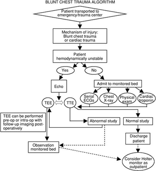
Mortality increases in thoracic trauma with increasing age, lower Glasgow coma scale scores, liver injury, splenic injury, more than five rib fractures, and long bone fractures. Mortality rates typically are between 9% and 20% in the United States (24). If the patient suffers an out-of-hospital cardiac arrest in relation to his or her trauma, the chances of survival diminish even further, with less than 10% of patients in this group surviving to hospital discharge (25). A proposed therapeutic algorithm is illustrated in Figure 79.1 and will be discussed below in detail.
Rib Fractures
Rib fractures are the most common type of chest trauma, with ribs five through nine being the most susceptible. Rib fractures by themselves are rarely life threatening but may serve as indicators for more severe intrathoracic or intra-abdominal injuries. Pain may be significant and impairs usual respiratory mechanics, leading to splinting, hypoventilation, atelectasis, and potentially pneumonia as pulmonary toilet is compromised. First and/or second rib fractures indicate a large transfer of energy to the thoracic cage and should raise further suspicion for other intrathoracic problems such as aortic rupture or tear (26). The elderly, defined in this instance as 65 years of age and older, pose a particular problem when faced with these types of injuries; mortality increases by 19% with each rib fracture and the risk of pneumonia by 27% (27). The implications of age begin at 45 years, given that those with four or more rib fractures in this group show more in-hospital complications, such as increased ventilator and ICU days (28).
One intervention, however, may prove valuable in improving the outcome of patients with multiple rib fractures. Provided patients are suitable candidates for epidural analgesia, this method of pain control is associated with a decreased incidence of nosocomial pneumonia and shorter duration of mechanical ventilation in those with three or more rib fractures (29).
Flail Chest
Flail chest occurs when two or more adjacent ribs are fractured at two or more sites. This leads to a paradoxical movement of that segment during inspiration, manifested as an inward collapse. As with rib fractures, pain control is important to avoid splinting and to facilitate pulmonary toilet. Positive pressure ventilation, whether invasive or noninvasive, may be required to stent open the affected lung region and thereby avoid atelectasis.
Hemothorax
Hemothorax is a collection of blood in the pleural cavity. Patients potentially experience chest pain, dyspnea, and tachycardia along with dullness to percussion and decreased or absent breath sounds to auscultation on the affected side. Chest radiographs help to confirm the diagnosis if the collection of blood is large enough (i.e., >200 mL) to be seen radiographically. The mainstays of therapy are ensuring adequate circulating blood volume and tube thoracostomy to drain blood from the pleural space. Thoracotomy is required if bleeding continues at a significant rate, defined as 1,500 mL of blood output with initial tube placement or continuous bleeding of at least 250 mL/hour for 4 hours or if the patient's vital signs suddenly decompensate (26). Failure to adequately drain the hemothorax potentially leads to a condition of retained clot. This is problematic, as it may progress to empyema or fibrothorax. Options for therapy include further large-diameter tube thoracostomy, open thoracotomy, video-assisted thoracoscopy (VATS), or intrapleural fibrinolytic therapy. Further tube thoracostomy likely has a limited role, as it does not consistently liberate clotted blood products, and early surgical drainage with VATS decreases the duration of tube drainage, length of hospital stay, and hospital cost (30). VATS is the surgical intervention of choice because it is less invasive than open thoracotomy and just as effective, unless there are extensive adhesions (31). Fibrinolytic therapy offers a possible alternative to thoracoscopy or decortication, especially if the time from injury to therapy is delayed, with a response rate of up to 92% in terms of resolution (32).
|
|
|
Figure 79.1. Therapeutic algorithm. ECGs, electrocardiograms; TEE, transesophageal echocardiography; TTE, transthoracic echocardiography. |
Pneumothorax
Pneumothorax is the accumulation of air, originating from the lung, between the visceral and parietal pleura, and is the most common intrathoracic finding following blunt or penetrating trauma. The size of the pneumothorax is expressed as a percentage, determined by its size relative to the entire lung on an anterior-posterior chest radiographic film. Treatment with tube thoracostomy is indicated when the size is >20%, the patient is on positive pressure ventilation, and there are signs and symptoms of hypoxia and dyspnea. If following tube placement the lung does not completely re-expand and there is a persistent air leak, it is important to search for a more severe tracheal or bronchial injury, as this situation would require surgical intervention.
Tension Pneumothorax
Tension pneumothorax is a life-threatening condition requiring immediate therapy. It occurs when air accumulates in a hemithorax under pressure, causing impaired venous return and cardiac output. The absence of breath sounds on the affected side, along with deviation of the trachea and mediastinum away from that side, are the hallmarks of this condition, especially in the presence of severe hypotension. Initial therapy includes relief of the pressure by placing a large-bore (14-gauge) intravenous catheter into the second intercostal space in the midclavicular line on the affected side, followed by tube thoracostomy to treat the pneumothorax.
Pulmonary Contusion
Pulmonary contusion is a result of blunt force transmitted across the thorax. The mechanism for its development is not completely understood but is felt to be related to compression and re-expansion of the lung tissue, leading to capillary disruption with interstitial and intra-alveolar edema, decreased compliance, and hypoxemia due to a shunt physiology (26). Care is largely supportive in this population, with close attention to pain management and pulmonary toilet. Typical chest radiographic findings are shown in Figure 79.2.
|
|
|
Figure 79.2. There are multiple fractured ribs on the right with several fractured in more than one place. This leads to a flail segment with inspiration. |
Cardiac Contusion
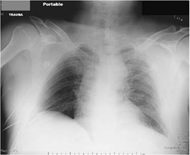
Cardiac contusion is a potential complication of blunt chest trauma (Fig. 79.3.) Its exact incidence is unclear, as different studies used varying criteria to make the diagnosis (33). It is typically well tolerated in mildly injured patients but may lead to fatal arrhythmias or cardiogenic shock if severe. Rapid deceleration, as occurs in motor vehicle accidents, is the most common cause since, in this situation, the heart moves freely and can strike the internal sternum with a substantial amount of force (33). Electrocardiography findings in cardiac contusion are summarized in Table 79.5. Biomarkers, such as creatine kinase (CK) or troponin I and T, are potentially helpful in the diagnosis, as the histologic changes associated with contusion are similar to those seen with infarction. Troponin I and T are specific to the myocardium and may avoid false positives by relying solely on CK, as trauma patients often have diffuse muscular damage leading to massive CK release from many tissues. Electrocardiography may show nonspecific findings, such as sinus tachycardia and premature atrial or ventricular systoles, and not provide further clarification of the diagnosis (33). Echocardiography, whether transthoracic (TTE) or transesophageal (TEE), offers the best insight into cardiac damage from contusion due to its ability to directly visualize wall motion abnormalities (Table 79.6). Treatment involves cardiac monitoring and stabilization of the traumatically induced injuries, supporting blood pressure and cardiac output as indicated. (See algorithm, Fig. 79.1.)
|
|
|||||
|
Figure 79.3. Diffuse infiltrative pattern on the left side is consistent with findings seen following pulmonary contusion. |
|||||
|
Table 79.5 Electrocardiographic Findings in Cardiac Contusion |
|||||
|
Aortic Disruption
Aortic disruption leads to a high mortality, as only 13% to 15% of these patients reach the hospital alive. Those surviving that long have a 30% chance of subsequent rupture and death (34). Diagnostic modalities include findings on chest radiograph of a widened mediastinum, aortography (the gold standard), CT scanning, and TEE. Treatment is immediate surgery by a cardiothoracic surgeon to repair the injury.
|
Table 79.6 Echocardiographic Findings in Acute Cardiac Contusion |
||
|
Posttraumatic empyema occurs in up to 2% of the thoracic trauma population and is most often caused by Staphylococcus aureus (35). Factors increasing the likelihood of developing empyema include retained hemothorax, pulmonary contusion, and multiple chest tube placement (36). Of note, since most empyemas are of a parapneumonic etiology (37), pneumonia has little impact on the development of pleural space infection in this population. Treatment includes removal of the infected fluid or collection and may be accomplished by guided drainage, VATS, or open thoracotomy.
Lung Volume Reduction Surgery
Lung volume reduction surgery (LVRS) is used in an effort to improve the pulmonary dynamics of patients with severe emphysema by palliating dyspnea and improving functional status. This population is prone to more postsurgical complications than most other thoracic surgery patients due to their underlying fragile nature. Anticipated complications include arrhythmias, prolonged respiratory failure, air leaks, pneumonia, and ICU readmission. Air leaks occur in 90% of patients and rarely require surgical intervention (38). The prevalence of air leaks correlates with a more prolonged, complicated hospital course, and severity is predicted by patient characteristics such as worsened pulmonary function, use of inhaled steroids, distribution of disease (lower lobe disease is less frequent and has shorter duration), and presence of adhesions (38). Management involves chest tube drainage with efforts to minimize or eliminate suction so the tissue has the greatest chance to heal.
Esophageal Surgery
Esophageal surgery patients undergo procedures that traumatize the lung, the interposed stomach, and the diaphragm. Patients who undergo esophageal resection for carcinoma tend to be malnourished. Pulmonary complications, including atelectasis, pneumonia, aspiration, and retained secretions, are all possible. Up to 29% of patients experience respiratory complications (39) with increasing patient age and decreasing performance on spirometry, which is predictive of increased risk (40). Aspiration risk is minimized by having patients undergo a thorough swallowing evaluation, including radiographic testing, prior to initiating oral intake.
The most dangerous complication of esophageal surgery is leakage from the surgical site. Anastomotic leak occurs in as many as 11% of patients, and factors impacting the incidence include high estimated intraoperative blood loss, cervical location of the anastomosis, and the development of postoperative ARDS (41). Interestingly, the use of thoracic epidural analgesia is associated with a decreased occurrence of anastomotic leakage (41). Mortality associated with anastomotic leaks is historically high, but with improved surgical techniques, the patients now face a more promising outcome. One center showed a reduction in mortality with intrathoracic leaks from 43% to 3.3% over a 34-year period (42). Early identification is important, and endoscopy provides a safe method for determining the integrity of the graft and whether surgery is necessary to avoid loss of the graft (43).
Respiratory Therapy
Thoracic surgical patients often have significant underlying COPD, impaired mucociliary clearance, excessive secretions, and/or increased closing volumes, all of which predispose to atelectasis. The respiratory therapist plays an important role in providing secretion management and chest physiotherapy (percussion and vibration). Other modalities supporting recovery include adequate hydration, aerosolized bronchodilators, humidified oxygen, and early identification and treatment of infection of the tracheobronchial tree. Chest physiotherapy should begin as soon as the patient has recovered sufficiently from anesthesia to cooperate. Mucolytic agents (such as N-acetylcysteine) are helpful in solubilizing thick secretions, but may cause bronchospasm. Oral or nasotracheal suctioning is used in selected extubated patients, but discomfort and the possibility of complications (hypoxemia, vagal-mediated bradycardia, or cardiac arrest) limit routine use. A mini-tracheostomy (bedside percutaneous cricothyroidotomy for suctioning) can provide access to the lower airway in patients with thick secretions. Inadequate clearance of secretions often requires flexible bronchoscopy, which is of greatest benefit in the extubated patient who cannot adequately be suctioned. If pulmonary parenchymal involvement is confined to one lung, altering body position can improve gas exchange by changing the relationships between ventilation and perfusion. The lateral decubitus position, with the uninvolved lung down, allows maximal blood flow to ventilated areas during spontaneous ventilation. This relationship may be altered with mechanical breaths and application of PEEP. Specialized beds can be set to supine, lateral, or rotating modes to optimize oxygenation (44).
Complications
Complications common to all thoracic surgical patients are listed in Table 79.7. Those more likely to occur following specific procedures are listed in Table 79.8.
Airway complications can be precipitated by prolonged intubation with large or double-lumen endotracheal tubes, passage of bronchial blockers, use of rigid bronchoscopes, or frequent reintubation. Edema of the larynx or trachea can substantially narrow the cross-sectional area of the airway. Assessing the patient for an air leak around an occluded endotracheal tube just prior to extubation may identify significant laryngeal and supraglottic edema. Upright or sitting position, intravenous corticosteroids (45), and racemic epinephrine respiratory treatments are the mainstay of edema reduction. A critical airway may be converted to an adequate airway by the administration of Heliox, a helium and oxygen mixture (46). Helium, being less dense and less viscous than nitrogen, allows maintenance of laminar flow through a critically swollen upper airway. Prolonged endotracheal intubation or temporary tracheostomy may be required to allow resolution of airway edema.
|
Table 79.7 Postoperative Complications following Any Thoracic Procedure |
||
|
The recurrent laryngeal nerves branch from the vagus nerves as they enter the chest. The right recurrent laryngeal nerve arises high in the apex of the right chest and loops around the right subclavian artery to travel back to the larynx in the tracheoesophageal groove. The left recurrent laryngeal nerve, which is more susceptible to injury, wraps around the aortic arch in the left chest before it enters the tracheoesophageal groove. Injury can result from excessive traction, aggressive dissection about these nerves, or the operative sacrifice of these nerves. Mediastinoscopy, anterior mediastinotomy, left pulmonary resection with subaortic exenteration, and resections of mediastinal tumors are common operations in which the recurrent laryngeal nerve may be damaged. Associated airway and laryngeal edema may allow for adequate coaptation of the vocal cords for the first days postextubation, often preventing identification of cord injury until after discharge from the ICU, when ineffective cough or aspiration of secretions will become apparent. If there is permanent damage or division of the recurrent laryngeal nerve, injection of the vocal cord with a long-lasting substance such as Teflon may be considered. In many instances, aggressive chest physiotherapy, careful airway management, and temporary avoidance of oral feeding may eliminate the need for any intervention until recovery of the nerve function has occurred. Intermittent noisy inspiration and painful swallowing suggest arytenoid dislocation, an uncommon cause of postextubation respiratory failure (47). Treatment consists of surgical reduction, which must be accomplished before the cricoarytenoid joint becomes fibrosed in poor position.
Retained secretions and blood in the airway are especially common if the airway was opened, such as during a bronchoplastic procedure or closure of a bronchial stump. Mechanical airway obstruction secondary to secretions may be aggravated by bronchospasm, and preoperative bronchodilators should be continued in patients with reactive airways, as secretions can precipitate coughing and bronchospasms.
Postoperative air leaks most often result from very distal fistulae between tiny bronchioles or respiratory units and the pleural cavity. One of the main functions of the chest tube is to evacuate air from these small air leaks to ensure complete expansion of the lung and coaptation of the cut surface of the lung to the parietal pleura, which will seal these leaks. Repositioning of the chest tubes or insertion of further chest tubes into undrained spaces, adequate suction applied to the pleural cavity, and full expansion of the lung with vigorous chest physiotherapy help to close these small distal fistula. A substantial persistent air leak from the chest tube, or incomplete expansion of the lung, suggests a significant bronchopleural fistula. Major proximal airway problems such as failure of the anastomosis, disruption of a bronchial closure, or retained secretions or foreign bodies can be identified by bronchoscopy. Within the first 7 postoperative days, any fistula is likely to be due to a technical problem. More than 1 week after the operation, but usually within the first 6 weeks, fistulae are more often due to an empyema or local peribronchial abscess. Late occurrence of a bronchopleural fistula, (more than 6 months after the operation) is frequently due to recurrent lung carcinoma.
Early postoperative bronchopleural fistula in a pneumonectomy patient is a surgical emergency. The typical presentation is sudden expectoration of copious amounts of pink, frothy sputum, which may be misdiagnosed as pulmonary edema. The patient should be positioned with the operated or pneumonectomy side down to trap remaining fluid in the pneumonectomy space and prevent drowning. A chest radiograph will show loss of fluid from the pneumonectomy space. Further management will likely include both bronchoscopy to assess the stump closure and immediate reoperation.
Empyema is initially treated with closed tube drainage and antibiotic therapy. After the patient has been stabilized and any bronchopleural fistula identified and treated, drainage of the empyema cavity is converted from closed tube drainage to open tube drainage. A chest radiograph helps to determine if the mediastinum is fixed or whether it has shifted and compressed the contralateral remaining lung. If the mediastinum is stable, drainage of the cavity may be permanently converted to open drainage. This may take the form of rib resection and marsupialization of the pneumonectomy cavity (Clagett window or Eloesser flap). With time, the pneumonectomy cavity shrinks in size, and the window or flap may be closed.
Postoperative hypoxemia is common and may be due to sepsis, ARDS, pneumonia, or pulmonary embolization. If pulmonary emboli are suspected, ventilation/perfusion scanning, spiral computed tomography, and pulmonary angiography should be done, and treatment initiated with anticoagulation or lytic therapy, depending on timing and indications. If these measures are contraindicated, an inferior vena caval filter should be placed. Systemic tumor emboli, though uncommon, may be seen after pulmonary resections for primary bronchogenic carcinomas or metastatic sarcomas.
|
Table 79.8 Complications of Specific Thoracic Procedures |
||||||||||||||||||||||||||||||||||||||||||||||||||
|
||||||||||||||||||||||||||||||||||||||||||||||||||
Massive postoperative hemorrhage can present as significant shock and occasionally occurs during transfer of the patient to the recovery room or ICU. This potentially lethal condition can be the result of a slipped tie from the pulmonary vein—or, less commonly, the pulmonary artery—and requires emergent reoperation. Slower postoperative hemorrhage usually results from small bleeding arteries or veins in the mediastinum or chest wall. Reoperation is required for control of bleeding and to evaluate the hemothorax to prevent future fibrothorax or restrictive lung disease.
Pulmonary Parenchymal Complications
Tracheoesophageal Fistula
A communication may occur between the anterior esophageal wall and membranous (posterior) wall of the trachea following prolonged intubation due to pressure exerted from the cuff of the endotracheal or tracheostomy tube, which can lead to potential tissue necrosis. Tracheoesophageal fistula should be suspected when feedings are aspirated from the airway. Surgical revision of the damaged area is the definitive therapy but may not be practical in patients still requiring positive pressure ventilation, and may need to be delayed until the patient has been weaned off mechanical ventilation (48). Stenting of the esophagus, performed at the bedside in the ICU, allows for temporary sealing of the fistula until the patient is a suitable candidate for final surgical repair (49).
Tracheoinnominate Fistula
Bleeding seen 48 hours or greater following tracheostomy raises concern about the presence of a tracheoinnominate fistula. Even though this occurs rarely, it is a life-threatening complication and requires urgent action. Most cases arise when pressure necrosis on the posterior aspect of the blood vessel occurs due to overinflation of the endotracheal cuff. Initial steps include hyperinflation of the tracheostomy cuff or direct arterial compression with a finger to tamponade the bleeding. Bronchoscopy is the diagnostic procedure of choice, followed by surgery to divide the innominate artery, with subsequent separation of the trachea from the divided artery by viable tissue (50).
Atelectasis
The most common complication following thoracic surgery is atelectasis. Potential contributors include hypoventilation, splinting, bronchospasm, poor cough, retained secretions, pneumothorax, and trauma to the lung during the surgical procedure. Most patients in the ICU will tolerate some degree of atelectasis with symptoms including fever, tachypnea, arrhythmias, hypoxia, or respiratory failure. More severe atelectasis or a compromised host can present with respiratory failure requiring mechanical ventilation. Auscultation of the lungs may identify the impaired areas due to the diminished air movement, and a chest radiograph may further highlight the portions of collapsed lung. Atelectasis is not limited to the lung on the operative side, although it is more likely to occur in that parenchyma.
Efforts to avoid atelectasis include frequent suctioning of the tracheobronchial tree, chest percussion and postural drainage, humidification of inspired gases, frequent patient rotation, bronchodilators, pain control, coughing, deep breathing, bronchoscopy for removal of mucus plugs or secretions, invasive or noninvasive positive pressure ventilation, and early ambulation (14). Chest physiotherapy uses physical therapy such as clapping and vibration to stimulate coughing and thereby move secretions out of the lungs. Incentive spirometry, often used following surgical procedures, does not provide the same benefit of chest physiotherapy in the recovery phase, as physiotherapy potentially decreases hospital costs due to shorter lengths of stay (51).
Lobar Collapse and Torsion
Lobar collapse most commonly occurs in either the right upper lobe following surgery that uses a double-lumen endotracheal tube or in the right middle lobe after right upper lobectomy, which alters the anatomy, allowing the horizontal fissure to rise. This results in compression on the right middle lobe bronchus and subsequent occlusion.
Lobar torsion occurs as the result of a lung segment twisting about its hilar structures. This twisting occludes the bronchial, arterial, and venous supply to the affected segment, with infarction occurring if the process is not recognized and treated. The right middle lobe and lingula are at greatest risk following thoracotomy (14). The patient may initially present as simply having atelectasis, but over time, the chest radiograph shows opacification of the affected lobe. As ischemia progresses to infarction, there will be signs of tissue necrosis, such as bloody or malodorous secretions and foul chest tube drainage. Bronchoscopy is the initial tool for evaluation, followed by thoracotomy for an urgent detorsion and lobectomy if necrotic tissue is found.
Pleural Complications
Pneumothorax is the second most frequent postoperative complication after atelectasis (14). Signs and symptoms of a pneumothorax range from subtle to severe and include increased work of breathing, decreased breath sounds and chest movement, wheezing, hypoxia, increased airway pressures if still on the ventilator, and hemodynamic instability if tension pneumothorax occurs. A chest radiograph is the first modality used in the diagnosis of pneumothorax, save for the instance when tension is present and chest tube placement must occur immediately to prevent further clinical deterioration of the patient. If a pneumothorax develops on the surgical side and chest tubes are already present, it is necessary to ensure that the tubes are functioning properly. This involves inspecting the entire system for any evidence of leaks, inadequate suction, or loss of tubing patency due to blood clots. It is possible to declot chest tubes with vigorous stripping or by placing a balloon occlusion catheter to physically remove the obstruction.
Pleural effusions persisting in the postoperative period should be evaluated via thoracentesis to determine if there is an intrathoracic or extrathoracic cause. If the fluid is transudative, the underlying cause needs to be addressed. If the fluid is exudative, a high suspicion for empyema must be maintained. If pleural effusions and pneumothoraces do not resolve over time, the decision for more definitive therapy, such as pleurodesis, needs to be entertained.
Thoracic duct injury is a known complication following any surgery involving dissection in the posterior mediastinum and may result in chylothorax. Confirmation of the diagnosis includes testing the pleural fluid which is high in triglycerides and chylomicrons. The leakage site is localized with lymphangiography or CT scanning, and the clinical situation will dictate the type of management. It occurs in approximately 4% of patients undergoing esophageal surgery with the transthoracic approach as compared to transhiatal; an increased number of positive nodes are predictive of its incidence (52). Management initially includes chest tube drainage and parenteral nutrition to decrease the thoracic duct output. If the drainage resolves with conservative management, surgical intervention can likely be reserved for those not decreasing their chylous output (53).
Other Complications
Postoperative Infections
The incidence of developing nosocomial infection following lung surgery increases with a history of COPD, duration of surgery (with an increased risk for each additional minute), and ICU admission (54). Surgical site infection occurs rarely due to the use of prophylactic perioperative antibiotics. Factors increasing the risk of infection at the incision site include emergent thoracotomy in the trauma patient and procedures for treatment of empyema, lung abscess, mediastinitis, or perforated esophagus. Any unexplained fever requires careful inspection of the surgical site.
When air tracks into the subcutaneous space via the path of least resistance, it generates subcutaneous emphysema. The air is forced along these pathways with positive pressure ventilation and instances of increased intrathoracic pressure during spontaneous breathing. Coughing and forced exhalation are two examples. Subcutaneous air may be striking in appearance but rarely affects patient outcome in a detrimental way. If the collection of air is massive enough to compromise the airway, endotracheal intubation may be indicated, realizing the pitfalls of instituting positive pressure ventilation in such a situation. Following sterile preparation, puncturing the skin with small-gauge (no. 25) needles provides a conduit for the subcutaneous air to escape and decrease the cosmetic deformation.
Summary
The thoracic patient presents unique management issues due to the complexities of fluid management, preoperative morbidities, the need for specialized pain control to preserve respiratory function, and nature of drainage devices left in place postoperatively. As a critical care physician, it is important to understand the interplay of these circumstances when anticipating the patient's postoperative needs in the ICU. The pulmonary system is more susceptible to adverse events if fluid management is not judiciously managed. Patients undergoing thoracotomies for lung or esophageal surgery tend to have an impaired functional status and nutrition at baseline, negatively impacting their postoperative course. If pain control is inadequate, the patient will have impaired pulmonary function, which potentially delays recovery. Chest tubes require extra vigilance due to the potential complications should they malfunction.
With a well thought out care plan, the outcome for thoracic surgery patients will be positive and the road to recovery smooth.
References
1. Nunn JF, Miledge S, Chen D, et al. Respiratory criteria for fitness for surgery and anesthesia. Anaesthesia. 1988;43:543–611.
2. Garibaldi RA, Britt MR, Coleman ML, et al. Risk factors for postoperative pneumonia. JAMA. 1981;70:677–680.
3. Wahba WM. Influence of aging on lung function—clinical significance of changes from age twenty. Anesth Analg. 1983;62:764–776.
4. Warner MA, Offord KP, Warner ME, et al. Role of preoperative cessation of smoking and other factors in postoperative pulmonary complications: a blinded, prospective study of coronary artery bypass patients. Mayo Clin Proc. 1989;64:609–616.
5. Arozullah AM, Daley J, Henderson WG, et al. Multifactorial risk index for predicting postoperative respiratory failure in men after major noncardiac surgery. Ann Surg. 2000;332:242–253.
6. Palesty JA, McKelvey AA, Dudrick SJ. The efficacy of x-rays after chest tube removal. Am J Surg. 2000;179:13–16.
7. Collier B, Kolff J, Devineni R, et al. Prophylactic positive end-expiratory pressure and reduction of postoperative blood loss in open-heart surgery. Ann Thorac Surg. 2002;74(4):1191–1194.
8. Van den Berghe G, Wouters P, Weekers F, et al. Intensive insulin therapy in critically ill patients. N Engl J Med. 2001;345:1359–1367.
9. DeHaven CB, Hurst JM, Branson RD. Evaluation of two different extubation criteria: attributes contributing to success. Crit Care Med. 1986;14:92.
10. The Acute Respiratory Distress Syndrome Network. Ventilation with lower tidal volumes as compared with traditional tidal volumes for acute lung injury and the acute respiratory distress syndrome. N Engl J Med. 2000;342:1301–1308.
11. Baumann MH, Sahn SA. Medical management and therapy of bronchopleural fistulas in the mechanically ventilated patient. Chest. 1990;97:721.
12. Slinger PD. Perioperative fluid management for thoracic surgery: the puzzle of postpneumonectomy pulmonary edema. J Cardiothorac Vasc Anesth. 1995;9(4):442–451.
13. van der Werff YD, van der Houwen HK, Heijmans P, et al. Postpneumonectomy pulmonary edema, a retrospective analysis of incidence and possible risk factors. Chest. 1997;111:1278–1284.
14. Amini S, Gabrielli A, Caruso LJ, et al. The thoracic surgical patients: initial postoperative care. Semin Cardiothorac Vasc Anesth. 2002;6(3):169–188.
15. Jordan S, Mitchell JA, Quinlan GJ, et al. The pathogenesis of lung injury following pulmonary resection. Eur Respir J. 2000;15:790–799.
16. Loan WB, Morrison JD. The incidence and severity of postoperative pain. Br J Anaesth. 1967;39:695–698.
17. Katz J, Jackson M, Kavanagh BP. Acute pain relief after thoracic surgery predicts long term post-thoracotomy pain. Clin J Pain. 1996;12(1):50–55.
18. Yee JP, Koshiver JE, Allbon C, et al. Comparison of intramuscular ketorolac tromethamine and morphine sulfate for analgesia of pain after major surgery. Pharmacotherapy. 1986;6(5):253–261.
19. Lardinois D, Vogt P, Yang L, et al. Non-steroidal anti-inflammatory drugs decrease the quality of pleurodesis after mechanical pleural abrasion. Eur J Cardiothorac Surg. 2004;25(5):865–871.
20. Ballantyne JC, Carr DB, deFerranti S, et al. The comparative effects of postoperative analgesic therapies on pulmonary outcome: cumulative meta-analyses of randomized, controlled trials. Anesth Analg. 1998;86:598–612.
21. Yegin A, Erdogan A, Kayacan N, et al. Early postoperative pain management after thoracic surgery; pre and postoperative versus postoperative epidural anesthesia: a randomized study. Eur J Cardiothorac Surg. 2003;24(3):420–424.
22. Peeters-Asdourian C, Gupta S. Choices in pain management following thoracotomy. Chest. 1999;115:122S–124S.
23. Herr DL, Sum-Ping ST, England M. ICU sedation after coronary artery bypass graft surgery: dexmedetomidine-based versus propofol-based regimens. J Cardiothoracic Vasc Anesth. 2003;17(5):576–584.
24. Kulshrestha P, Munshi I, Wait R. Profile of chest trauma in a level I trauma center. J Trauma. 2004;57:576–581.
25. Lockey D, Crewsdon K, Davies G. Traumatic cardiac arrest: who are the survivors? Ann Emerg Med. 2006;48(3):240–244.
26. Munshi IA. Thoracic trauma. In: Higgins TL, ed. Cardiopulmonary Critical Care. Oxford, UK: BIOS Scientific Publishers Ltd.; 2002.
27. Bulger EM, Arneson MA, Mock CN, et al. Rib fractures in the elderly. J Trauma. 2000;48(6):1040–1046.
28. Holcomb JB, McMullin NR, Kozar RA, et al. Morbidity from rib fractures increases after age 45. J Am Coll Surg. 2003;196(4):549–555.
29. Bulger EM, Edwards T, Klotz P, et al. Epidural analgesia improves outcome after multiple rib fractures. Surgery. 2004;136(2):426–430.
30. Meyer DM, Jessen ME, Wait MA, et al. Early evacuation of traumatic retained hemothoraces using thoracoscopy: a prospective, randomized trial. Ann Thorac Surg. 1997;64:1396–1400.
31. Navsaria PH, Vogel RJ, Nicol AJ. Thoracoscopic evacuation of retained posttraumatic hemothorax. Ann Thorac Surg. 2004;78:282–285.
32. Inci I, Ozcelik C, Ulku R, et al. Intrapleural fibrinolytic treatment of traumatic clotted hemothorax. Chest. 1998;114:160–165.
33. Sybrandy KC, Cramer MJM, Burgersdijk C. Diagnosing cardiac contusion: old wisdom and new insights. Heart. 2003;89:485–489.
34. Fabian TC, Richardson JD, Croce MA, et al. Prospective study of blunt aortic injury: multicenter trial of the American Association for the Surgery of Trauma. J Trauma. 1997;42(3):1128–1143.
35. Mandal AK, Thadepalli H, Chettipalli U. Posttraumatic empyema thoracis: A 24-year experience at a major trauma center. J Trauma. 1997;43(5):764–771.
36. Aguilar MM, Battistella FD, Owings JT, et al. Posttraumatic empyema. Risk factor analysis. Arch Surg. 1997;132(6):647–650.
37. Hoth JJ, Burch PT, Bullock Tk, et al. Pathogenesis of posttraumatic empyema: the impact of pneumonia on pleural space infections. Surg Infect. 2003;4(1):29–35.
38. DeCamp MM, Blackstone EH, Naunheim KS, et al. Patient and surgical factors influencing air leak after lung volume reduction surgery: lessons learned from the national Emphysema Treatment Trial. Ann Thorac Surg. 2006;82:197–207.
39. Atkins BZ, Shah AS, Hutcheson KA, et al. Reducing hospital morbidity and mortality following esophagectomy. Ann Thor Surg. 2004;78(4):1170–1176.
40. Ferguson MK, Durkin AE. Preoperative prediction of the risk of pulmonary complications after esophagectomy for cancer. J Thorac Cardiovasc Surg. 2002;123(4):661–669.
41. Michelet P, D'Journo KB, Roch A, et al. Perioperative risk factors for anastomotic leakage after esophagectomy: influence of thoracic epidural analgesia. Chest. 2005;128(5):3461–3466.
42. Martin LW, Swisher SG, Hofstetter W, et al. Intrathoracic leaks following esophagectomy are no longer associated with increased mortality. Ann Surg. 2005;242:392–402.
43. Maish MS, DeMeester SR, Choustoulakis E, et al. The safety and usefulness of endoscopy for evaluation of the graft and anastomosis early after esophagectomy and reconstruction. Surg Endosc. 2005;19(8):1093–1102.
44. Nelson LD, Anderson HB. Physiologic effects of steep positioning in the surgical intensive care unit. Arch Surg. 1989;124:352–355.
45. Cheng K-C, Hou C-C, Huang H-C, et al. Intravenous injection of methylprednisolone reduces the incidence of postextubation stridor in intensive care unit patients. Crit Care Med. 2006;34:1345–1350.
46. Skrinskas GJ, Hyland RH, Hutcheon MA. Using helium-oxygen mixtures in the management of acute upper airway obstruction. Can Med Assoc J. 1983;128:555–558.
47. Castella X, Gilabert J, Perez C. Arytenoid dislocation after tracheal intubation: an unusual cause of acute respiratory failure? Anesthesiology. 1991;74:613–615.
48. Athanassiadi K, Gerazounis M. Repair of postintubation tracheoesophageal fistula in polytrauma patients. Injury. 2005;36(8):897–899.
49. Eleftheriadis E, Kotzampassi K. Temporary stenting of acquired benign tracheoesophageal fistulas in critically ill ventilated patients. Surg Endosc. 2005;19(6):811–815.
50. Allan JS, Wright CD. Tracheoinnominate fistula: diagnosis and management. Chest Surg Clin N Am. 2003;13(2):331–341.
51. Varela G, Ballesteros E, Jimenez MF, et al. Cost-effectiveness of prophylactic respiratory physiotherapy in pulmonary lobectomy. Eur J Cardiothorac Surg. 2006;29:216–220.
52. Lagarde SM, Omloo JM, de Jong K, et al. Incidence and management of chyle leakage after esophagectomy. Ann Thor Surg. 2005;80(2):449–454.
53. Shimizu K, Yoshida J, Nishimura M, et al. Treatment strategy for chylothorax after pulmonary resection and lymph node dissection for lung cancer. J Thorac Cardiovasc Surg. 2002;124(3):499–502.
54. Nan DN, Fernandez-Ayala M, Farinas-Alvarez C, et al. Nosocomial infection after lung surgery. Chest. 2005;128:2647–2652.