Howard K. Song
Matthew S. Slater
Adult patients undergoing cardiovascular surgery procedures all pass through a period of critical illness during their recovery. These patients present intensivists with unique critical care management challenges because of the hemodynamic changes that occur as a result of the surgery itself and the broad inflammatory after-effects of cardiopulmonary bypass. The specialized nature of the critical care of adult cardiovascular surgery patients is apparent in the proliferation of heart centers, specialized units, treatment protocols, and pathways designed to optimize patient outcomes by standardizing care and focusing provider expertise. Optimal outcomes for adult cardiovascular surgery patients are most likely to occur where critical care systems are designed to support this specific patient population and practitioners have strong familiarity with normal recovery milestones and complications that can interrupt this process.
Cardiopulmonary Bypass
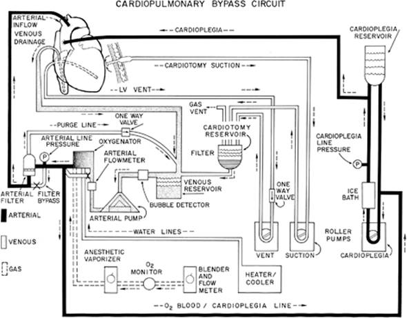
A significant portion of adult cardiovascular surgery procedures are performed with the use of cardiopulmonary bypass. The development of the heart–lung machine over the past 50 years represents one of the major achievements of modern medicine and has made complex repairs of intracardiac structures and the great vessels feasible. Cardiopulmonary bypass techniques allow the surgeon to interrupt blood flow through the heart and lungs, providing a relatively bloodless and motionless field for the conduct of the operation. The most basic components of a cardiopulmonary bypass system are a venous cannula, a venous reservoir, a pump, an oxygenator, a heat exchanger, a filter, and an arterial cannula (Fig. 80.1). Other components that are commonly used are a cardioplegia delivery system, an ultrafiltration unit, a cardiotomy suction system, a left ventricular vent, and a cell saver system.
Although cardiopulmonary bypass is an indispensable tool for the intraoperative care of the cardiovascular surgery patient, it incites a broad inflammatory cascade that can have far-reaching consequences on the subsequent intensive care unit (ICU) course of the patient. The primary trigger for this inflammatory cascade is the interaction between the patient's blood components and the large plastic surface area represented by the cardiopulmonary bypass circuit (1). This interaction activates plasma enzyme systems and blood cells, dilutes plasma proteins, causes coagulopathy, produces emboli, and leads to release of an array of vasoactive mediators that affect vascular motor tone and endothelial permeability (Table 80.1). Because of the systemic nature of this response, nearly every end organ is susceptible to at least temporary inflammatory-mediated dysfunction in the postoperative period.
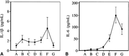
Fortunately, the post-cardiopulmonary bypass inflammatory cascade is typically self-limited (Fig. 80.2). Circulating levels of vasomotor mediators peak within 24 hours of surgery and subside over ensuing days (2). Although cardiovascular surgery patients frequently have temporary mild end organ dysfunction postoperatively, rates of major complications for elective routine cases are low. Much of the critical care of cardiovascular surgery patients is oriented toward supporting patients through the early period during which they are subject to the temporary effects of cardiopulmonary bypass.
Immediate Concerns
The patient's admission to the ICU is a hectic transition period during which the patient is physically transported, monitoring and mechanical ventilation is re-established, intravenous medications are titrated, diagnostic tests are performed, and patient information is transferred from providers on the perioperative team to providers on the postoperative ICU team. In the midst of all this activity, almost all patients arriving in the ICU after cardiovascular surgery are in a state of controlled shock because of fluid shifts and changes in vascular tone. Cardiovascular surgery patients are therefore especially vulnerable to lapses in monitoring or distractions that divert the attention of providers at the bedside. A checklist is useful to ensure that essential tasks are accomplished within the first 30 minutes of a patient's arrival in the ICU (Table 80.2).
The first step on the patient's arrival to the ICU should be a quick assessment of the ABCs (airway, breathing, circulation). The arterial pressure monitoring unit is transferred from the portable setup accompanying the patient to the ICU monitor. The systemic arterial pressure waveform allows you to assess adequate systemic perfusion pressure and provides a means to detect arrhythmias while the electrocardiogram (ECG) and other pressure measurement catheters are connected and calibrated. Next, connect the hemodynamic monitoring catheters that provide some measure of central circulatory volume status, including the central venous pressure (CVP) catheter, pulmonary artery catheter, or both. The ECG leads are then switched from the transport unit to the ICU system. After these monitors are established, others can be connected and calibrated including the mixed venous oxygen saturation (S[v with bar above]O2), and pulse oximetry waveform.
|
|
|
Figure 80.1. Schematic of a typical cardiopulmonary bypass circuit. (Reproduced from Wellons HA Jr, Zacour RK. Cardiopulmonary bypass. In: Kaiser LR, Kron IL, Spray TL, eds. Mastery of Cardiothoracic Surgery. 2nd ed. Philadelphia, PA: Lippincott Williams & Wilkins; 2007:312, with permission.) |
Mechanical ventilation is continued using the ICU ventilator. Significant arterial-alveolar (A-a) gradients are common following cardiopulmonary bypass so patients should routinely be given high concentrations of oxygen (70%–80%) until adequate oxygenation is confirmed. Tidal volume in the absence of acute lung injury is set at 8 mL/kg of predicted body weight and respiratory rate is set at 14 breaths per minute. If the patient is not synchronized with the ventilator, analgesia and sedation are assessed for adequacy.
|
Table 80.1 Vasoactive substances released following cardiopulmonary bypass |
||
|
Once stable hemodynamic function is confirmed, pressure monitoring can be interrupted to obtain blood samples for various tests, including arterial blood gas, complete blood count, serum potassium, ionized calcium, hematocrit, platelet count, and coagulation studies. Rezeroing of all pressure monitors and calibration of the venous saturation monitor should be performed. For patients with a pulmonary artery catheter, baseline measurements of cardiac output and calculations of systemic vascular resistance (SVR) and pulmonary vascular resistance (PVR) are obtained.
|
|
|
Figure 80.2. Changes in IL-1ß (A) and IL-6 (B) in 30 patients who had elective first-time myocardial revascularization. Letters on x-axis represent the following events: A, induction of anesthesia; B, 5 minutes after heparin; C, 10 minutes after starting cardiopulmonary bypass (CPB); D, end of CPB; E, 20 minutes after protamine; F, 3 hours after CPB; G, 24 hours after CPB. (Redrawn from Steinberg JB, Kapelanski DP, Olson JD, et al. Cytokine and complement levels in patients undergoing cardiopulmonary bypass. J Thorac Cardiovasc Surg. 1993;106:1008.) |
A physical exam is performed. Particular attention is given to adequacy of airway control and ventilation and assessment of peripheral pulses in all extremities. Although affected sometimes by the presence of severe atherosclerosis, skin temperature, pulse amplitude, and capillary refill time provide essential clinical information about the adequacy of cardiac output. Palpable peripheral pulses are an excellent indicator of systemic perfusion. Mental status and central nervous system assessment is also part of the initial exam.
|
Table 80.2 Tasks to be completed within the first 30 minutes of a patient's arrival to the ICU following cardiovascular surgery |
|||||
|
A portable chest radiograph is obtained to determine the proper placement of the endotracheal tube, monitoring catheters, and nasogastric tube. Examine this radiograph closely for evidence of a pneumothorax, hemopneumothorax, and areas of lung collapse or atelectasis.
A word of caution is necessary about obtaining chest radiographs immediately after the patient arrives in the ICU, particularly if the body temperature is below 35.5°C and hemodynamic instability is present. In this situation, the chest radiograph should be deferred until the temperature rises to above 35.5°C to avoid the occurrence of life-threatening arrhythmias during placement of the film cassette. Sudden movement may induce ventricular tachycardia or fibrillation resulting from the reduced fibrillation threshold caused by hypothermia and electrolyte imbalance, especially hypokalemia.
As the physical process of patient admission to the ICU is taking place, a transfer of responsibility for the patient's care is also occurring. This usually involves a verbal sign-out of patient information from providers on the perioperative team to providers on the ICU team. The patient's clinical history is reviewed, and the perioperative course is described. This information is particularly important to ensure the safe transition from perioperative to postoperative care. Important details may include information on ease of intubation, perioperative ventricular function, inotropic and vasopressor requirement, optimal volume status, completeness of revascularization, and suspected coagulopathy.
Initial Therapy
The first hour after admission to the ICU following cardiovascular surgery is a critical and unstable time. The goals in this first hour are to maintain adequate systemic perfusion, adequate oxygenation and respiration, and control cardiac rate and rhythm. Patients who experience intravascular volume shifts during transport to the ICU require intravascular volume expansion, bolus dosages of intravenous calcium chloride if ionized calcium is low, or both to maintain adequate systemic blood pressure and flow during the crucial 15 to 30 minutes required to settle the patient in the ICU.
In this early period of stabilization, especially when the patient is mildly to moderately hypothermic, serum potassium levels should be assessed rapidly and supplemented to maintain levels in the 4.5- to 5.0-mmol/L range. This intervention helps protect the heart against ventricular irritability. At times, patients arrive to the ICU in an acidemic state. Avoid attempting to correct acidosis with rapid intravenous infusions of sodium bicarbonate. Serum potassium can be acutely lowered, which may increase ventricular irritability. Once the potassium level is adequate, moderate to severe metabolic acidosis (base deficit >5 mmol/L) can be corrected with resuscitation and if necessary with sodium bicarbonate.
Urine output is monitored closely to detect inappropriate diuresis, which occurs commonly in patients after cardiopulmonary bypass. Patients exhibiting this may require especially rapid replacement of crystalloid and electrolyte losses. Mediastinal drain output is also closely observed to detect excessive blood loss. If rapid hemorrhage is suspected, transfusion therapy is initiated early to replace red blood cell mass and correct coagulopathy as directed by postoperative studies and clinical findings. The operating team should be made aware of any patient who has massive hemorrhage so that mediastinal exploration can be undertaken expeditiously when indicated.
The First 8 Hours after Cardiopulmonary Bypass
After initial stabilization, a period frequently follows when the patient's hemodynamics are adequate and the patient is relatively stable. This period of time is designated the golden period and lasts for approximately 8 hours after cessation of cardiopulmonary bypass (CPB). Pay careful attention to optimizing cardiac function, because the initial 8 to 14 hours are characterized by decreasing cardiac function. The nadir of this decline occurs approximately 12 hours after CPB. Particularly notable is a decrease in ventricular compliance leading to reduced cardiac output at any given filling pressure. This condition generally persists over the next 12 to 24 hours, followed by a gradual improvement in cardiopulmonary performance over the next 48 hours. In the golden period, the goal is to optimize cardiorespiratory performance so that the ensuing decrease in cardiac function from 8 to 24 hours after CPB does not jeopardize end organ perfusion. Caution should be used in weaning inotropes rapidly during this period despite adequate hemodynamics in anticipation of the deterioration that typically follows.
Despite this caution, stable patients undergoing non-emergent cardiovascular surgery can usually be extubated safely during this period. The major criteria for proceeding with extubation during this early period are adequate central nervous system function, stable hemodynamics without malignant arrhythmias, normothermia, low A-a gradient, adequate pulmonary mechanics, and no ongoing mediastinal hemorrhage. If these screening criteria are met, patients can be observed for a brief period on minimal ventilatory support followed by extubation. Intravenous narcotics are useful to treat incisional pain during the period that sedative drugs are weaned just prior to extubation. Anesthetic technique should also be tailored to allow patients who can reasonably be expected to be extubated early to emerge from the effects of anesthesia during this period. In our institution the majority of patients undergoing non-emergent cardiovascular surgery are extubated within the first 6 hours of admission to the ICU using this strategy. Patients who have undergone a period of deep hypothermic circulatory arrest or long cardiopulmonary bypass periods, patients with poor ventricular function or unstable hemodynamics, and patients with hemorrhage or coagulopathy and large anticipated transfusion requirement are not suitable for early extubation.
The Next 16 Hours after Cardiopulmonary Bypass
The next 16 hours is probably the most challenging time, especially for the patients whose cardiac performance was not optimized during the golden period. The cause of the decline in cardiac performance during this time period is unclear. It is notable that many of the vasoactive factors that are released after cardiopulmonary bypass reach their peak concentrations during this period (2). The effects of cardiac reperfusion injury peak during this period in patients who have had aortic cross-clamping and obligatory global myocardial ischemia during their operation (3).
Changes in Heart Compliance
Clinically, the heart seems to become noncompliant or “stiff.” In this situation, one usually observes a rise in pulmonary artery pressure and pulmonary artery occlusion pressure (PAOP), and a reduction in the systemic arterial pressure, cardiac output, and mixed venous oxygen saturation (4). If the patient is doing extremely well before the onset of compliance changes, alterations in these parameters may go unnoticed. However, patients with a low cardiac output syndrome before the onset of changes in compliance often develop hemodynamic instability.
Time Course
The usual time course is a steady deterioration in cardiac performance between the 8th to 12th hours following cardiopulmonary bypass. This condition seems to stabilize near the 12th hour and remains stable for the next 6 to 8 hours. Once a patient becomes unstable, far more interventions are required to return to a steady state than if problems are anticipated and appropriate changes made to help maintain stability. Even if patients remain in a relatively low cardiac output state during these 16 hours, they tolerate this condition better than those who are subjected to marked swings in hemodynamics.
Optimizing the Determinants of Cardiac Output
As patients enter this period, all parameters of cardiac performance should be evaluated individually. A strategy of optimizing cardiac rate and rhythm, preload, afterload, and contractility is useful. Heart rate and rhythm play a particularly important role in determining cardiac output in post-cardiopulmonary bypass patients. This is because the ventricles are stiff and noncompliant with limited capacity to increase stroke volume. In this situation, slower heart rates do not necessarily lead to increased filling and improved stroke volume. Rates of 90 to 110 beats per minute are typically necessary to optimize this component of cardiac output. In addition, atrial fibrillation may also be poorly tolerated during this period as the noncompliant ventricles may be dependent on atrial contraction to be adequately filled. Measures such as those described below should be taken to maintain sinus rhythm and restore it if atrial arrhythmias occur.
During this period, volume loading to achieve a PAOP beyond 12 to 14 mm Hg is unlikely to lead to significant increases in stroke volume due to noncompliance (unless the patient has an underlying stiff myocardium). Therefore, once a PAOP in this range is achieved, attention should be turned to other determinants of cardiac output to improve cardiac performance.
Afterload reduction is an efficient way to improve cardiac performance without increasing myocardial oxygen consumption (M[V with dot above]O2) (5). Afterload reduction should be instituted cautiously during this period to avoid hypotension. Renal function in older patients may be particularly sensitive to decreases in perfusion pressure. Patients with recent right ventricular infarcts and right ventricular stunning are also sensitive to hypotension.
Patients with a relatively normal cardiac output before surgery often poorly tolerate a cardiac index below 2.0 L/minute per m2, a venous PO2 below 30 mm Hg, or an S[v with bar above]O2 below 50% after surgery. Although costly to the heart in terms of M[V with dot above]O2, enhancement of cardiac contractility is an effective method to augment cardiac output by increasing the ejection fraction. In this period, inotropic support is a useful and frequently necessary means to achieve an adequate cardiac output until reversible cardiac dysfunction related to cardiopulmonary bypass and reperfusion effects have resolved. Moderate doses of inotropes are generally well tolerated by patients with ischemic heart disease who have been completely revascularized and by patients with valvular heart disease. Inotropes can cause myocardial ischemia in patients with ischemic heart disease who have been incompletely revascularized and should be used with caution in this setting.
Myocardial Ischemia
Increases in heart rate, filling pressures, and contractility to support patients during this period may cause myocardial ischemia from increased M[V with dot above]O2. An electrocardiogram should be obtained immediately after surgery and as clinically indicated thereafter. Continuous telemetry should be monitored closely for ischemic changes in the ST segment. Cardiac enzymes are usually not helpful in diagnosing myocardial ischemia during this early period because they typically rise in all cardiovascular surgery patients. Although an unusually high enzyme level or a sudden change in the usual pattern of enzyme leak may be useful. If acute graft closure is suspected following a coronary artery bypass procedure, echocardiography should show a new segmental wall motion abnormality. Comparison with intraoperative transesophageal studies, if performed, is particularly useful. If bypass graft failure of a large myocardial territory is suspected, coronary angiography with either percutaneous or open surgical reintervention may be indicated. In the absence of ischemia, cardiac performance will stabilize, usually by the 12th post-cardiopulmonary bypass hour.
The Second 24 Hours after Cardiopulmonary Bypass
During the second 24 postoperative hours, cardiovascular function typically improves. Small increases in the S[v with bar above]O2 and cardiac output, together with a noticeable decrease in fluid requirements, herald this recovery phase. Patients who preoperatively have normal systolic ventricular function usually tolerate weaning of inotropes. In these patients, the amount of active intervention is largely determined by the function of other organ systems such as the lungs and kidneys. If supraphysiologic cardiac output is desired to facilitate diuresis and optimize lung and kidney function, moderate inotropic support may be continued. For patients who preoperatively have decompensated left ventricular function, weaning of inotropic support should be slower. During this process, active diuresis and substitution of oral afterload-reducing drugs, particularly angiotensin-converting enzyme (ACE) inhibitors, may be useful.
Invasive monitoring with arterial and pulmonary artery catheters should be maintained until patients demonstrate recovery. If not extubated in the early postoperative period, the patient should be continuously re-evaluated for extubation to limit their risk for ventilator-associated complications. For patients who have undergone routine elective procedures, transfer to a step-down telemetry ward may be considered. Important criteria for transfer include adequate central nervous system (CNS) function, no or trivial requirement for inotropes, no dependence on temporary pacemaker leads, and good pulmonary toilet. Patients who have required maximum support during the first 24 hours after surgery may require an additional 24 hours to see progress, which warrants longer ICU stay.
The Third 24 Hours after Cardiopulmonary Bypass
During this period, patient care is focused on the transition from ICU care to ward care. Compliance in the left ventricle should improve rapidly accompanied by a decrease in interstitial pulmonary water. Lower pulmonary artery pressures and improved cardiac performance at lower filling pressure can be anticipated. Mobilization of third-spaced fluid occurs, and active diuresis should be instituted. Oral medications oriented more toward long-term cardiovascular risk reduction, such as beta-blockers, aspirin, and statins, are started. ACE inhibitors are favored in patients with valvular heart disease, particularly those who have had adverse left ventricular remodeling preoperatively. Physical rehabilitation with emphasis on pulmonary toilet and early ambulation is instituted. Intravenous insulin protocols are transitioned to long-acting subcutaneous regimens. Most patients who have undergone elective surgical procedures are able to be transferred to the step-down telemetry unit by the third postoperative day.
Catheters and Tubes
Mediastinal drains, pacing wires, arterial lines, Foley catheters, and pulmonary artery catheters often can be removed. If continuous invasive cardiac monitoring is still considered necessary, a CVP catheter positioned in the superior vena cava will provide a measurement of right ventricular filling pressure. Venous saturation readings from the superior caval–atrial junction correlate closely with the S[v with bar above]O2 in the absence of a left-to-right shunt (6,7). The association of the CVP and PAOP will be known by this point (although this relationship may change), and left heart filling pressures can thus be estimated.
Arrhythmias
Despite rapid improvements in the third 24-hour period, this is a time when atrial arrhythmias are prominent, including atrial flutter and atrial fibrillation. These may be significant problems because the atrial contribution to cardiac output remains high and a rapid heart rate may not be well tolerated.
Prophylaxis and treatment strategies for atrial fibrillation vary from institution to institution (8,9,10,11,12). Patients undergoing elective cardiovascular procedures frequently are already on beta-blockers preoperatively. At our institution, beta-blockers are instituted the week prior to surgery if patients are not already on them. Amiodarone and atorvastatin have also shown promise for prophylaxis against atrial fibrillation (9,11,12). Postoperatively, all patients are begun on low-dose metoprolol as soon as inotropes are discontinued. The dose of metoprolol is up-titrated as the patient's blood pressure and heart rate permit. For patients with depressed left ventricular function who require prolonged inotropic support, amiodarone may be used for arrhythmia prophylaxis.
The mainstays of atrial fibrillation treatment in the hemodynamically stable patient include diuresis, aggressive repletion of serum potassium and magnesium, intravenous amiodarone loading, and intravenous beta-blockers. Beta-blockers should be used with caution in patients still requiring inotropic support. Intravenous calcium channel blockers and procainamide are used occasionally in certain circumstances. For instance, procainamide is useful in heart transplant recipients who are still on inotropic support because of the desire to avoid long-acting agents such as amiodarone, which may have a prolonged slowing effect on sinus and atrioventricular node function. Of course, patients who deteriorate hemodynamically with the onset of atrial fibrillation should be treated with synchronized direct-current cardioversion. Whenever feasible, rapid atrial pacing should be attempted to suppress by overdrive pacing atrial flutter with a 2:1 block.
Monitoring and Managing Cardiovascular Performance
No single physiologic parameter reliably predicts adequacy of a patient's hemodynamic performance early in the postoperative course after cardiovascular surgery. We therefore rely on several physiologic measurements and clinical signs to assess hemodynamic performance and end organ perfusion.
Arterial Blood Pressure
For all critically ill patients, a minimum arterial blood pressure is required for overall systemic perfusion and especially for cerebral and renal perfusion. Most adults require a mean arterial pressure of at least 50 to 55 mm Hg (13). Older patients, particularly those with severe atherosclerotic disease, may require a mean arterial pressure of 65 mm Hg or greater depending on their baseline.
Pulse pressure is a useful indicator of systemic perfusion. We recommend display of the arterial waveform at full scale on the patient monitor. Determination of the systolic, diastolic, and mean pressures as well as the height of the dicrotic notch provides an indication of left ventricular ejection volume. Poorer ventricular ejection is observed when the dicrotic notch is at the base of the arterial pressure trace. Better ventricular output is found when the dicrotic notch is located midway up the down slope of the tracing, resulting in a greater area under the arterial pressure curve (14). The arterial pressure waveform also provides an indication of peripheral vascular tone. Harmonic augmentation in peripheral arteries suggests an increase in peripheral vascular tone and is characterized by a sharp, spiked arterial pressure tracing with virtually no dicrotic notch.
Normally, the arterial blood pressure is continuously monitored in a peripheral vessel such as the radial artery. However, any doubt concerning the pressure waveform from a peripheral artery should lead to measurement in a more central vessel, such as the femoral artery. A central arterial pressure reading provides better and more reliable assessment of overall hemodynamic function and cardiac output and is closer to the pressure that the heart sees.
Heart Rate and Rhythm
Sinus rhythm is extremely important in the first 24 hours, and efforts should be focused on maintaining sequential atrioventricular (AV) activity for better loading of the ventricles and optimal cardiac output. Normally, the atrial contraction or atrial “kick” contributes approximately 5% of the cardiac output. However, in the postoperative patient, the atrial contribution to cardiac output may be as high as 30%. Minimizing the incidence of atrial fibrillation using prophylaxis regimens as described above is an important component of rhythm management after cardiovascular surgery.
Pacemakers
Temporary epicardial atrial and ventricular pacing wires are commonly placed at the completion of cardiovascular procedures and can be invaluable both diagnostically and therapeutically. Heart rates of 90 to 110 beats per minute are generally well tolerated, even in patients who have undergone revascularization procedures. This is an important strategy to optimize cardiac output because of the poor compliance of the ventricles immediately following cardiovascular surgery. In addition, premature ventricular beats can frequently be suppressed by pacing to this heart rate range. Patients with temporary heart block following surgery can have A-V synchrony restored with dual-mode pacing/sensing (DDD) pacemakers.
In certain situations, an atrial pacing wire may be needed to determine the cardiac rhythm and differentiate supraventricular tachycardia. To perform an atrial ECG, simply connect the atrial pacing wire to the right arm ECG limb lead and observe the rhythm of leads I, II, or III on the patient monitor. Observation of the contour of the left and right atrial waveforms is another useful way of determining sinus rhythm.
Ventricular Preload
Ventricular preload, the load on the muscle that determines resting muscle length, is primarily manipulated in the postoperative period by fluid administration. Generally, we try to maintain cardiac preload at an adequate but not excessive level. For the left ventricle, this means maintaining the left ventricular end-diastolic pressure between 12 and 14 mm Hg as assessed by PAOP.
Fluid Administration
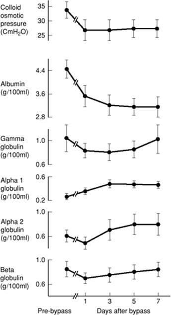
A common tendency is the desire to increase right and left ventricular filling pressures to their upper limits (16 to 20 mm Hg) to ensure adequate volume resuscitation. This approach exacerbates several problems that are difficult to manage later in the postoperative period, especially pulmonary dysfunction. Immediately after cardiopulmonary bypass, colloid oncotic pressure is reduced by as much as 50% and returns to normal over a 2-week period of time (Fig. 80.3) (15). In addition, neutrophil and endothelial activation following cardiopulmonary bypass increase pulmonary vascular permeability, resulting in a higher loss of intravascular fluids at lower hydrostatic pressures, promoting increased pulmonary interstitial water accumulation and pulmonary edema (16). We therefore caution against trying to maximize cardiac output by moving toward the upper pressure–volume limits along the Starling curve. Attempts to reach venous pressures greater than 16 mm Hg result in administration of excess fluids accompanied by increased pulmonary interstitial water.
Crystalloids and Colloids
Controversy persists concerning which type of fluids are most effective for volume resuscitation in post–cardiopulmonary bypass patients. The focus of this debate concerns the administration of crystalloid versus colloid (blood products, albumin, or hydroxyethyl starch) solutions (17). We have used multiple fluid combinations for maintaining adequate preload, all of which have advantages and disadvantages. Sole reliance on crystalloid solutions such as Ringer lactate for volume expansion may result in an inordinate amount of fluid administration to maintain adequate preload. Our current practice for volume resuscitation in patients without ongoing hemorrhage is to use up to 2 L of crystalloid solution after which 5% albumin may be used.
When the hematocrit is low (<22%) and the patient is bleeding or hemodynamically unstable, blood products are indicated. If red cell mass is adequate but coagulation factors are abnormal and bleeding continues, fresh frozen plasma and platelets should be considered. The threshold to give blood is lowered in patients with incomplete revascularization, low cardiac output, and signs of inadequate oxygen delivery such as low mixed venous oxygen saturation (S[v with bar above]O2).
Ventricular Afterload
Afterload reduction offers the most efficient means to improve cardiac output at little or no expense to myocardial oxygen demand. Following afterload reduction, ventricular volume, wall tension, and M[V with dot above]O2 usually are not increased. In some patients, the ejection fraction can be dramatically improved by a reduction in systemic vascular resistance (SVR) and peripheral vascular resistance (PVR). Afterload reduction should be performed after judicious volume loading to avoid hypotension and hemodynamic collapse. Afterload reduction typically begins in the operating room after the cessation of cardiopulmonary bypass (CPB) with the release of vasoactive mediators and with systemic rewarming. The extent of afterload reduction that is necessary greatly depends on ventricular performance. Patients with severely depressed systolic ventricular function benefit the most from afterload reduction.
|
|
|
Figure 80.3. The relationship between colloid osmotic (oncotic) pressure and plasma proteins during and after cardiopulmonary bypass. (Reproduced from Webber CE, Garnett ES. The relationship between colloid osmotic pressure and plasma proteins during and after cardiopulmonary bypass. J Thorac Cardiovasc Surg. 1973;65:234, with permission.) |
Vasodilators
Nicardipine hydrochloride, a peripheral acting calcium channel blocker, acts as a direct systemic arterial vasodilator. Its dose range is 1 to 15 mg per hour. It is our primary afterload-reducing agent because it is reasonably evanescent and avoids problems with cyanide toxicity associated with sodium nitroprusside. In our experience, it is less associated with pulmonary artery shunting and ventilation–perfusion (V-Q) mismatch (resulting in low PaO2) than sodium nitroprusside.
Sodium nitroprusside remains as an alternative agent for afterload reduction. It increases venous capacitance and directly vasodilates the systemic and pulmonary arterioles. Its advantage is its fast onset of action (15–20 seconds), allowing rapid titration (0.3–10 µg/kg per minute). It can be associated with cyanide toxicity as well as pulmonary artery shunting and V-Q mismatch.
Nitroglycerin (NTG) is a venous and arterial vasodilator but is primarily a preload-reducing agent with mild afterload-reducing effects including on the coronary arterial tree. An additional advantage in patients with right ventricular failure is its vasodilatory effects on the pulmonary vasculature. The intravenous dosage of NTG ranges between 0.5 and 2 µg/kg per minute. It decreases myocardial oxygen consumption and may be especially useful in patients with incomplete revascularization.
Pulmonary Artery Vasodilators
Certain patients benefit from vasodilators that act specifically on the pulmonary artery tree. Right ventricular failure is common in patients who have suffered right ventricular myocardial infarction and in patients who have undergone orthotopic heart transplantation as the implanted organ adjusts to its new setting of increased pulmonary artery vascular resistance. Patients who have had pulmonary endarterectomy may have reactive pulmonary artery constriction. Patients with a long-standing history of congestive heart failure also may have a component of reactive pulmonary artery hypertension following cardiopulmonary bypass in addition to fixed changes. In these scenarios, isolated right ventricular failure may be the primary cause of low cardiac output because of inadequate left ventricular filling.
The inhaled prostacyclin analogue iloprost is our preferred specific pulmonary artery vasodilator. An important advantage is its ease of administration. It can be delivered intermittently via a nebulizer to either intubated or extubated patients. Inhaled nitric oxide also has vasodilatory activity restricted to the pulmonary artery tree because of its short half-life. It requires continuous delivery via a specialized system and can be used only in intubated patients.
It is important to keep in mind the pulmonary vasoconstrictive effects of hypercapnia and hypoxia when managing patients with severe right ventricular dysfunction. Hypercapnia and hypoxia should be strictly avoided in patients with marginal right ventricular function, even if this requires prolonging the period of intubation. High FiO2 can be considered a form of pulmonary vasodilator in this subset of patients.
Vasoconstrictors
Cardiopulmonary bypass causes release of numerous vasoactive substances, many of which have vasodilatory effects (1). Vasodilation following cardiovascular surgery is especially common in patients who have had prolonged cardiopulmonary bypass, a period of circulatory arrest, or who have been treated with ACE inhibitors preoperatively (18,19). Various agents, including steroids and methylene blue, have been studied for the treatment of vasoplegic shock following cardiopulmonary bypass (20). Vasoconstrictors remain the first-line treatment for patients with low SVR and adequate volume status and contractile state, however (21). It is crucial that prior to instituting vasoconstrictor treatment, patients must be adequately volume resuscitated and their hypotension determined to be related to vasodilation rather than pump failure. Treatment of hypotension related to pump failure with a pure vasoconstrictor can lead to permanent end organ damage.
Patients ideally treated with vasoconstrictors are in a high cardiac output state with low SVR.
Phenylephrine hydrochloride, an α-specific catecholamine, acts as a pure peripheral vasoconstrictor. Its dose range is 20 to 180 µg/minute. It has a relatively short half-life, and its primary advantage is that it can be easily titrated for goal parameters, such as mean arterial blood pressure.
Vasopressin also acts as a pure peripheral vasoconstrictor. Because of its longer half-life, it is less suited for minute-to-minute changes in response to blood pressure changes. It may be particularly useful in patients treated preoperatively with ACE inhibitors. Its usual dose range is 1 to 10 units/hour. Its mechanism of action is synergistic with norepinephrine. Vasopressin in combination with norepinephrine is therefore useful in patients who have profound vasodilatory shock.
Contractility
The contractile state of the heart is a determinant of cardiac output, and patients with reversible myocardial stunning after aortic cross-clamping and cardiopulmonary bypass or pre-existing decompensated left ventricular function frequently benefit from a period of inotropic support. When required, inotropes are usually begun in the operating room at the time of weaning from CPB under guidance of intraoperative transesophageal echocardiography and are continued into the postoperative period. In the ICU, if cardiac performance is marginal and does not respond to other measures, we quickly institute inotropes and continue them throughout the first 24 to 48 hours. Remember that the specific conditions under which the operation was conducted, including repairs performed, completeness of revascularization, length of cardiopulmonary bypass, and quality of myocardial preservation can profoundly influence postoperative myocardial contractility.
Inotropes
Few pharmacologic agents affect only one determinant of cardiac performance, and this is especially true of inotropes. When selecting drugs, individually or in combination, bear in mind that a major goal after CPB is to gain cardiac output with as little increase in myocardial oxygen consumption as possible. The choice of drugs varies with the patient's condition.
Epinephrine, in the 0.01 to 0.10 µg/kg per minute range, is our principle choice. These dosage levels are not exceeded unless absolutely necessary to maintain an acceptable mean arterial perfusion pressure. Epinephrine also has vasoconstrictor and chronotropic effects; however, its inotropic effects predominate in this dose range. Epinephrine can be particularly useful when used to support right ventricular function.
Norepinephrine, also in the 0.01 to 0.10 µg/kg per minute range, is also frequently used. Because of increased α agonist, norepinephrine has stronger vasoconstrictor effects in this range and has been associated with improved coronary and splanchnic blood flow (22,23). Because of its vasoconstrictor properties, it can be especially useful in patients with vasodilatation after cardiopulmonary bypass who also require inotropic support.
Isoproterenol is a relatively weak inotrope with primarily chronotropic properties. In our ICU, its primary use is in heart transplant recipients in whom we are trying to cause relative tachycardia because of the normal coronary arteries, diastolic dysfunction, and right ventricular distention commonly encountered in this population. It can be used in dosages between 0.005 and 0.02 µg/kg per minute to drive heart rates into the 100 to 120 beats per minute range.
Milrinone is a phosphodiesterase inhibitor with potent inotropic and vasodilatory effects. It is commonly used in patients with pre-existing ventricular systolic dysfunction. It is given as a loading dose of 50 µg/kg (optional) followed by a continuous infusion of 0.1 to 0.75 µg/kg per minute. Because of its vasodilatory properties, it is important that patients be adequately volume resuscitated prior to its use. It frequently is given in combination with agents that have vasoconstrictor properties, such as norepinephrine, phenylephrine, or vasopressin.
Dopamine hydrochloride at dosages between 3 and 5 µg/kg per minute can be used to decrease SVR and augment cardiac contractility when a more potent catecholamine is not required. In this dosage range, dopamine also stimulates dopaminergic receptors in the kidney, thereby enhancing renal perfusion, although prevention of renal failure has not been documented.
Dobutamine in dosages between 3 and 5 µg/kg per minute has inotropic and vasodilatory effects similar to milrinone. At higher doses, it has α stimulatory effects and acts as a vasoconstrictor. Our practice has increasingly relied on epinephrine and milrinone as first-line inotropes rather than dopamine and dobutamine because of their perceived proarrhythmic effects. When selecting an inotropic regimen, remember that catecholamines at varying doses stimulate both β and α receptors. When we wish to avoid α receptor activity, multiple catecholamines are used synergistically to increase β receptor activity without stimulating α receptor activity.
Cardiac Output and Cardiac Index
A pulmonary artery catheter allows cardiac output measurement by the thermodilution method as well as facilitates calculation of SVR and PVR, pulmonary and intracardiac shunts, and estimation of left ventricular end-diastolic pressure (LVEDP) by PAOP. Cardiac output readings should always be normalized by conversion to cardiac index (cardiac output divided by the body surface area). This adjusts cardiac output for the size of the individual patient and therefore is a better estimate of adequacy of tissue perfusion. A cardiac index of 2.0 L/minute per m2 or greater is usually sufficient to maintain end organ performance. Exceptions exist in certain pathologic conditions, however. For example, patients with long-standing mitral valve disease and in a low cardiac output state preoperatively may well tolerate a cardiac index of less than 2.0 L/minute per m2 in the immediate postoperative period. In contrast, higher body temperature and agitation, or other concomitant conditions such as sepsis, may lead to cardiac index requirements of greater than 2.0 L/minute per m2. Bear in mind that these indices are at the lower limits for providing adequate organ function and that postoperative stress normally produces cardiac indices approaching 3.5 to 4.0 L/minute per m2. Cardiac output should not be the sole criterion used to determine adequacy of end organ oxygen delivery.
Mixed Venous Saturation
Continuous display of the S[v with bar above]O2 affords one the opportunity to observe acute as well as gradual changes in cardiac performance, increasing oxygen consumption ([V with dot above]O2), or both. Based on the Fick method of determining cardiac output, changes in the S[v with bar above]O2 reflect changes in systemic blood flow as long as [V with dot above]O2 remains stable. Acute changes in [V with dot above]O2 and cardiac performance are especially demonstrable during episodes of agitation, tracheal suctioning, and rapid extracellular volume shifts. S[v with bar above]O2 monitoring is particularly helpful in tracking the gradual deterioration of cardiac performance during the second 8-hour period after cardiopulmonary bypass. S[v with bar above]O2 varies with cardiac output, hemoglobin, SaO2, and oxygen consumption and is a balance between oxygen delivery and oxygen consumption.
Equipment used to measure physiologic parameters such as the arterial pressure and S[v with bar above]O2 must be calibrated regularly. S[v with bar above]O2 monitors are calibrated using venous saturation readings from a mixed venous blood sample from the pulmonary artery. The accuracy of any single measure of cardiac performance should always be evaluated in the context of other measures. For example, if the arterial pressure waveform is dampened, it may be compared against readings from an oscillometric, Doppler, or return-to-flow technique. Incongruent readings should always be evaluated with a suspicion for the accuracy of measurement.
Serum Lactate
As an indicator of anaerobic metabolism, serum lactate can provide information about the adequacy of cardiac performance in meeting the body's oxygen requirement. Several problems, however, limit the usefulness of serum lactate as a means to assess cardiac performance. On arrival to the ICU, lactate values may range between normal (<2.0 mmol/L) to as high as 8 to 9 mmol/L. As body temperature rises to normal, serum lactate may actually rise despite improving hemodynamics because underperfused vascular beds open and release lactate into the systemic circulation. The difficulty in this situation is whether to attribute a rise in serum lactate to a washout phenomenon or to a diminution of cardiac performance. Evaluation of the cardiac index, S[v with bar above]O2, and SVR in conjunction with serial serum lactate determinations helps to resolve this dilemma. Lactate levels should decrease after 8 hours if perfusion is adequate.
Serum Ionized Calcium and Potassium
Rapid access (5–10 minutes) to laboratory determinations or bedside point-of-care testing is essential for guiding therapy, especially in the first 24 hours. Important serum values include ionized calcium (Ca2+), potassium, hematocrit, glucose, and sodium. Serial measurement of Ca2+ is especially helpful when solutions containing protein such as blood, fresh frozen plasma, albumin, or plasma protein solutions are administered. Protein binds calcium and may diminish that which is available for myocardial function. Clinically, this phenomenon can be recognized during the rapid infusion of protein-containing solutions by noting a rise in cardiac filling pressures, a drop in blood pressure, and a drop in the S[v with bar above]O2, all indicative of worsening performance despite volume resuscitation. In certain situations, particularly in hemodynamically unstable patients, simultaneous infusion of calcium chloride markedly improves cardiac performance.
Postoperative maintenance of adequate serum potassium is crucial, especially after the usually brisk diuresis following CPB and in conjunction with diuretic therapy. Potassium chloride should be considered a first-line agent for prevention and treatment of arrhythmias after cardiovascular surgery. Potassium chloride should be given as a standing medication to actively diuresing patients with normal renal function, and serum potassium values should be measured frequently to guide replacement therapy toward a normal to high-normal range (4.5–5 mmol/L).
Urine Output
In the initial postoperative period, urine output is typically brisk but then diminishes dramatically between the 8th and 12th post-CPB hours. Initially, urine output alone should not be relied on to judge cardiac performance, because it is influenced by numerous variables including high serum glucose, denatured plasma protein fractions, and diuretics such as furosemide and mannitol used during cardiopulmonary bypass. In later stages of the postoperative period, urine output may be influenced by various stress hormones such as antidiuretic hormone, aldosterone, and cortisol, which generally cause conservation of intravascular volume. Similarly, conservation of intravascular volume occurs in patients who enter surgery with high ventricular end-diastolic filling pressures but return to the ICU after surgery with lower filling pressures. In patients who are conditioned to higher filling pressures, the sudden reduction in atrial pressure usually sets off a strong antidiuretic hormone–mediated response designed to correct perceived hypovolemia and leads to fluid retention and low urine output.
Almost all patients return from the operating room with excess total body water because of the hemodilution and obligate volume load caused by the bypass circuit. These effects have been partially ameliorated by cell saver systems and ultrafiltration units that reconcentrate red cell mass. Despite the usual excess in total body water, intravascular volume status is variable because of third-spacing related to increased capillary permeability and post-cardiopulmonary bypass inflammation. Diuretic therapy should therefore be guided by cardiac filling pressures. Our practice is to diurese patients to low normal filling pressures as long as hemodynamics are not compromised to minimize interstitial lung water and improve pulmonary function. Diuresis should be performed cautiously during the early postoperative period when ventricular compliance is poor and cardiac function may be especially preload dependent. This is particularly true in patients with concentric ventricular hypertrophy, such as those with long-standing hypertension or aortic stenosis.
Special Concerns
Bleeding
Bleeding after cardiovascular operations with cardiopulmonary bypass is a major destabilizing complication. Severe postoperative bleeding occurs in 3% to 5% of patients who undergo cardiopulmonary bypass (24). Significant progress has been made in understanding its causes and optimal treatment.
Causes
Although inadequate surgical hemostasis should always be suspected, at least half of patients with severe postoperative bleeding have acquired hemostatic defects (25,26). These include activation of fibrinolysis, decreased levels of clotting factors due to dilution and consumption, and transient platelet dysfunction. Therapeutic protocols may influence postoperative bleeding. For example, chronic preoperative use of antiplatelet agents such as clopidogrel and aspirin are associated with increased postoperative transfusion requirements.
Treatment
Antifibrinolytic agents, such as epsilon aminocaproic acid and aprotinin, have been shown to decrease the need for transfusion in high-risk subpopulations (27,28). Aprotinin is currently not available until further studies are done to determine its side effects. These agents are begun intraoperatively and discontinued shortly on arrival to the ICU. There is no evidence supporting initiation of their use in the postoperative period. When bleeding does occur, blood component replacement is guided by laboratory studies. Red blood cell transfusions should be initiated promptly when massive hemorrhage is suspected. Clotting factor deficiencies demonstrated by prolonged prothrombin and activated partial thromboplastin times are treated with fresh frozen plasma. Cryoprecipitate is usually reserved for patients with low fibrinogen levels. Factor replacement therapy with recombinant clotting factors such as activated factor VII and factor XIII is an emerging strategy under current study (29,30).
Isolated elevation of the activated partial thromboplastin time may indicate heparin rebound and should be reversed with protamine sulfate administration (25). Circulating heparin levels are also useful in guiding protamine administration (31).
Platelet counts of greater than 100,000 may seem reassuring; however, the function of these platelets is frequently inadequate in patients with clinical coagulopathy. Point-of-care platelet function assays may be useful for directing platelet transfusion in patients without thrombocytopenia.
Our threshold for returning the patient to the operating room is a blood loss rate through the chest tubes of greater than 3 mL/kg per hour for several consecutive hours. Patients who experience bleeding at this rate cannot be stabilized hemodynamically, irrespective of the amount of volume replacement. Patients with brisk hemorrhage should be returned to the operating room even before coagulopathy is corrected to stop presumed surgical bleeding prior to development of cardiac tamponade and hemodynamic collapse. Even when no source of surgical bleeding is identified on mediastinal re-exploration, clinical coagulopathy frequently improves after evacuation of the mediastinal hematoma.
Cardiac Tamponade
Mediastinal drains may fail to adequately evacuate blood after cardiovascular surgery, leading to cardiac tamponade. Abrupt cessation of bleeding from the mediastinal drains warrants close attention to the cardiac filling pressures and to overall cardiac performance. When flexible drains have been used, they should be periodically milked to prevent clotting off.
Two fairly reliable signs of cardiac tamponade are increased or exaggerated cycling of the systolic blood pressure during positive pressure ventilation, and equalization of right and left atrial and pulmonary artery diastolic pressures. A reduction in S[v with bar above]O2 and a decrease in urine output will also be observed. The diagnosis may be confirmed by echocardiography.
In cardiac tamponade, equalization of the atrial pressures does not always occur. Blood clots on the acute margin of the right ventricle may substantially affect cardiac performance without atrial pressure equalization. In severe low-output states, cardiac tamponade must be ruled out by either mediastinal exploration in the operating room or a limited opening of the lower portion of the chest incision at the bedside. Percutaneous drainage is not useful in the immediate postoperative period because the hematoma is generally clotted.
Thrombocytopenia
Thrombocytopenia is common following cardiovascular surgery (25). Causes include platelet activation and consumption during cardiopulmonary bypass, mechanical destruction by intravascular devices including prosthetic heart valves and intra-aortic balloon pumps (IABPs), inadequate production caused by malnutrition and drugs such as milrinone, and heparin-induced thrombocytopenia. Platelet counts of less than 100,000/µl are quite typical and are generally managed conservatively by removal of offending agents as soon as is feasible. If the platelet count falls by greater than 50% or a thrombotic event occurs between 4 and 14 days of heparin exposure, a diagnosis of heparin-induced thrombocytopenia (HIT) should be excluded by testing for HIT antibody seroconversion. If HIT is diagnosed or is strongly suspected, anticoagulation with a nonheparin anticoagulant such as lepirudin or bivalirudin should be considered because of the risk of thrombotic complications (32). Patients should be screened for deep vein thrombosis (DVT) with ultrasonography. For patients who require long-term anticoagulation for DVT, warfarin can be started once platelet levels return to a normal range. Warfarin is not recommended for initial therapy of HIT due to the risk of thrombotic complications.
Ventilator Management
Ventilator management after cardiac surgery is directed toward optimizing pulmonary function without compromising hemodynamics. On patient admission to the ICU in our institution, the synchronous intermittent mandatory ventilation (SIMV) rate is set at 14 breaths per minute with a tidal volume of 8 mL/kg of predicted body weight. We do not pressure-limit the ventilator, and we carefully monitor hemodynamic function throughout the period of assisted ventilation. Acute hemodynamic changes may be indicative of patient distress, a blocked endotracheal tube, the need for suctioning, or a bronchodilator such as albuterol. Patient dyssynchrony with the ventilator should prompt assessment of the patient's level of sedation and analgesia.
Caution is advised when using positive end-expiratory pressure (PEEP) above 8 to 10 cm H2O, because cardiac chamber filling may be affected causing cardiac performance to decrease. When high PEEP is used, intravascular volume augmentation may be necessary to raise the effective LVEDP. Although some use the estimate that one half the PEEP subtracted from the PAOP is the effective LVEDP, this assumption may not be correct since the amount of pressure transmitted through diseased lungs may vary.
Administration of nitroprusside and to a lesser extent nicardipine to patients who are heavy smokers or have severe chronic lung disease may increase the amount of pulmonary shunting and cause hypoxia due to V-Q mismatch. Bronchodilators and PEEP as high as 15 to 20 cm H2O rarely correct this problem. In such cases, these vasodilators should be discontinued and substituted with another drug such as nitroglycerin (3 to 5 µg/kg per minute) or hydralazine.
The appearance of the acute respiratory distress syndrome (ARDS) after cardiac surgery is much less frequent due, in part, to shorter CPB times and the use of membrane oxygenators. Unfortunately, when ARDS does occur in the postoperative period, it may be accompanied by multiple organ dysfunction syndrome or sepsis (33).
Diuretics
Diuresis is desirable during the recovery period to reverse hemodilution and improve pulmonary function, but should always be closely monitored. Excessive diuretic therapy rapidly depletes intravascular volume if the renal response to these drugs is brisk. Forced diuresis that results in a urine output of 500 to 1,500 mL over several hours may significantly reduce cardiac performance and promote increased SVR.
Smaller and more frequent doses of diuretics produce a more constant diuresis and the ability to maintain a gradual negative fluid balance throughout the second 24-hour postoperative period. A sustained diuresis that prevents large hourly urine loss spares the patient rapid volume shifts. To accomplish a stable diuresis in patients with normal ventricular and renal function, our practice is to use frequent doses of relatively small doses of furosemide (10–20 mg) every 4 to 8 hours as needed. In patients with marginal ventricular or renal function, particularly those who have been treated with furosemide chronically, more aggressive treatment is necessary. In this situation we use a continuous furosemide infusion at doses of 0 to 20 mg/hour. We state a 24-hour diuresis goal and titrate the infusion to meet this goal over the course of the day with reassessments to avoid hypovolemia. Intermittent doses of thiazide diuretics are a useful adjunct in patients who have an inadequate response to the infusion.
Renal Failure
Patients who experience an extremely low cardiac output or hypotension during the first 24 hours after cardiopulmonary bypass may develop acute tubular necrosis. Most respond to adequate volume maintenance and diuretic therapy and can be maintained in a nonoliguric renal failure. However, some, especially those with a preoperative creatinine clearance of less than 50 mL/minute, may develop oliguric or anuric renal failure.
Diuretics
Patients with oliguria should be given a test bolus of furosemide (60–120 mg) and then be evaluated for a urine response of greater than 50 mL/hour. A continuous furosemide infusion at doses of 0 to 20 mg/hour can then be maintained with intermittent doses of thiazide diuretics as necessary. This regimen usually maintains urine output and avoids gross volume overload after surgery, even if acute tubular necrosis is present. Avoiding hypovolemia is also essential.
Ultrafiltration
Patients with renal failure and volume overload may be candidates for ultrafiltration to remove excess salt and water (34). There are several advantages to this approach for patients who fail initial diuretic therapy. Commercially available systems are highly automated and allow device-based removal of salt and water from the circulation without clinically significant effects on hemodynamics or electrolyte balance. Standard central or peripheral venous catheters can be used for access, frequently avoiding the need for invasive procedures to place specialized hemodialysis catheters. We reserve this approach for patients with adequate electrolyte and urea clearance who require removal of excess water.
Dialysis
Intravascular volume overload and inadequate clearance of potassium levels are the principal indications for dialyzing a patient with acute renal failure in the immediate postoperative period. The overall goal of dialysis is to remove excess water and solutes while maintaining cardiovascular stability. For the postoperative cardiovascular surgery patient, this is best achieved with the use of continuous venovenous hemodialysis (CVVHD). This technique is easily initiated, offers good clearance, and allows accurate control of ultrafiltration that is adaptable to the patient's hemodynamic requirements. Fluid removal can therefore usually be accomplished while maintaining stable hemodynamics. CVVHD does require placement of a dedicated double-lumen central venous catheter. If postoperative bleeding is not a concern, heparin should be administered to prolong filter life. Without heparin, the average filter life ranges from 16 to 24 hours.
Patients should be transitioned to conventional hemodialysis only after a significant period of myocardial recovery. Conventional hemodialysis requires blood flow rates of at least 200 mL/hour and results in large volumes of fluid removal over a several-hour period. The fluid shifts associated with this technique are inappropriate for patients early in their recovery from cardiopulmonary bypass.
Central Nervous System Complications
After operations with cardiopulmonary bypass, the historical incidence of cerebrovascular accidents (CVAs) with focal neurologic sequelae is 2% to 5% (35). The contemporary rate may be lower because of advancements in intraoperative technique including routine use of epiaortic ultrasound (36). Advanced age, history of CVA, peripheral vascular disease, valvular heart surgery, and procedures requiring a period of circulatory arrest are associated with a higher incidence of central nervous system events. Patients with a history of CVA whose focal deficits have resolved prior to operation may have unmasking of their old symptoms in the immediate postoperative period. Untoward psychological and cognitive sequelae are more prevalent than focal CVAs (37,38). The precise cause of neuropsychological complications remains unclear, however recent data suggest that microemboli, especially air, are related to both severe and subtle postoperative neuropsychological deficits (39,40).
Patients should be continuously monitored for the appearance of neuropsychological complications in the postoperative period. From the time of their emergence from anesthesia, patients should be assessed for focal motor deficits and the ability to comprehend and follow simple commands. Focal deficits should be evaluated promptly with noncontrast computed tomography (CT) of the brain to rule out hemorrhagic mass lesions that may require intervention. Magnetic resonance imaging (MRI) may be useful to identify smaller embolic foci that are not apparent on head CT. Fresh post-cardiopulmonary bypass patients are usually poor candidates for thrombolysis because of their risk for bleeding. The treatment for major embolic CNS events is usually supportive. Higher blood pressures may lead to greater perfusion via collaterals in patients with severe occlusive cerebrovascular disease.
Delirium
Postcardiotomy delirium is typically preceded by a lucid interval, usually 36 to 48 hours from the time patients awaken from anesthesia. This syndrome is characterized by confusion, disorientation, and disordered thinking and perception (37). Severe manifestations may include visual and auditory illusions, hallucinations, and paranoid ideation (41). Treatment consists of undisturbed rest, frequent reorientation, ensuring patient safety, reassurance to both the patient and family, and treatment with haloperidol. It is our practice to avoid the use of benzodiazepines in this situation because of their paradoxical excitatory response seen occasionally in the elderly population. Subtle changes in mentation and thinking processes occur frequently during the first few weeks after surgery. They may be frightening to the patient and may interfere with the immediate recovery process. However, most of these problems are temporary and usually resolve by the sixth postoperative month (37,38,42).
Sedation and Paralysis
In addition to the pain and discomfort characteristic of any major surgical procedure, several conditions occurring after cardiac operation warrant sedation and, in rare instances, complete paralysis. As the patient emerges from anesthesia, a perceived reduction in cardiac performance may result, in part, from shivering, agitation, or both. Shivering can produce a marked increase in [V with dot above]O2, a reduction in S[v with bar above]O2, and an overall imbalance of oxygen supply and demand in the body, especially in low cardiac output states. Once gross neurologic function is assessed (e.g., movement and sensation in the extremities and the ability to follow simple commands), shivering or agitation in the immediate postoperative period is best treated with narcotic agents. Our preference is to use fentanyl to control shivering or agitation, although these conditions can be managed with morphine or other narcotics. Meperidine is particularly efficacious, although the mechanism by which it acts is unknown.
For patients who have undergone elective procedures and are expected to be extubated early in the postoperative course, a propofol infusion is started soon on arrival to the ICU. This agent is useful in this setting because once hemodynamics and bleeding are assessed and the patient is warm, the patient's sedation can be stopped quickly because of the agent's short half-life. The patient's neurologic function can be assessed, and the patient can be extubated after a brief spontaneous breathing trial.
When a patient is expected to require a prolonged period of mechanical ventilation, we typically use a combination of narcotic and benzodiazepine infusions. Fentanyl is our preferred narcotic because of its limited effects on myocardial contractility and the peripheral vasculature. In rare instances, such as severe pulmonary artery hypertension and right heart failure, a paralytic agent may be indicated to prevent any respiratory effort by the patient, which can trigger increased pulmonary artery pressures and precipitate hemodynamic collapse. Because of their association with critical illness neuropathy, we prefer to avoid paralytic agents and treat these patients with deep sedation. When paralytics are required, our practice is to use vecuronium, which has a lesser chronotropic effect than pancuronium.
Mechanical Cardiac Assistance
Mechanical cardiac assist devices include IABPs, centrifugal blood pumps, ventricular assist devices, and total artificial hearts. In some cases, extracorporeal membrane oxygenation (ECMO) is indicated for profound cardiorespiratory failure.
Intra-aortic Balloon Pumps
An IABP should be considered when patients fail to respond to moderately high-dose inotropic drug combinations such as epinephrine and milrinone. The IABP improves cardiac performance primarily by afterload reduction and augmentation of systemic diastolic pressure, thereby increasing coronary artery perfusion pressure. IABP therapy should be considered earlier rather than later and can be instituted quickly at the bedside via a percutaneous technique. Its main contraindications are aortic insufficiency and aortoiliac occlusive disease. Complications and adverse effects include platelet consumption, catheter sepsis, and lower extremity ischemic complications.
Ventricular Assist Devices and Total Artificial Hearts
Ventricular assist devices (VADs) replace the pumping function of the right or left ventricle, or both. VADs may be used to support patients until myocardial recovery occurs or until cardiac transplantation can be performed. For patients with end-stage congestive heart failure who are not candidates for transplantation, a VAD may be placed as destination therapy. Several different VAD systems are FDA-approved and marketed for these various applications. Currently, total artificial heart therapy remains experimental and is limited to patients who are treated on research protocols.
Blood Glucose Control
There has been increasing emphasis on maintaining tight blood glucose control in critically ill patients because of demonstrated reductions in morbidity and mortality. The benefits of tight glucose control have been demonstrated in cardiac surgery patients specifically (43,44,45). Maintenance of tight glucose control reduces the incidence of mediastinitis, atrial fibrillation, ischemia, length of stay, hospital costs, and mortality and has been shown to benefit both diabetic and nondiabetic patients following cardiovascular surgery. In our practice, we have instituted an intensive intravenous insulin therapy protocol with a goal of maintaining normoglycemia (blood glucose 80–110 mg/dL). A continuous insulin infusion is typically started intraoperatively, and we have found all adult patients to require intravenous insulin at least through the immediate postoperative period if normoglycemia is to be maintained.
The requirement for intravenous insulin is reduced as inotropes, particularly epinephrine, are weaned. Once patients begin taking an oral diet, we supplement the intravenous insulin infusion with a fast-acting insulin formulation given subcutaneously. The dose of insulin aspart is based on the patient's oral intake and intravenous insulin requirement over the preceding 12 hours. Patients are subsequently transitioned to a long-acting insulin formulation, such as NPH, given subcutaneously three times a day to cover basal needs in addition to fast-acting insulin, which is still based on percentage of meals consumed. Nondiabetic patients typically have a progressively decreasing insulin requirement during their hospital recovery and are not discharged on any insulin. We have found this regimen to provide improved blood glucose control while also decreasing the frequency and severity of episodes of hypoglycemia and hyperglycemia.
Catheter Sepsis
Bloodstream infections have particularly significant implications in cardiovascular surgery patients as prosthetic intravascular devices, such as heart valves and arterial conduits, are frequently implanted during surgery. Patients typically return from the operating room following cardiovascular surgery with multiple invasive monitoring catheters including central lines, pulmonary artery catheters, arterial lines, and urinary catheters. The incidence of sepsis increases when invasive monitoring catheters are left in place for longer than 72 hours (46). For straightforward cases, our goal is to have all monitoring catheters removed by this time. All peripheral intravenous lines are also removed at the time of the patient's transfer to the intermediate care floor and a new peripheral intravenous line is placed. If a patient remains critically ill and still requires invasive monitoring, we observe the patient closely for signs of sepsis such as fever and leukocytosis, and have a low threshold to remove old lines. We avoid rewiring catheters and prefer to insert new catheters at a different site. Cultures are drawn at this time, and vancomycin is started if clinical suspicion for catheter sepsis is high. Antibiotic coverage is narrowed or discontinued based on subsequent culture results.
Clinical Pathways
Clinical pathways have been increasingly used by health care systems to improve the continuity and coordination of care for patients being treated for many diseases. They frequently are developed around procedures, and patients undergoing coronary artery bypass grafting (CABG) procedures were among the first to benefit from their implementation. CABG procedures are common, usually elective, have typical recovery milestones, and require coordination of multidisciplinary provider teams. These characteristics make CABG pathways extremely useful to support both clinical and administrative management.
Many institutions have implemented clinical pathways for other cardiovascular procedures including heart valve repair or replacement and heart transplantation. Clinical pathways typically are based around a timeline, categories of care such as nursing and physical therapy, and a variance record that allows deviations to be incorporated. When successfully implemented, they encourage the use of clinical guidelines, improve multidisciplinary teamwork, reduce variations in patient care, and improve clinical outcomes. It is important that clinical pathways not discourage personalized care and that they function well even in the face of unexpected changes in a patient's condition.
Cardiac Surgery Databases
Institutional and multi-institutional databases are useful tools with which to track outcomes following cardiovascular operations, conduct clinical research, and guide continuous quality improvement. These data are also used by various regulatory bodies and third-party payers to monitor health care quality and costs. It will also be used for pay-for-performance initiatives in the future. The data collected in these databases is clinical, administrative, or a combination of both. The Society of Thoracic Surgeons Cardiac Database is the largest clinical cardiothoracic surgery database in the world and currently has more than 500 participating sites. The large volume of clinical information collected allows risk modeling for common procedures and provides individual institutions with comparative outcomes data and national benchmarks. All cardiac databases are limited by the variable quality of collected data, which in many instances is self-reported. Comparison of outcomes is also subject to selection bias not corrected by imprecise risk models.
Special Clinical Scenarios
Off-pump Coronary Artery Bypass Surgery
Over the last 10 years, there has been increasing interest in performing CABG surgery without the use of cardiopulmonary bypass. This growth has been largely driven by increasing recognition of the deleterious effects of cardiopulmonary bypass and the desire to avoid the diffuse inflammatory response, multiorgan dysfunction, and neurocognitive complications that may follow (Table 80.3) (47,48,49,50,51,52,53,54,55). Approximately 25% of CABGs performed in the United States are performed off-pump, and some centers report a significantly higher percentage of off-pump coronary artery bypass procedures (OPCABs). Even among surgeons not routinely performing coronary revascularization off-pump, there are newly recognized clinical scenarios, such as a patient with severe atherosclerosis of the ascending aorta, for whom use of OPCAB techniques is strongly favored. OPCAB itself is a facilitating technology for surgeons developing minimally invasive approaches to coronary revascularization. OPCAB has therefore evolved into a requisite component of modern cardiovascular surgery practice.
|
Table 80.3 Benefits of Off-Pump Coronary Artery Bypass Grafting Demonstrated in Prospective, Randomized Studies |
|||||||||
|
The postoperative care of patients who have undergone OPCAB surgery does not differ in many respects from that of patients undergoing conventional CABG surgery. There are several important differences, however, that providers must be aware of to take advantage of the opportunity for expedited care that OPCAB surgery can offer. Patients who have undergone OPCAB surgery have a decreased need for inotropic support in the postoperative period, most likely because of avoidance of global ischemia and reduced myocardial stunning. The intravenous fluid requirement for patients who have undergone OPCAB surgery is also reduced because the systemic inflammatory response and capillary leak related to CPB is avoided. Massive volume resuscitation in the early postoperative period should be avoided in favor of low-dose vasopressors that may be necessary secondary to intravenous sedation. OPCAB patients typically are volume loaded intraoperatively and are more likely to be euvolemic in the immediate postoperative period than conventional CABG patients.
Postoperative hemorrhage and transfusion requirements are reduced in OPCAB patients because of reduced fibrinolytic pathway activation and coagulation factor and platelet consumption. We do not routinely check platelet counts or clotting times in these patients in the immediate postoperative period for this reason. Persistent or massive hemorrhage in the postoperative period should prompt early evaluation for surgical bleeding because this is unlikely to be related to factor or platelet deficiency in an OPCAB patient.
Patients who have undergone OPCAB surgery are in a relatively hypercoagulable state as opposed to manifesting the coagulopathy that is typical after CPB. This state has the potential to adversely affect graft patency in the postoperative period. After surgery we continue aspirin administration daily as with patients undergoing conventional CABG surgery. In addition, we start clopidogrel, 75 mg/day, in the immediate postoperative period once chest tube drainage has been low for 3 consecutive hours.
One of the major benefits of OPCAB surgery to the healthcare system is the potential for OPCAB patients to have reduced resource use. An area where this advantage can be exploited is in reducing length of mechanical ventilation and ICU stay. To realize this benefit for the patient and the health system, providers should be cognizant of this and be immediately prepared to wean and extubate patients as their need for mechanical assistance is diminished. With appropriate anesthesia planning and staffing, patients can generally be extubated in the operating room after an OPCAB procedure or within 30 minutes of arrival in the ICU. When this is not feasible, clinical pathways that set objective criteria and goals facilitate the timely progression of ventilator weaning and extubation that minimize patient exposure to ventilator-related complications and maximize efficiency and cost-effectiveness.
Thoracic Aortic Surgery
Patients undergoing surgery for disorders of the thoracic aorta present critical care providers with several unique challenges, including neurologic complications, hemodynamic disturbances, and malperfusion syndromes. Several of these conditions are caused by the period of circulatory arrest that the patient undergoes during the conduct of the surgical repair whereas others are caused by the specific anatomy of the underlying thoracic aortic pathology.
Circulatory Arrest Sequelae
Patients undergoing aortic arch procedures usually have a period of circulatory arrest during their operation. This increases their risk for neurologic complications, particularly psychological and cognitive disorders. It is typical for elderly patients who require prolonged hypothermic circulatory arrest (greater than 30 minutes) to have neurologic dysfunction resulting in agitation and delirium. Although this finding is usually transient, it may necessitate a several-day period of supportive care during which the patient requires mechanical ventilation.
Circulatory arrest is also associated with increased bleeding and transfusion requirement related to profound coagulopathy (56,57). Profound coagulopathy is especially problematic in this patient group because of the long suture lines used in large-vessel surgery. Patients presenting with acute aortic dissections have attenuated and friable aortic tissue, making vascular anastomoses potentially tenuous. Coagulopathy following emergency thoracic aortic surgery for dissection should be treated aggressively as described earlier in this chapter. Surgical bleeding should always be suspected in patients with brisk hemorrhage. Avoidance of hypertension is desirable in patients with long suture lines and friable aortic tissue.
Patients who have undergone complex vascular repairs with long bypass times and circulatory arrest also may develop profound vasodilatory shock that is recalcitrant to combination therapy with multiple vasopressors. The cause for this is unclear but may be related to bacterial translocation of intestinal flora (58,59,60). Patients who exhibit this should be adequately volume resuscitated and supported with vasopressors and inotropes if poor myocardial contractility is contributing to the hypotension. We have also used intravenous steroids and methylene blue in patients with profound vasodilation (20,61,62,63). Addition of these agents has allowed reduction of high doses of vasopressors that can lead to ischemic complications.
Spinal Cord Ischemia
Spinal cord ischemia and paralysis is a particularly devastating complication following thoracoabdominal aneurysm repair. Spinal cord ischemia occurs at the time of thoracoabdominal aneurysm repair and is exacerbated by postoperative hypoperfusion, particularly when the variable origin of the artery of Adamkiewicz is not adequately reconstituted. This results in an anterior spinal artery syndrome with impaired lower extremity motor function and, to a lesser degree, impaired sensory function. Patients should be monitored closely for these findings in the immediate postoperative period. When spinal cord ischemia is suspected, a lumbar drain should be placed if one was not already placed intraoperatively. Cerebrospinal fluid (CSF) should be drained for a CSF pressure of 10 mm Hg or greater, and the mean arterial blood pressure should be raised to 85 to 100 mm Hg using vasopressors to increase the perfusion pressure to the spinal cord. These maneuvers have led to reversal of acute lower extremity neurologic findings in patients following thoracoabdominal aneurysm repair. MRI is useful for confirming the diagnosis of spinal cord ischemia. Treatment as outlined above should not be delayed until after MRI scanning if spinal cord ischemia is suspected.
Malperfusion Syndromes
Malperfusion syndromes occur in patients with aortic dissection as a result of obstruction of arterial branch orifices by the dissected intimal flap or inadequate perfusion via the false lumen of the aorta. Patients presenting with aortic dissection can develop malperfusion of virtually any limb or end organ, depending on the anatomic extent of the dissection. Patients with ascending aorta involvement (Stanford type A) typically undergo emergency surgery whereas patients with only descending aorta involvement (Stanford type B) are treated initially with medical therapy. Even patients who have undergone repair of ascending aortic dissection are frequently left with residual dissection involving the descending aorta. Therefore, providers must remain vigilant for the development of malperfusion syndromes in both patient groups. Patients admitted to the ICU with dissection should undergo serial peripheral pulse, neurologic, and abdominal exams for evidence of malperfusion. Serial arterial blood gases, liver enzymes, and lactate levels are useful when visceral malperfusion is suspected.
We have a low threshold for performing angiography when malperfusion is suspected. If confirmed by this study, interventional techniques such as fenestration or stenting can frequently be used to treat malperfusion during the same procedure. Laparotomy may be occasionally required for patients with visceral malperfusion who progress to bowel infarction.
References
1. Hall RI, Smith MS, Rocker G. The systemic inflammatory response to cardiopulmonary bypass: pathophysiological, therapeutic, and pharmacological considerations. Anesth Analg. 1997;85:766.
2. Steinberg JB, Kapelanski DP, Olson JD, et al. Cytokine and complement levels in patients undergoing cardiopulmonary bypass. J Thorac Cardiovasc Surg. 1993;106:1008.
3. Miller BE, Levy JH. The inflammatory response to cardiopulmonary bypass. J Cardiothor Vasc Anesth. 1997;11:355.
4. Lobato EB, Gravenstein N, Martin TD. Milrinone, not epinephrine, improves left ventricular compliance after cardiopulmonary bypass [see comment]. J Cardiothor Vasc Anesth. 2000;14:374.
5. Kouchoukos NT, Karp RB. Management of the postoperative cardiovascular surgical patient. Am Heart J. 1976;92:513.
6. Reinhart K, Kersting T, Fohring U, et al. Can central-venous replace mixed-venous oxygen saturation measurements during anesthesia? Adv Exp Med Biol. 1986;200:67.
7. Reinhart K, Rudolph T, Bredle DL, et al. Comparison of central-venous to mixed-venous oxygen saturation during changes in oxygen supply/demand. Chest. 1989;95:1216.
8. Halonen J, Hakala T, Auvinen T, et al. Intravenous administration of metoprolol is more effective than oral administration in the prevention of atrial fibrillation after cardiac surgery. Circulation. 2006;114:I1.
9. Katariya K, DeMarchena E, Bolooki H. Oral amiodarone reduces incidence of postoperative atrial fibrillation. Ann Thor Surg. 1999;68:1599.
10. Kurz DJ, Naegeli B, Kunz M, et al. Epicardial, biatrial synchronous pacing for prevention of atrial fibrillation after cardiac surgery. Pacing Clin Electrophysiol. 1999;22:721.
11. Patti G, Chello M, Candura D, et al. Randomized trial of atorvastatin for reduction of postoperative atrial fibrillation in patients undergoing cardiac surgery: results of the ARMYDA-3 (Atorvastatin for Reduction of Myocardial Dysrhythmia After cardiac surgery) study. Circulation. 2006;114:1455.
12. Stamou SC, Hill PC, Sample GA, et al. Prevention of atrial fibrillation after cardiac surgery: the significance of postoperative oral amiodarone. Chest. 2001;120:1936.
13. Spaan JA, Piek JJ, Hoffman JI, Siebes M. Physiological basis of clinically used coronary hemodynamic indices. Circulation. 2006;113:446.
14. Kouchoukos NT, Sheppard LC, McDonald DA, et al. Estimation of stroke volume from the central arterial pressure contour in postoperative patients. Surg Forum. 1969;20:180.
15. Webber CE, Garnett ES. The relationship between colloid osmotic pressure and plasma proteins during and after cardiopulmonary bypass. J Thor Cardiovasc Surg. 1973;65:234.
16. Ng CS, Wan S, Yim AP, et al. Pulmonary dysfunction after cardiac surgery. Chest. 2002;121:1269.
17. Gallagher JD, Moore RA, Kerns D, et al. Effects of colloid or crystalloid administration on pulmonary extravascular water in the postoperative period after coronary artery bypass grafting. Anesth Analg. 1985;64:753.
18. Morales DL, Garrido MJ, Madigan JD, et al. A double-blind randomized trial: prophylactic vasopressin reduces hypotension after cardiopulmonary bypass. Ann Thorac Surg. 2003;75:926.
19. Weis F, Kilger E, Beiras-Fernandez A, et al. Association between vasopressor dependence and early outcome in patients after cardiac surgery. Anaesthesia. 2006;61:938.
20. Shanmugam G. Vasoplegic syndrome—the role of methylene blue. Eur J Cardiothorac Surg. 2005;28:705.
21. Masetti P, Murphy SF, Kouchoukos NT. Vasopressin therapy for vasoplegic syndrome following cardiopulmonary bypass. J Cardiac Surg. 2002;17:485.
22. Di Giantomasso D, Morimatsu H, May CN, et al. Increasing renal blood flow: low-dose dopamine or medium-dose norepinephrine. Chest. 2004;125:2260.
23. Nikolaidis LA, Trumble D, Hentosz T, et al. Catecholamines restore myocardial contractility in dilated cardiomyopathy at the expense of increased coronary blood flow and myocardial oxygen consumption (MvO2 cost of catecholamines in heart failure). Eur J Heart Fail. 2004;6:409.
24. Eagle KA, Guyton RA, Davidoff R, et al; American Heart Association Task Force on Practice, and American Society for Thoracic Surgery and the Society of Thoracic Surgeons. ACC/AHA 2004 guideline update for coronary artery bypass graft surgery: summary article: a report of the American College of Cardiology/American Heart Association Task Force on Practice Guidelines (Committee to Update the 1999 Guidelines for Coronary Artery Bypass Graft Surgery) [erratum in Circulation. 2005 Apr 19;111(15):2014]. Circulation. 2004;110:1168.
25. Linden MD. The hemostatic defect of cardiopulmonary bypass. J Thromb Thrombolys. 2003;16:129.
26. Woodman RC, Harker LA. Bleeding complications associated with cardiopulmonary bypass. Blood. 1990;76:1680.
27. Slaughter TF, Faghih F, Greenberg CS, et al. The effects of epsilon-aminocaproic acid on fibrinolysis and thrombin generation during cardiac surgery. Anesth Analg. 1997;85:1221.
28. Smith PK, Datta SK, Muhlbaier LH, et al. Cost analysis of aprotinin for coronary artery bypass patients: analysis of the randomized trials. Ann Thorac Surg. 2004;77:635.
29. Bishop CV, Renwick WE, Hogan C, et al. Recombinant activated factor VII: treating postoperative hemorrhage in cardiac surgery. Ann Thorac Surg. 2006;81:875.
30. Halkos ME, Levy JH, Chen E, et al. Early experience with activated recombinant factor VII for intractable hemorrhage after cardiovascular surgery. Ann Thorac Surg. 2005;79:1303.
31. Hayward CP, Harrison P, Cattaneo M, et al. The Platelet Physiology Subcommittee of the Scientific and Standardization Committee of the International Society on Thrombosis and Haemostasis. Platelet function analyzer (PFA)-100 closure time in the evaluation of platelet disorders and platelet function. J Thromb Haemost. 2006;4:312.
32. Holmes-Ghosh E. Heparin-induced thrombocytopenia and thrombosis syndrome after cardiopulmonary bypass. Am J Crit Care. 2000;9:276.
33. Asimakopoulos G, Smith PL, Ratnatunga CP, et al. Lung injury and acute respiratory distress syndrome after cardiopulmonary bypass. Ann Thorac Surg. 1999;68:1107.
34. Jaski BE, Miller D. Ultrafiltration in decompensated heart failure. Curr Heart Fail Rep. 2005;2:148.
35. Gardner TJ, Horneffer PJ, Manolio TA, et al. Stroke following coronary artery bypass grafting: a ten-year study. Ann Thorac Surg. 1985;40:574.
36. Hogue CW Jr, Palin CA, Arrowsmith JE. Cardiopulmonary bypass management and neurologic outcomes: an evidence-based appraisal of current practices. Anesth Analg. 2006;103:21.
37. Gao L, Taha R, Gauvin D, et al. Postoperative cognitive dysfunction after cardiac surgery. Chest. 2005;128:3664.
38. O'Brien DJ, Bauer RM, Yarandi H, et al. Patient memory before and after cardiac operations. J Thorac Cardiovasc Surg. 1992;104:1116.
39. Clark RE, Brillman J, Davis DA, et al. Microemboli during coronary artery bypass grafting. Genesis and effect on outcome [see comment]. J Thorac Cardiovasc Surg. 1995;109:249.
40. Stump DA. Embolic factors associated with cardiac surgery. Semin Cardiothor Vasc Anesth. 2005;9:151.
41. Vasquez E, Chitwood WR Jr. Postcardiotomy delirium: an overview. Int J Psych Med. 1975;6:373.
42. Townes BD, Bashein G, Hornbein TF, et al. Neurobehavioral outcomes in cardiac operations. A prospective controlled study. J Thorac Cardiovasc Surg. 1989;98:774.
43. Ouattara A, Lecomte P, Le Manach Y, et al. Poor intraoperative blood glucose control is associated with a worsened hospital outcome after cardiac surgery in diabetic patients [see comment]. Anesthesiology. 2005;103: 687.
44. Rassias AJ, Givan AL, Marrin CA, et al. Insulin increases neutrophil count and phagocytic capacity after cardiac surgery [see comment]. Anesth Analg. 2002;94:1113.
45. Shann KG, Likosky DS, Murkin JM, et al. An evidence-based review of the practice of cardiopulmonary bypass in adults: a focus on neurologic injury, glycemic control, hemodilution, and the inflammatory response. J Thor Cardiovasc Surg. 2006;132:283.
46. Eggimann P, Sax H, Pittet D. Catheter-related infections. Microbes Infect. 2004;6:1033.
47. Angelini GD, Taylor FC, Reeves BC, et al. Early and midterm outcome after off-pump and on-pump surgery in Beating Heart Against Cardioplegic Arrest Studies (BHACAS 1 and 2): a pooled analysis of two randomised controlled trials [see comment]. Lancet. 2002;359:1194.
48. Ascione R, Lloyd CT, Gomes WJ, et al. Beating versus arrested heart revascularization: evaluation of myocardial function in a prospective randomized study. Eur J Cardiothorac Surg. 1999;15:685.
49. Ascione R, Lloyd CT, Underwood MJ, et al. Economic outcome of off-pump coronary artery bypass surgery: a prospective randomized study. Ann Thor Surg. 1999;68:2237.
50. Ascione R, Williams S, Lloyd CT, et al. Reduced postoperative blood loss and transfusion requirement after beating-heart coronary operations: a prospective randomized study. J Thorac Cardiovasc Surg. 2001;121:689.
51. Puskas JD, Williams WH, Duke PG, et al. Off-pump coronary artery bypass grafting provides complete revascularization with reduced myocardial injury, transfusion requirements, and length of stay: a prospective randomized comparison of two hundred unselected patients undergoing off-pump versus conventional coronary artery bypass grafting. J Thorac Cardiovasc Surg. 2003;125:797.
52. Puskas JD, Williams WH, Mahoney EM, et al. Off-pump vs conventional coronary artery bypass grafting: early and 1-year graft patency, cost, and quality-of-life outcomes: a randomized trial [see comment]. JAMA. 2004;291:1841.
53. Van Dijk D, Jansen EW, Hijman R, et al. Cognitive outcome after off-pump and on-pump coronary artery bypass graft surgery: a randomized trial [see comment]. JAMA. 2002;287:1405.
54. van Dijk D, Nierich AP, Jansen EW, et al. Early outcome after off-pump versus on-pump coronary bypass surgery: results from a randomized study [see comment]. Circulation. 2001;104:1761.
55. Zamvar V, Williams D, Hall J, et al. Assessment of neurocognitive impairment after off-pump and on-pump techniques for coronary artery bypass graft surgery: prospective randomised controlled trial. BMJ. 2002;325:1268.
56. Westaby S. Coagulation disturbance in profound hypothermia: the influence of anti-fibrinolytic therapy. Semin Thorac Cardiovasc Surg. 1997;9:246.
57. Wilde JT. Hematological consequences of profound hypothermic circulatory arrest and aortic dissection. J Cardiac Surg. 1997;12:201.
58. Rossi M, Sganga G, Mazzone M, et al. Cardiopulmonary bypass in man: role of the intestine in a self-limiting inflammatory response with demonstrable bacterial translocation. Ann Thorac Surg. 2004;77:612.
59. Ryan T, Mc Carthy JF, Rady MY, et al. Early bloodstream infection after cardiopulmonary bypass: frequency rate, risk factors, and implications. Crit Care Med. 1997;25:2009.
60. Tsunooka N, Maeyama K, Hamada Y, et al. Bacterial translocation secondary to small intestinal mucosal ischemia during cardiopulmonary bypass. Measurement by diamine oxidase and peptidoglycan. Eur J Cardiothorac Surg. 2004;25:275.
61. Callister ME, Evans TW. Haemodynamic and ventilatory support in severe sepsis. J R Coll Physicians Lond. 2000;34:522.
62. Maslow AD, Stearns G, Batula P, et al. The hemodynamic effects of methylene blue when administered at the onset of cardiopulmonary bypass. Anesth Analg. 2006;103:2.
63. Sparicio D, Landoni G, Zangrillo A. Angiotensin-converting enzyme inhibitors predispose to hypotension refractory to norepinephrine but responsive to methylene blue [comment]. J Thorac Cardiovasc Surg. 2004;127:608.